I. INTRODUCTION
One-dimensional (1D) titanium dioxide (TiO2) nanostructures have received a great deal of research interest in recent years because of their promising applications in many fields such as solar cells, biomaterials, optical devices, sensors and photocatalysts.Reference Liu and Yang1–Reference Chong, Xia, Suresh, Yamaki and Kadowaki8 Much research has demonstrated that 1D nanowires or nanobelts have advantages over commonly used nanospheres or nanoparticles: in the first place, visible light scattering and absorption are highly enhanced in nanowires duo to their high length-to-diameter ratioReference Zhu, Neale, Miedaner and Frank9; second, the 1D geometry facilitates rapid, diffusion-free electron transport along the longest directionReference Adachi, Murata, Takao, Jiu, Sakamoto and Wang10–Reference Varghese, Gong, Paulose, Ong, Dickey and Grimes13; moreover, the high degree of crystallinity of the nanowires/nanobelts and the low number of grain boundaries may exhibit lower recombination rates.Reference Mor, Shankar, Paulose, Varghese and Grimes14, Reference Leschkies, Divakar, Basu, Enache-Pommer, Boercker, Carter, Kortshagen, Norris and Aydil15 Therefore, numerous approaches have been developed to prepare TiO2 nanowires/nanobelts with high surface-to-volume ratio that can offer more active sites.
Metal-doped TiO2 nanobelts are expected to have effective visible light response, high surface-to- volume ratio and fast charge transfer, and thereby high visible photocatalytic activity. Recently, Wang et al.Reference Wang, Zhou, Wang and Liu7 reported that silver nanoparticles-loaded TiO2 nanobelts were prepared via hydrothermal processing and subsequent heat treatment, and found that silver (Ag) particles could be absorbed onto nanobelts in high density to produce stable Ag/TiO2 nanobelt composites, which hold application potentials in photocatalysis and photoelectronics. Anatase TiO2 nanobelts doped with rare earth ions of ytterbium or erbium, Yb3+, Er3+ or Yb3+/Er3+ by Ji et al.Reference Ji, Liu, Zhao, Du, Sun and Ge6 show strong visible up-conversion fluorescence under 980 nm excitation. However, Li+/TiO2 photocatalystsReference Brezová, Blažková, Karpinský, Grošková, Havlinova, Jorik and Čeppan16, Reference López, Hernandez-Ventura, Gómez, Tzompantzi, Sánchez, Bokhimi and Garcıa17 prepared by the same sol-gel technique indicate good phenol decomposition activity, but worse 2, 4-dinitroaniline decomposition activity even more than that of pure TiO2. The contradictory experimental phenomenon renders any explanation of the photocatalysis mechanism of Li+/TiO2 photocatalysts inconclusive. Apart from this, the visible light photocatalytic activity of mesoporous Li-doped TiO2 nanobelts has not been reported, the morphology evolution and the crystalline structure of TiO2 nanobelts have not been studied in depth either. In this work, mesoporous TiO2 and Li-doped TiO2 nanobelts have been synthesized via a facile solvothermal process. The crystalline structure and morphology of the nanobelts have been studied in detail and the photocatalytic degradation of Malachite green over these Li-doped TiO2 nanobelts has been carried out under visible light irradiation.
II. EXPERIMENTAL
Analytical reagent (AR) grade sodium hydroxide (NaOH), lithium nitrate (LiNO3), absolute ethyl alcohol and commercial P25 powders with a particle size of about 20 nm, were used as source raw materials for fabrication.
A. Preparation of TiO2 nanobelts
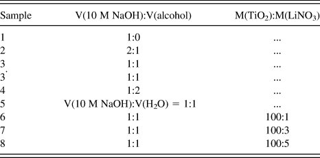
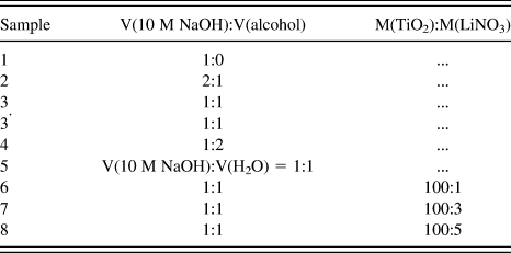
TiO2 nanobelts were fabricated through a solvothermal reaction between NaOH, LiNO3 solution, ethyl alcohol and P25 powders. The detail preparation conditions are shown in Table I. In a typical experiment, 2.00 g P25 was added to 40 mL 10 M NaOH solution under stirring. The turbid liquid was mixed with equal-volume absolute ethyl alcohol and then transferred to a 100 mL Teflon-lined stainless-steel autoclave. The autoclave was sealed and heated at 180 °C for 12 h. After the solvothermal treatment, the sample was immersed in 1 M hydrochloric acid solution for 24 h under stirring. The resulting product was separated by centrifugation, washed with distilled water and absolute ethyl alcohol for several times, respectively, and dried at 80 °C for 6 h. After calcination for 2 h at 400 °C, the TiO2 nanobelts were finally obtained.
The preparation conditions for TiO2 and Li-doped TiO2 nanobelts.
B. Preparation of Li-doped TiO2 nanobelts
The prepared TiO2 nanobelts were mixed with different concentrations of LiNO3 aqueous solution, transferred to a 100 mL Teflon-lined stainless-steel autoclave and then treated in purely deionized water at 160 °C for 6 h to prepare 1%, 3%, and 5% (theoretical concentrations) Li-doped TiO2 nanobelts. After the treatment, the resulting product was separated by centrifugation, washed with distilled water and alcohol for several times, respectively, and dried at 80 °C for 6 h. For comparison, the TiO2 nanobelts without any Li+ ions were also synthesized under the same preparation process (Sample 3’).
C. Evaluation of photocatalytic activity
0%, 1%, 3%, and 5% Li-doped mesoporous TiO2 nanobelts (sample 3’, 6–8) were synthesized for studying their photocatalytic degradation of Malachite green under visible light irradiation. The photocatalytic reactions were carried out at room temperature under atmospheric pressure in a closed reactor system. The rate of photocatalytic degradation was determined in an aqueous solution (200 mL) containing 0.25 g catalyst and 4 mg Malachite green. Before the photocatalytic reactions, the aqueous solution was sonicated for 15 min and then placed in the dark for 1 h. After irradiation under stirring for 0, 0.5, 1, 2, 4, and 8 h, the centrifuged aqueous solution was measured via UV–Vis spectrophotometer to get the corresponding absorbance An (n = 0, 1, 2, 3, 4, 5, 6) at 617 nm which is the maximum absorption wave length of Malachite green. The ratio An/A0 represents the rate of photocatalytic degradation C/C0.
D. Characterization
The structural properties of the samples were analyzed with a Shimadzu XRD-6000 x-ray diffractometer (Tokyo, Japan) using Cu Kα irradiation with a scan rate of 6° per min. Scanning electron microscope (SEM) data were taken on JEOL JSM-6700F (Tokyo, Japan), transmission electron microscopy (TEM) and high-resolution TEM images were measured on JEOL JEM-2010 (Tokyo, Japan) at an acceleration voltage of 200 kV. Fourier transform infrared (FTIR) spectra were measured on Nicolet Avata370. UV–Vis diffuse reflection spectra of samples were recorded on a Lambda 35 UV–Vis scanning spectrophotometer (Perkin Elmer Waltham, MA).
III. RESULTS AND DISCUSSION
A. Structure and phase
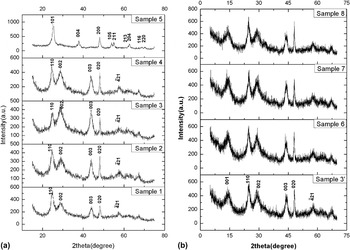
X-ray diffraction (XRD) experiments have been performed to investigate the structure and phase of the as-prepared TiO2 nanobelts. As for sample1-4, 3’ and 6–8 in Fig. 1, the XRD patterns can be well indexed to β-TiO2 (PDF No. 35-0088) which is monoclinic, space group C2/m (12), while that of sample 5 can be well indexed to Anatase (syn, PDF No.84-1286). With the increase in the volume fraction of alcohol, the intensities of sample 1, 2, 4 change a little but no phase transition. The molar concentration of NaOH in sample 3 is about 5 M, which is similar to that of sample 5, however, the crystalline phases are changed from monoclinic (sample 3) to tetragonal (sample 5). Besides, the intensities of (002) and (003) peaks of sample 3 surpass that of (110) and (020) peaks, which is not found in sample 1, 2, and 4. So, we can believe that the alcohol plays a key role in the process of crystal growth and the crystalline phase transition. About this issue, there are two reasons we can take into consideration. On the one hand, in the process of crystal growth, the adsorption of alcohol on some surfaces of TiO2 can reduce the barrier arising from the grains gathering together, which will result in priority growth of some crystal planes. In the case of nanobelts, these planes are {001}. On the other hand, the vapor pressure of alcohol is much larger than that of water at the same temperature. Larger pressure is favorable for the formation of monoclinic β-TiO2, which can be demonstrated by its larger density of 3.99 g/cm3 (the density of Anatase is 3.79 g/cm3). As a result, the appropriate volume fraction of alcohol, 0.5 in Sample 3, is the key factor for the uniform belt-like geometry.
XRD patterns of TiO2 nanobelts (Sample 1–5) and Li-doped TiO2 nanobelts (Sample 3’ 6–8).

From the XRD patterns of sample 3’, 6–8, we can come to the conclusion that the doping of lithium does not cause crystalline phase transition or significant intensity changes.
B. Morphology evolution
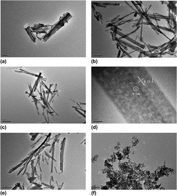
The volume ratio of the reaction medium has a great influence on the morphologies of TiO2 nanostructures. As can be seen from Fig. 2(a), when the medium is 10 M NaOH solution, we get TiO2 nanostructures with granular, linear and lamellar geometries; when alcohol is added, in Fig. 2(b), nanobelts with a length of several micrometers and a width of tens of nanometers are prepared; Figs. 2(c) and 2(d) show the TEM and the corresponding HRTEM images of the uniform TiO2 mesoporous nanobelts obtained under the condition of V (10 M NaOH): V (alcohol) = 1:1. The length is 3–4 μm and the width is 40–200 nm. The shapes and sizes of the pores are also uniform with 5–7 nm in diameter. The lattices are parallelly oriented to the wall with the interlattice distance of 4.12 Å, which is assigned to the interplane distance of β-TiO2 (110). When the volume fraction of alcohol is over 50% [Fig. 2(e)], however, the geometry of TiO2 nanostructures return to be diverse, similar to that of sample 2. Besides the crystalline phase, the geometry of sample 5 is also different from others. Figure 2(f) reveals that the as-prepared TiO2 nanostructures are composed of numerous cymbals-like anatase nanoparticles.
TEM and HRTEM images of TiO2 nanobelts (a). Sample 1, (b). Sample 2, (c) and (d). Sample 3, (e). Sample 4, (f). Sample 5.

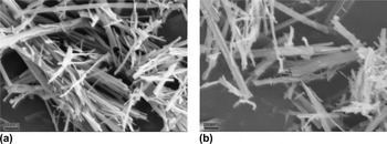
For studying the influence of pressure on the morphologies of TiO2 nanostructures obtained under the condition of V (10 M NaOH): V (alcohol) = 1:1, we reduced the total volume of the reaction medium (VT) to 20 mL and 40 mL. The SEM images of TiO2 nanostructures are shown in Figs. 3(a) and 3(b), respectively. In Fig. 3(a), the vast majority are TiO2 nanowires with a length of 1–2 μm, few are TiO2 nanobelts. However, the proportion of TiO2 nanobelts rises to a considerable amount in Fig. 3(b). When the total volume of the reaction medium is 80 mL, uniform mesoporous TiO2 nanobelts can be acquired, as seen in Fig. 2(c). It seems that the greater the pressure, it is more beneficial to the formation of TiO2 nanobelts.
SEM images of TiO2 nanostructures (a). VT = 20 mL, (b). VT = 40 mL.

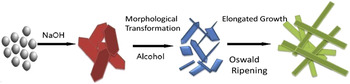
It is generally accepted that NaOH treatment plays important roles in the conversion of titania into 1D nanomaterials (wires, tubes, belts). During the reaction process, titania reacts with NaOH, forming layered alkali titanate having the general formula of M2TinO2n+1(M: Na+ or H+, 3 ≤ n ≤ 6). They have a monoclinic lattice, with parallel kinked layers of edge-sharing TiO6 octahedra. The adsorption of alcohol on the surfaces of TiO2 will change their rates of growth, resulting in priority growth of {001}. The nanostructures under appropriate pressure finally turn into nanobelt geometry. During the washing process, the Na+ cations are replaced gradually by H+ while the morphology remaining satin nanobelts. After being annealed at a high temperature, the layer-like nanobelts dehydrate to form mesoporous β-TiO2 nanobelts. The growth mechanism of TiO2 nanobelts is revealed by Fig. 4.
Growth mechanism of TiO2 nanobelts.

C. Photocatalytic properties
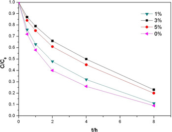
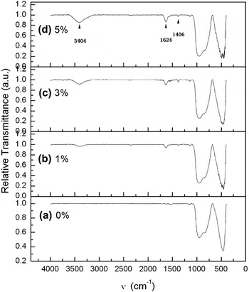
Figure 5 shows the photocatalytic activity of the 0%, 1%, 3%, 5% Li-doped mesoporous TiO2 nanobelts. It is obvious that the doping of lithium reduce the photocatalytic activity of TiO2 nanobelts. The ionic radius of Li+ (0.07 nm) is close to that of Ti4+ (0.061 nm),Reference Bessekhouad, Robert, Weber and Chaoui18 which makes it possible for Li+ to substitute Ti4+ or enter into the lattice space of TiO2. As the doping concentration of lithium increases, more and more lattices of TiO2 are decomposed, resulting in the depression of the photocatalytic activity. It is confirmed by Fig. 6, in which the vibration absorption peaks at 3404 cm−1 and 1642 cm−1 for Ti-O as well as vibration absorption peak at 1406 cm−1 for Li–O increase along with the increase of lithium concentration. On the other hand, the electronegativity of mesoporous TiO2 nanobelts is strengthened as the doping concentration of lithium increases, because Li+ has less positive charges than Ti4+. So it is favorable to adsorb electropositive Malachite green in the solution on the surface of TiO2 nanobelts and thus enhance the photocatalytic activity. That is why the activities of sample 6–8 are lower than that of sample 3’ but the activity of sample 8 is higher than that of sample 7. In summary, the lattice breaking and the electronegativity have an important but contradictory influence on the photocatalytic activity of the mesoporous Li-doped TiO2 nanobelts.
The degradation rate of Malachite green over Li-doped mesoporous TiO2 nanobelts.

The IR spectrums of Li-doped mesoporous TiO2 nanobelts.

IV. CONCLUSIONS
In this work, mesoporous TiO2 and Li-doped TiO2 nanobelts have been synthesized via a facile solvothermal process. The crystalline structure and morphology of the nanobelts strongly depend on the volume ratio of the reaction medium and the pressure. The appropriate volume ratio for the formation of uniform TiO2 nanobelts is 1:1, and greater pressure is also beneficial to the TiO2 nanobelts. The doping of lithium does not change the crystalline phase of the nanobelts but reduces the photocatalytic activity. The accurate quantity of Li+ in the doped TiO2 nanobelts is hard to obtain by the methods applied as discussed in this article; in further research, we plan to get theory support from computer simulation.
ACKNOWLEDGMENTS
The work was supported by the National Natural Science Foundation of China (No.50702053), Natural Science Foundation of Shandong Province (Y2008B46, ZR2009FZ003) and Key Foundation of Qingdao (09-1-3-39-jch, 10-3-4-1-10-jch).