INTRODUCTION
Neospora caninum was originally identified as a Toxoplasma gondii-like parasite that predominantly caused neuromuscular disorders in dogs (Bjerkas and Presthus, Reference Bjerkas and Presthus1989), or abortion and stillbirth in cattle (Dubey and Lindsay, Reference Dubey and Lindsay1993; Gottstein et al. Reference Gottstein, Hentrich, Wyss, Thur, Busato, Stark and Muller1998). N. caninum infection induces cell-mediated immunity, which is an important host defence mechanism. While classical CD8+ T cells and CD4+ T cells are required for the acquisition of protective immunity against N. caninum infection (Marks et al. Reference Marks, Lundén, Harkins and Innes1998; Baszler et al. Reference Baszler, Long, McElwain and Mathison1999; Nishikawa et al. Reference Nishikawa, Tragoolpua, Inoue, Makala, Nagasawa, Otsuka and Mikami2001b, Reference Nishikawa, Zhang, Ibrahim, Yamada, Nagasawa and Xuan2010), different types of innate cells such as macrophages, natural killer (NK) cells and natural killer T (NKT) cells exert distinct roles during this immune response. However, in order to control host immunity, N. caninum can infect different immune cell types including NK cells, macrophages and dendritic cells (DC) (Boysen et al. Reference Boysen, Klevar, Olsen and Storset2006; Hemphill et al. Reference Hemphill, Vonlaufen and Naguleswaran2006; Strohbusch et al. Reference Strohbusch, Müller, Hemphill, Margos, Grandgirard, Leib, Greif and Gottstein2009). Therefore, N. caninum can modulate cell migration to enhance parasitic invasion and proliferation during the early phases of infection. Previous studies have suggested that N. caninum might possess an ability to enhance interferon-gamma (IFN-γ) production and CC-chemokine receptor 5 (CCR5)-dependent cell migration (Tuo et al. Reference Tuo, Fetterer, Jenkins and Dubey2005b; Mineo et al. Reference Mineo, Oliveira, Silva, Oliveira, Abatepaulo, Ribeiro, Ferreira, Mineo and Silva2010).
Toxoplasma gondii, which is closely related to N. caninum, stimulates interleukin-12 (IL-12) production and cell migration via a unique pathway that involves the triggering of CCR5 in DCs and macrophages by secreted T. gondii cyclophilin 18 (TgCyp18) (High et al. Reference High, Joiner and Handschumacher1994; Aliberti et al. Reference Aliberti, Valenzuela, Carruthers, Hieny, Andersen, Charest, Reis e Sousa, Fairlamb, Ribeiro and Sher2003; Ibrahim et al. Reference Ibrahim, Bannai, Xuan and Nishikawa2009, Reference Ibrahim, Xuan and Nishikawa2010). Cyclophilins are ubiquitous cytosolic proteins and have been described in both prokaryotic and eukaryotic organisms (Hamilton and Steiner, Reference Hamilton and Steiner1998). Cyclophilins possess peptidyl-prolyl cis-trans isomerase (PPIase) activity and high binding affinity to cyclosporin, an immunosuppressant drug (Handschumacher et al. Reference Handschumacher, Harding, Rice, Drugge and Speicher1984). TgCyp18-mediated IL12 production and cell migration were also blocked by cyclosporin A (CsA) (Aliberti et al. Reference Aliberti, Valenzuela, Carruthers, Hieny, Andersen, Charest, Reis e Sousa, Fairlamb, Ribeiro and Sher2003; Ibrahim et al. Reference Ibrahim, Bannai, Xuan and Nishikawa2009, Reference Ibrahim, Xuan and Nishikawa2010).
Tuo et al. (Reference Tuo, Fetterer, Davis, Jenkins and Dubey2005a) identified a potential Neospora cyclophilin (NcCyp) candidate from tachyzoite lysate using mass spectrometry that was associated with T-cell antigenic stimulatory activity. The ability of excreted/secreted antigens from N. caninum to attract monocytic cells to the site of primary infection was also reported (Mineo et al. Reference Mineo, Oliveira, Silva, Oliveira, Abatepaulo, Ribeiro, Ferreira, Mineo and Silva2010). These activities were inhibited by CsA (Tuo et al. Reference Tuo, Fetterer, Jenkins and Dubey2005b; Mineo et al. Reference Mineo, Oliveira, Silva, Oliveira, Abatepaulo, Ribeiro, Ferreira, Mineo and Silva2010), suggesting the presence of NcCyp. A cyclophilin-like protein (Gene Accession no. CF422590) was identified by mass spectrometric analysis (Tuo et al. Reference Tuo, Fetterer, Jenkins and Dubey2005b). Therefore, the aim of this study was to examine the NcCyp activity of cell migration to better understand parasite and host interactions.
MATERIALS AND METHODS
Parasite and cell cultures
N. caninum (NC1 isolate) tachyzoites were propagated in monkey kidney adherent fibroblasts (Vero cells) cultured in Eagle's minimum essential medium (EMEM, Sigma, St Louis, USA) supplemented with 8% heat-inactivated fetal bovine serum (FBS). Purification of tachyzoites was conducted as previously described (Nishikawa et al. Reference Nishikawa, Xuan, Nagasawa, Igarashi, Fujisaki, Otsuka and Mikami2000a). The final pellet was resuspended in cold phosphate-buffered saline (PBS) and passed through a 27-gauge needle and a 5 μm-pore filter (Millipore, Bedford, USA).
Animals
Female C57BL/6J and BALB/c mice (6–8 weeks of age) and a female Japanese white rabbit were obtained from Clea Japan (Tokyo, Japan). CCR5 knockout mice (CCR5−/−, B6.129P2-Ccr5tm1Kuz/J, Stock No. 005427) were purchased from the Jackson Laboratory (Bar Harbor, Maine, USA). Animals were housed under specific pathogen-free conditions in the animal facility of the National Research Center for Protozoan Diseases at the Obihiro University of Agriculture and Veterinary Medicine, Obihiro, Japan. Animals used in this study were treated and used according to the Guiding Principles for the Care and Use of Research Animals published by the Obihiro University of Agriculture and Veterinary Medicine.
Three-month old, male Holstein cattle (n=2) were housed at the Animal Research Center, Hokkaido Research Organization (Shintoku, Hokkaido, Japan). To confirm any evidence of N. caninum infection, serological testing using an enzyme-linked immunosorbent assay (ELISA) was performed as described previously (Huang et al. Reference Huang, Liao, Zhang, Lee, Nishikawa and Xuan2007).
Construction and expression of recombinant NcCyp
Complementary deoxyribonucleic acid (cDNA) was synthesized from ribonucleic acid isolated with TRI reagent (Sigma) using a SuperScript™ First-strand Synthesis System for reverse transcription-polymerase chain reaction (RT-PCR) (Invitrogen, Carlsbad, USA). cDNA was used as a template to amplify the coding region of wild-type NcCyp. NcCyp DNA and amino acid sequences were submitted to the GenBank database (Accession number CF422590). To clone the recombinant NcCyp without the signal peptide (from amino acids 1 to 17) that consisted of 161 amino acids (aa), a set of oligonucleotide primers that included a BamH I restriction enzyme site in the forward primer (5′-TAG GAT CCA TGG AAA ACG CCG GAG TCC AG-3′) and an EcoR I site in the reverse primer (5′-GCG AAT TCT TAC AAC AAA CCA ATG TCC GT-3′), were designed. PCR products were digested with BamH I and EcoR I and then ligated into the glutathione S-transferase (GST)-fusion protein in the Escherichia coli expression vector pGEX-4T1 (GE Healthcare, Buckinghamshire, UK), which had been digested with the same set of restriction enzymes (pGEX-NcCyp). Plasmid nucleotide sequences were determined using an ABI 3100 DNA sequencer (Applied Biosystems, Foster City, CA, USA). Recombinant NcCyp was expressed as glutathione S-transferase (GST) fusion proteins in the E. coli DH5α strain (Takara Bio Inc., Japan). GST tags of the recombinant proteins were removed with thrombin protease (GE Healthcare) according to the manufacturer's instructions.
Recombinant NcCyp (rNcCyp) was dialysed in PBS and purified with Detoxi-Gel™ Endotoxin Removing Gel (Pierce Biotechnology Inc., Rockford, USA) to remove endotoxins. Proteins used for cell culture were filtered using a 0·45 μm low protein binding Supor membrane (Pall Life Sciences, Ann Arbor, MI, USA). Protein purity and quantity were detected as a single band with sodium dodecyl sulfate polyacrylamide gel electrophoresis (SDS-PAGE) followed by Coomassie Brilliant Blue R250 staining (MP Biomedicals Inc., France). Protein concentration was measured using a BCA protein assay kit (Pierce Biotechnology Inc.).
Production of anti-rNcCyp serum and purification of immunoglobulin-G (IgG)
Fifty micrograms of rNcCyp in Freund's complete adjuvant (Sigma) were intraperitoneally injected into 6-week-old female BALB/c mice (n=5) on day 0. rNcCyp in Freund's incomplete adjuvant (Sigma) was also intraperitoneally injected into the mice on days 14, 28, and 42. Anti-rNcCyp sera were collected from the mice at day 56. Serum that exhibited a high sensitivity and specificity to N. caninum was used in this study. Three hundred micrograms of the rNcCyp in Freund's complete adjuvant were also intradermally injected into a female Japanese white rabbit on day 0. rNcCyp in Freund's incomplete adjuvant was further intradermally injected into the rabbit on days 14, 28, and 42. IgG was purified from 2 ml of serum collected 7 days after the last immunization using Protein A Chromatograhy Columns (Bio-Rad Laboratories, CA, USA) following the manufacturer's instructions. Fractions containing IgG were pooled and resolved by SDS-PAGE to test protein purity and quantity. Protein concentrations of purified rabbit anti-rNcCyp polyclonal IgG were measured using a BCA protein assay kit (Thermo Fisher Scientific, Inc. Rockford, USA).
Indirect fluorescent antibody test (IFAT)
NcCyp localization was confirmed by microscopic examination of N. caninum-infected Vero cells on coverslips. Coverslips were collected 24 h after parasite inoculation, washed twice with PBS supplemented with 1 mm CaCl2 and MgCl2 (PBS++), and then fixed with 3% paraformaldehyde (PFA) in PBS++. After washing twice with PBS++, the cells were permeabilized with 0·3% Triton X-100 in PBS++ for 5 min at room temperature. After washing, the coverslips were incubated with 3% bovine serum albumin (BSA) in PBS++ at room temperature for 30 min. Coverslips were incubated with anti-rNcCyp mouse serum diluted at 1:100 in 3% BSA in PBS++ for 1 h at room temperature. After washing 3 times with PBS++, the coverslips were incubated with Alexa fluor 488-conjugated goat anti-mouse IgG (Sigma) diluted at 1:400 in 3% BSA in PBS++ for 1 h at room temperature and then washed again. The coverslips were placed on a glass slide coated with Mowiol (Calbiochem, San Diego, USA) and examined using a fluorescence microscope (Nikon, Tokyo, Japan).
Sample preparation of secreted NcCyp
NcCyp secretion from extracellular parasites was determined as described previously (Ibrahim et al. Reference Ibrahim, Xuan and Nishikawa2010). Purified N. caninum tachyzoites (3×10⁶) were kept in 250 μl of GIT medium (Nihon Pharmaceutical Co., LTD, Tokyo, Japan) on ice. Before parasite suspensions were transferred from ice to 37°C for the secretion assay, samples were removed and processed (designated as the time zero reading). The parasite suspension was then incubated at 37°C in a water bath. After 15, 30, 60 and 120 min, the parasite suspensions were centrifuged sequentially (760 g for 10 min at 4°C then 7000 g for 10 min at 4°C), and the supernatant fractions were subjected to a sandwich ELISA.
Sandwich ELISA for the detection of secreted NcCyp
Rabbit anti-rNcCyp IgG was diluted to 20 μg/ml in a 0·05 m carbonate buffer (pH 9·6). One microgram of the IgG per well was used as the capture antibody to coat microtitre plates at 4°C overnight. Blocking was performed with a blocking solution (3% skimmed milk in PBS) at 37°C for 2 h. Plates were incubated at 37°C for 30 min with each supernatant in quadruplicate. After washing 6 times with a washing solution (0·05% Tween 20 in PBS), anti-rNcCyp mouse serum diluted at 1:500 in blocking solution was added to each well and the plate was incubated at 37°C for 1 h. After a further 6 washes, the plates were incubated with HRP-conjugated goat anti-mouse IgG (Amersham Pharmacia Biotech. Uppsala, Sweden) diluted at 1:10 000 in blocking solution at 37°C. Binding was visualized with substrate solution (0·3 mg/ml 2,2′-azino-bis(3-ethlbenzothiazoline-6-sulphonic acid), 0·1 m citric acid, 0·2 m sodium phosphate, 0·003% H2O2). Absorbance at 415 nm was measured using a MTP-500 microplate reader (Corona Electric, Tokyo, Japan). NcCyp concentration for each sample was calculated by standardization with rNcCyp.
Isolation of mononuclear cells
Isolation of spleen cells was conducted as previously described (Nishikawa et al. Reference Nishikawa, Kousaka, Fukumoto, Xuan, Nagasawa, Igarashi, Fujisaki, Otsuka and Mikami2000b). Naïve C57BL/6J and CCR5−/− mice were sacrificed and their spleens were aseptically removed. A single cell suspension was prepared from macerated spleen tissue and haemolysed in lysing buffer (0·83% NH4Cl, 0·01 m Tris-HCl, pH 7·2). After washing with PBS, mixed spleen cells from 5 mice were then suspended in RPMI 1640 medium. Blood taken from the cattle was treated with sodium citrate and was then centrifuged at 1000 g for 30 min at room temperature with the brake off. Buffy coat fractions were suspended in RPMI 1640 medium. Mononuclear cells were isolated from spleen cells and the buffy coat using HISTOPAQUE®-1077 (Sigma) according to the manufacturer's instructions.
Cell migration assays
Cell migration was assessed using 24-well chemotaxis chambers and inserts with 8 μm pore polycarbonate filters (Falcon, Beckton Dickinson, Franklin Lakes, NJ, USA). Mononuclear cells (1×10⁶) suspended in 0·4 ml of RPMI 1640 supplemented with 10% heat-inactivated FBS, were applied to the upper compartment of the chamber. In the lower chamber, rNcCyp in 1·2 ml of RPMI 1640 supplemented with 10% heat-inactivated FBS was added as a chemoattractant. Chambers were incubated at 37°C for 24 h. Cells that migrated into the lower compartment were counted by microscopy. Cyclosporin A (CsA) from Tolypocladium inflatum (Sigma, 1 mm stock solution in 0·01% ethanol/RPMI 1640 medium) was used to inhibit rNcCyp activity. Each treatment was performed in triplicate wells. Results were expressed as the percentage of mononuclear cells in the lower compartment in relation to the total number of mononuclear cells used in the experiment as follows: Percentage of migrating cells=(number of cells in the lower chambers/total cell number)×100.
Clinical findings of N. caninum-infected mice
Ten-week old, female BALB/c mice (n=5) were interperitoneally infected with 106N. caninum tachyzoites. Two mice exhibited clinical signs of neosporosis such as head tilting, limb paralysis, circling motion and febrile response (stary stiff coat). In addition, a decrease in body weight of 10–15% was observed. At 30 days after the infection, the mice were sacrificed and their brains extracted for immunohistochemistry (IHC).
IHC
After fixation in 10% formalin solution, brain tissues were routinely embedded in paraffin wax and sectioned (4 μm) serially. Sections were stained with haematoxylin and eosin (HE). N. caninum IHC was performed with anti-N. caninum goat serum (210-70 NC, VMRD Inc., Pullman, WA, USA) as the primary antibody, and a secondary antibody conjugated with horseradish peroxidase-labelled streptavidin biotin (LSAB™+ Kit, Universal, Dako, USA). Briefly, after deparaffinization, tissue sections were subjected to antigen retrieval with pronase (AR6542-01, ImmunoBioScience Corp, Mukilteo, USA), and then were blocked for endogenous peroxidase with 3% hydrogen peroxide and nonspecific protein binding with 2% BSA. Sections were subsequently incubated with the primary antibody diluted 1:2000 for 45 min at 37°C. After washing, sections were incubated with the biotin-conjugated second antibody, followed by horseradish peroxidase-labelled streptavidin, each for 20 min at 37°C. To absorb the activity of mouse immunoglobulin in the secondary antibody, normal mouse serum (X0910, Dako) was added to the biotin-conjugated secondary antibody at a ratio of 1:10 and incubated for 45 min at room temperature before it was used according to the manufacturer's instructions. Chromogen was developed with 3,3′-diaminobenzidine (DAB; Impact DAB®, Vector Laboratories Inc., Burlingame, USA).
IHC for NcCyp was performed with anti-rNcCyp rabbit serum as the primary antibody, and a secondary antibody conjugated with horseradish peroxidase-labelled polymer (Envision+ kit, Dako). Normal rabbit serum was used as a negative control. Briefly, after deparaffinization, tissues were placed in TE buffer (10 mm Tris, 1 mm EDTA, pH 9·0) and heated with a microwave oven (200 W for 5 min, twice) for antigen retrieval. After blocking of endogenous peroxidase and nonspecific protein binding with 10% normal goat serum (Histofine SAB-PO kit, Nichirei Corp, Tokyo, Japan) for 30 min at room temperature, sections were incubated with the primary antiserum diluted at 1:4000 for 45 min at 37°C. After washing, sections were incubated with the secondary antibody for 40 min at 37°C. Chromogen was developed as described above.
Statistical analysis
Statistical significance was calculated as a comparison to the medium control (0 μg/ml) group using one-way ANOVA followed by Tukey's multiple comparison tests. Values of P<0·05 were considered significant (*).
RESULTS
Expression and secretion of NcCyp
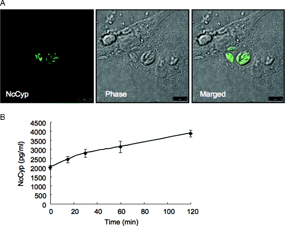
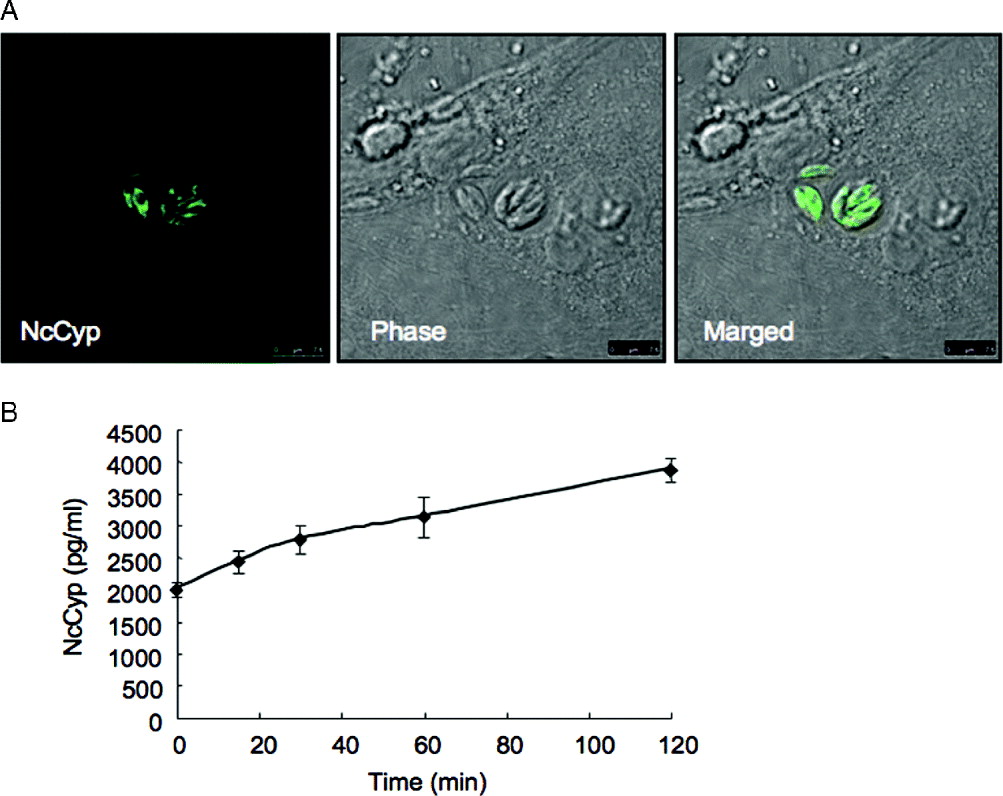
To confirm the localization of native NcCyp in N. caninum, we conducted IFAT using anti-rNcCyp serum. The entire parasite body was faintly recognized (Fig. 1A), indicating that the protein was located within the cytoplasm. Even after proliferation of the parasites, the localization of the protein did not change substantially. Next, we further performed sandwich ELISA to detect the release of NcCyp from extracellular parasites (Fig. 1B). Extracellular tachyzoites released NcCyp in a time-dependent manner. NyCyp was detectable at time 0, suggesting constitutive release of NcCyp during the sample preparation.
(A) IFAT images of NcCyp. Neospora caninum-infected Vero cells on coverslips fixed and immunostained with anti-Cyp18 mouse serum and Alexa fluor 488-conjugated goat anti-mouse IgG. (B) NcCyp release from extracellular parasites. After 15, 30, 60 and 120 min incubations, extracellular parasite supernatants were prepared and subjected to sandwich ELISA. Each value represents the mean±the standard deviation of quadruplicate samples. Results are representative of 2 repeated experiments with similar results.
NcCyp-induced cell migration in a CCR5-dependent manner
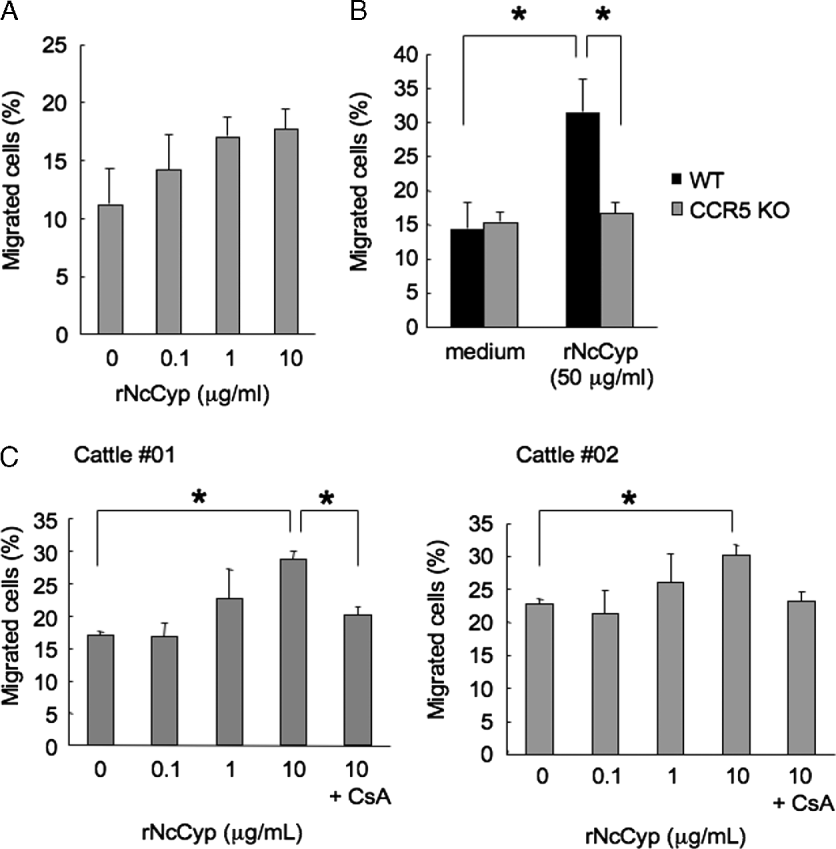
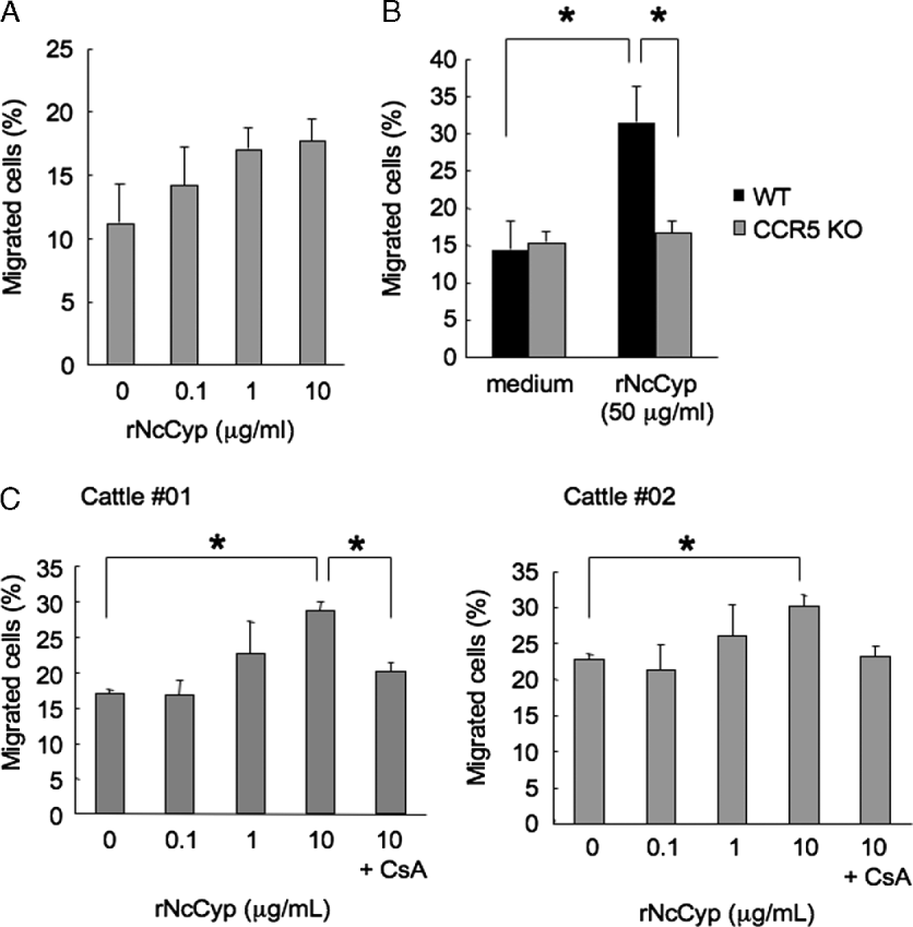
To investigate the ability of NcCyp to chemoattract mononuclear cells and spleen cells of mice, cells were placed in the upper chemotaxis chamber and permitted to migrate to a different rNcCyp concentration (Fig. 2A and B). rNcCyp elicited a typical chemotactic response compared to the negative control (no rNcCyp). To determine the importance of the CCR5 receptor for the chemotactic effect of rNcCyp on spleen cells, we evaluated the ex vivo migration of mouse wild-type and CCR5−/− cells. Significant migratory responses were observed after incubation of wild-type cells with rNcCyp (P<0·05) as compared to the control (Fig. 2B). In contrast, CCR5−/− cells exhibited an absence of migratory responses upon incubation with rNcCyp. Moreover, we tested the chemotactic response of rNcCyp to bovine peripheral blood mononuclear cells (PBMCs) (Fig. 2C). rNcCyp exhibited the ability to chemoattract PBMCs in a dose-dependent manner. Moreover, rNcCyp migratory responses were blocked by CsA and suggested CCR5 participation within bovine PBMCs.
Effect of rNcCyp on spleen cell and PBMC migration. (A) Spleen cells (1×10⁶) mixed from 5 wild-type mice were applied to the upper compartment of a chemotaxis chamber. In the lower compartment, serial concentrations of rNcCyp (0, 1, and 10 μg/ml) were added as chemoattractants. (B) Spleen cells (1×10⁶) mixed from 5 wild-type or 5 CCR5−/− mice were applied to the upper compartment of a chemotaxis chamber. In the lower compartment, 50 μg/ml rNcCyp was added as a chemoattractant. (C) PBMCs (1×10⁶) from 2 cows (#01 and #02) were applied to the upper compartment of a chemotaxis chamber. In the lower compartment, serial concentrations of rNcCyp (0·1, 1, and 10 μg/ml) and 1 μ m cyclosporin A (CsA) were added. Chambers were incubated at 37°C for 24 h. Cells that migrated into the lower compartment were counted by microscopy. The percentage of cell migration represents the mean±the standard deviation of triplicate wells.
IHC using anti-N. caninum and anti-rNcCyp antibodies
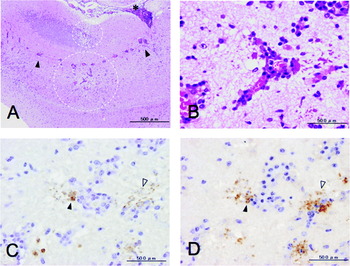
To detect NcCyp in N. caninum infected hosts, we performed IHC on the brain of the two mice infected with neosporosis using an anti-rNcCyp antibody. Most major brain lesions included multifocal necrosis, malacia and inflammatory cell infiltration that consisted of macrophages, lymphocytes and polymorphonuclear cells. Perivascular cuffing was frequently observed, with varying degrees of mononuclear cell infiltration within the perivascular space and meninges (Fig. 3A and B). IHC for N. caninum tachyzoites revealed that most tachyzoites were located within or near the lesions; in particular, clusters of tachyzoites and free extracellular tachyzoites were observed adjacent to vessels with perivascular cuffing (Fig. 3C). NcCyp was consistently detected in the tachyzoites distributed within or around the lesions (Fig. 3D).
Immunohistochemistry of mouse brain tissue infected with Neospora caninum. At 30 days after the infection, the two mice that exhibited signs of neosporosis were sacrificed and their brains extracted. (A) HE staining. Focal malacia (dotted line circles), cuffing (arrowheads), and mononuclear cell infiltration into the meninges (asterisk) were observed. (B–D) Serial sections of mouse brain infected with N. caninum. (B) Higher magnification of (A). Mononuclear cells infiltrated in perivascular space. (C) Immunohistochemistry with anti-N. caninum goat serum. Clusters of tachyzoites (black arrowhead) and extracellular tachyzoites (open arrowhead) were observed adjacent to vessels. (D) Immunohistochemistry with anti-rNcCyp rabbit serum. NcCyp was consistently detected in tachyzoite clusters (black arrowhead) and extracellular tachyzoites (open arrowhead). No positive staining was detected in negative control rabbit serum. Results are representative of 1 mouse, and similar results were observed for the second mouse.

DISCUSSION
An important function of cyclophilin is its ability to induce cell migration. Mineo et al. (Reference Mineo, Oliveira, Silva, Oliveira, Abatepaulo, Ribeiro, Ferreira, Mineo and Silva2010) reported that N. caninum excreted/secreted antigens triggered cell migration in a CCR5-dependent manner. In our study, rNcCyp possessed the ability to trigger CCR5-dependent cell migration although it lacked the important 17GEH19 domain that is necessary for interactions with CCR5. Our previous study reported that the recombinant TgCyp18 mutant, which lacks a CCR5 binding domain due to changes in amino acids (17GEH19 to 17AAA19 and 149RP150 to 149YV150) located in the N and C termini of the protein (Yarovinsky et al. Reference Yarovinsky, Andersen, King, Caspar, Aliberti, Golding and Sher2004), showed significant migratory responses (Ibrahim et al. Reference Ibrahim, Xuan and Nishikawa2010). These results are consistent with our present data for NcCyp. Furthermore, NcCyp might enhance partial binding with receptors other than CCR5. This partial binding might be responsible for the migratory properties of the TgCyp18 mutant and NcCyp.
Cell migration induced by high NcCyp doses (μg/ml order) in our study is potentially questionable because 3·85±0·02 ng/ml NcCyp was detected from 3×10⁶ tachyzoites after a 120 min incubation. We consequently interpreted that the activity of our recombinant NcCyp protein relative to that of native NcCyp might be reduced. Therefore, higher doses of rNcCyp expressed in E. coli would have been equivalent to lower doses of native protein with high activity. In the case of TgCyp18, low IL-12-inducing activity of recombinant TgCyp18 was observed compared to the native TgCyp18 in the soluble tachyzoite extract (Aliberti et al. Reference Aliberti, Valenzuela, Carruthers, Hieny, Andersen, Charest, Reis e Sousa, Fairlamb, Ribeiro and Sher2003). Such reduced activity of recombinant TgCyp18 expressed in E. coli relative to native TgCyp18 resulted from a missing co-factor rather than improper expression (Aliberti et al. Reference Aliberti, Valenzuela, Carruthers, Hieny, Andersen, Charest, Reis e Sousa, Fairlamb, Ribeiro and Sher2003). Thus, these lower levels compared to the doses used in our study suggest that exceedingly high numbers of tachyzoites are required to secrete a threshold dose of NcCyp capable of having effects on cell migration. Therefore, NcCyp might exhibit paracrine effects (locally or systemically) at the site of infection.
Our study indicated that NcCyp is a cytosolic protein that is released from motile tachyzoites in a time-dependent manner. Previous studies also suggest that active secretion of TgCyp18 is both time and temperature-dependent (Aliberti et al. Reference Aliberti, Valenzuela, Carruthers, Hieny, Andersen, Charest, Reis e Sousa, Fairlamb, Ribeiro and Sher2003; Ibrahim et al. Reference Ibrahim, Xuan and Nishikawa2010). We subsequently investigated the antigenicity of NcCyp in mice and cattle because N. caninum was found to release NcCyp. Although antibodies against N. caninum surface antigen 1 (NcSAG1) were produced in mice and cattle after becoming infected with this parasite, antibodies against NcCyp were not detectable in both animals (data not shown). This result suggests that NcCyp has a low antigenicity and is not inhibited by specific antibodies in infected host animals.
In conclusion, NcCyp production potentially functions as a survival strategy for N. caninum, as it is beneficial for a parasite not to kill its host. Our data suggested that N. caninum, through producing NcCyp, has the ability to recruit immune cells to the site of infection, invade these cells and subsequently take advantage of their migratory properties. Migratory immune cells, such as lymphocytes, then attack parasite-infected cells, thereby potentially enhancing the spread of parasites into additional host cells, as is the case for T. gondii infection (Persson et al. Reference Persson, Agnarson, Lambert, Hitziger, Yagita, Chambers, Barragan and Grandien2007). This is further supported by reports that treatment of N. caninum-infected cells with IFN-γ induced host-cell apoptosis (Nishikawa et al. Reference Nishikawa, Mishima, Nagasawa, Igarashi, Fujisaki, Otsuka and Mikami2001a, Reference Nishikawa, Makala, Otsuka, Mikami and Nagasawa2002). In addition, the parasite may manipulate lymphocyte migration to facilitate infection and protect the host. Thus, N. caninum survival may be dependent upon the balance between parasite immune evasion and the host immune response. Our data therefore provide new evidence for the existence of complex parasite-host interactions via the function of NcCyp.
ACKNOWLEDGEMENT
We thank Dr Dubey (United States Department of Agriculture, Agriculture Research Service, Livestock and Poultry Sciences Institute, and Parasite Biology and Epidemiology Laboratory) for the gift of Neospora caninum, NC-1 isolate.
FINANCIAL SUPPORT
This research was supported by the Japan Society for the Promotion of Science (JSPS) through the ‘Funding Program for Next Generation World-Leading Researchers (NEXT Program)’, initiated by the Council for Science and Technology Policy (CSTP) (2011/LS003) and by the Ministry of Education, Culture, Sports, Science and Technology, Grant-in-Aid for Young Scientists (B) (2009-2010/21780262).