NEOSPORA CANINUM, NEOSPOROSIS AND HOST-PARASITE INTERACTIONS
Neospora caninum (Apicomplexa: Eimeriina: Sarcocystidae) was initially reported as an unidentified protozoan in dogs with encephalomyelitis and myositis (Bjerkas et al. 1984). Dubey et al. (1988a,b) were the first to isolate and name the parasite. Meanwhile, N. caninum infection has been reported in various species of livestock, including sheep, goats, horses and deer (reviewed by Dubey and Lindsay, 1996; Hemphill, 1999; Dubey, 2003). However, most importantly, the current evidence strongly suggests that infection with N. caninum represents a major cause of reproductive failure in cattle worldwide (Hemphill and Gottstein, 2000; Dubey et al. 2002; Gondim et al. 2005; Innes et al. 2005). The exact phylogenetical relationship of N. caninum to other members of the Apicomplexa has been, and still is, under controversy (Tenter and Johnson, 1997; Dubey et al. 2002; Heydorn and Mehlhorn, 2002). McAllister et al. (1998) were the first to show that the dog is a definitive host for N. caninum, and this was later confirmed by Lindsay et al. (1999b). Gondim et al. (2004) demonstrated that coyotes also shed oocysts, indicating that other final hosts cannot be ruled out.
There are different routes of transmission. One relevant route, but probably not the most important one, is through the oral uptake of sporozoite-containing oocysts (Trees et al. 2002; Gondim et al. 2005). Infection of an immunocompetent host with oocysts will not cause clinical disease, but liberated sporozoites, by analogy with other coccidians such as Toxoplasma gondii, are thought to infect the intestinal tissue, cross the epithelium, reach blood and lymphatic vessels, and infect other nucleated cells, including macrophages and lymphocytes. In the initial phases of the infection, parasites disseminate throughout the body, and transform to the rapidly proliferating tachyzoite stage. Subsequently, the stress caused by the host immune response is believed to be one of the factors that trigger stage conversion to the slowly proliferating N. caninum bradyzoites (reviewed by Buxton et al. 2002; Lyons et al. 2002). Bradyzoites represent a quiescent stage of the parasite, which forms intracellular tissue cysts, surrounded by a cyst wall that protects the parasite from immunological and physiological reactions on part of the host. N. caninum tissue cysts have been identified almost exclusively within the central nervous system, with few exceptions (Peters et al. 2001). Bradyzoites can survive within a latently infected, but immunocompetent, animal for many years without causing any clinical signs. However, during pregnancy bradyzoites can get reactivated, and the partial immune-incompetence of a pregnant dam, namely the decrease of an efficient Th-1 response and shift to Th-2, leads to limited suppression of the cell-mediated immunity which normally keeps tachyzoite proliferation in check (Innes et al. 2000, 2002, 2005; Quinn et al. 2002). This can contribute to the reactivation of bradyzoites and re-conversion to tachyzoites, and to infection of the placenta and possibly the fetus with N. caninum tachyzoites. Transmission of N. caninum from mother to fetus in utero is highly efficient, and acute fetal neosporosis is a major cause for abortion, stillbirth, or at least clinical or subclinical disease in newborn calves (Williams et al. 2000; Trees et al. 2002; Gondim et al. 2005). Vertical transmission in cattle can occur over successive pregnancies, and in cases where congenitally infected but clinically healthy heifers are used for breeding, parasite spreading within a herd can take place very efficiently (Björkman et al. 1996). In the USA and the EU, neosporosis is reported as the leading cause of abortions in cattle (Hemphill and Gottstein, 2000; Dubey et al. 2002).
Since its discovery, the realization of the economical significance of this parasite has lead to increased efforts in elucidating how N. caninum interacts with its host. These interactions occur on two levels. On the cellular level, N. caninum can only survive, proliferate and proceed during most stages of its life-cycle as an intracellular parasite. Thus the processes which lead to host cell invasion and intracellular development are of crucial importance (Hemphill, 1999; Hemphill et al. 2004). On the other hand, it is the complex relationship with the host immune system, which decides the fate of this parasite once it enters the host organism. In this review we will focus on the use of cell culture approaches and laboratory animals to elucidate how N. caninum interacts with its host on the cellular and immunological level.
NEOSPORA CANINUM AND ITS INTERACTIONS WITH THE HOST CELL
N. caninum, similar to T. gondii, is capable of actively invading a large variety of target cells. This process has been initially investigated in vitro using bovine aorta endothelial cell monolayers (Hemphill et al. 1996), and later for many other cell types (reviewed by Hemphill et al. 2004). While human cells or cell lines such as human foreskin fibroblasts, Hela cells and Caco2 (colon cancer) cells are readily infected in vitro, there is no conclusive evidence that N. caninum actually infects humans (Peterson et al. 1999; Tranas et al. 1999; Graham et al. 1999; Dubey, 2003; Omata et al. 2005).

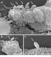
Fig. 1. Scanning electron microscopy of Neospora caninum tachyzoites in the process of invading Vero host cells. (A) Parasites are establishing contact with their host cells, and once dedicated to invade their host cells, they rearrange themselves perpendicularly to the host cell surface membrane (B), and move into the host cell with the apical end first (C).
Surface constituents of N. caninum tachyzoites
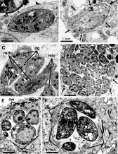
The first step in the physical relationship between the parasite and the host cell is the establishment of a low-affinity contact between tachyzoite and host cell surface membrane, followed by the actual adhesion process, namely a more stable association between tachyzoites and the host cell surface (Fig. 1). In order to initiate host cell invasion, tachyzoites re-orientate themselves perpendicularly to the host cell surface membrane, and enter the host cell cytoplasm, by advancing anterior end first, until they are located in the cytoplasm, enclosed by a parasitophorous vacuole (PV) (Fig. 2A, D). Invasion is an active process requiring metabolic energy solely on the part of the parasite, but not on the part of the host cell. This is highlighted by earlier findings that N. caninum can even infect formaldehyde-fixed host cells (Hemphill et al. 1996). However, by far not all N. caninum tachyzoites adhering to the host cell surface will actually achieve host cell entry (Naguleswaran et al. 2003). Thus, specific signals and/or receptor-ligand interactions are required that enable tachyzoites to exploit their invasive capacity, and it will be a challenge in the future to elucidate those processes involved. Recently, comparative studies on host cell invasion by T. gondii and N. caninum using dog fibroblasts, cat kidney cells and Vero cells showed that T. gondii invaded all cell types with greater efficiency (Lei et al. 2005a).
On the molecular level, the initial low-affinity host-parasite contact is mediated, at least in part, through the 2 major immunodominant surface antigens of N. caninum tachyzoites, NcSAG1 and NcSRS2, which are both inserted into the plasma membrane by a GPI-anchor (Sonda et al. 1998; Howe et al. 1998; Schares et al. 2000). Polyclonal and monoclonal antibodies directed against these 2 surface antigens inhibit host cell adhesion and invasion (Hemphill, 1996; Nishikawa et al. 2000; Haldorson et al. 2005). Monoclonal antibodies directed against a 73 kDa Neospora surface antigen have also been shown to inhibit host cell invasion (Uchida et al. 2004). Specific in vivo-metabolic labelling of GPI-anchored proteins in N. caninum tachyzoites using radioactive 3H-ethanolamine identified at least 7 protein bands carrying a GPI anchor, the most prominent of which largely co-migrated with NcSRS2 and NcSAG1, and the others were of 80 and 100 kDa (Fuchs et al. 1999). Thus there remain a number of surface antigens to be investigated. A possible approach to this has been undertaken by Lei et al. (2005b), who isolated pellicle and plasmalemma fractions of N. caninum tachyzoites using biochemical subcellular fractionation. They found that pellicle fractions contained several major proteins, and analyses of plasmalemma of N. caninum revealed the presence of 2 abundant proteins in addition to other lower abundance antigens, some of which were detectable by monoclonal antibodies (Lei et al. 2005b).
The surface of N. caninum tachyzoites exhibits considerable differences from T. gondii with regard to surface carbohydrate content. Baszler et al. (1996) were the first to demonstrate that the surface of N. caninum is glycosylated, and they showed that a monoclonal antibody directed against a periodate-sensitive epitope on a 65 kDa surface protein is useful for serological diagnosis of N. caninum infection by competitive ELISA (Baszler et al. 2001). Subsequently, Hemphill et al. (1997) reported that NcSAG1-epitopes recognized by polyclonal anti-SAG1 antibodies could be glycosylated. Fuchs et al. (1999) then demonstrated the presence of surface carbohydrates on N. caninum tachyzoites by ruthenium red labelling, and the absence of glycans on the T. gondii surface. The same group showed that glycoproteins largely co-migrated with GPI-anchored proteins following SDS-PAGE separation. In addition, they used a panel of lectins for the identification of surface-associated glycoproteins in N. caninum tachyzoites, and showed that ConA stained the surface of N. caninum, but not of T. gondii tachyzoites. ConA binding sites were found to be localized to the surface and the dense granules in N. caninum tachyzoites. Therefore, an important feature that defines the differences between N. caninum and T. gondii could be post-translational modifications. The presence of carbohydrate modifications on antigenic proteins could potentially have implications regarding host-parasite interactions, especially with regard to masking of epitopes, and the immunological consequences thereof. Glycosylation could thus serve as a possible explanation for the lack of extensive cross-antigenicity between the two closely related species.
Secretory proteins and proteases
Once a low-affinity contact between N. caninum and its host cell is achieved, N. caninum tachyzoites discharge secretory organelles named micronemes, rhoptries and dense granules, either prior, during or following host cell invasion. Using T. gondii as a model, there have been extensive advances during the last few years on the molecular composition of these secretory organelles and how these secretion processes are accomplished (for reviews refer to Soldati et al. 2001; Tomley and Soldati, 2001; Carruthers, 2002; Mercier et al. 2005). In this context, proteases have been shown to be critical for assembly and trafficking of microneme and rhoptry proteins, and parasite proteases are being considered as potential targets for chemotherapeutical intervention (Kim, 2004). Invasion of host cells by T. gondii takes place through the formation of a moving junction, which selectively excludes host cell plasma transmembrane proteins on the basis of their membrane anchoring (Mordue et al. 1999), and this mechanism most likely applies to all members of the apicomplexans. In addition, it was shown that T. gondii tachyzoite penetration of the host cell surface membrane is dependent on, and powered by, a parasite actin/myosin system, as determined by the use of specific inhibitors and parasite mutants (reviewed by Keeley and Soldati, 2004). More recently, it was shown that invasion itself is accompanied by proteolytic cleavage and shedding of secreted proteins during host cell invasion (Kim, 2004; Binder and Kim, 2004; Carruthers and Blackmann, 2005). Among the proteases involved in these processes, subtilisin-like serine proteases have essential roles in processing of secretory components. Other studies have demonstrated the role of cysteine proteases and shown that rhomboid proteases, a newly described class of serine proteases, are also important (Dowse and Soldati, 2005). It is conceivable that similar mechanisms would account for N. caninum invasive stages, although the respective key players have not been identified nor studied so far.
However, there is some experimental evidence that N. caninum and T. gondii differ with regard to their susceptibility to protease inhibitors (Naguleswaran et al. 2003). Comparative quantification of host cell invasion events has shown that T. gondii invasion of host cells is impaired by the serine protease inhibitor PMSF, while inhibitors affecting other protease classes (e.g. phenanthrolin, E64, pepstatin) affect adhesion, but not invasion. In contrast, inhibition of serine-, metallo- and cysteine-proteases did not affect N. caninum adhesion nor invasion, but pepstatin, an inhibitor of aspartyl proteases, did have a profound impact on N. caninum invasion (Naguleswaran et al. 2003). The molecular nature of the aspartyl protease activity has not been elucidated to date. The serine protease NcSUB1, formerly known as NC-p65 (Louie and Conrad, 1999) is the first proteolytic enzyme of N. caninum that was described at the molecular and functional level. Antibodies generated against an internal fragment of NcSUB1 (amino acids 649–783) labelled primarily the microneme organelles of the parasite. Analysis of secreted parasitic proteins indicated that a protein of 65 kDa (reduced) or 55 kDa (non-reduced) was recognized by the antibody, and the same secreted proteins were shown to contain major proteolytic activity by zymography (Louie et al. 2002). More recently, we have identified a metalloprotease activity present in a fetuin-binding fraction of N. caninum tachyzoite extracts, and antibody inhibition studies showed that this metalloprotease activity could also be implicated in host cell invasion (Vonlaufen et al., manuscript submitted for publication). The importance and exact functional relevance of this fetuin-binding fraction needs to be further investigated.
Micronemal proteins and host cell surface receptors
The first organelles to be secreted by N. caninum tachyzoites at the onset of adhesion are the micronemes. Microneme proteins include potentially adhesive soluble components such as NcMIC1 (Keller et al. 2002), NcMIC2 (Lovett et al. 2000), NcMIC4 (Keller et al. 2004), and membrane-bound microneme proteins such as NcMIC3 (Sonda et al. 2000; Naguleswaran et al. 2001). While proteolytic processing of the soluble microneme proteins has been shown to occur, there is no indication so far that the membrane-bound NcMIC3 is undergoing modification. Secretion of microneme contents is initiated in vitro by simply incubating N. caninum tachyzoites in medium at 37 °C. This indicates that microneme secretory processes take place as soon as parasites egress from the host cells (Naguleswaran et al. 2001; Keller et al. 2002, 2004). Most likely, Neospora microneme proteins, by analogy with T. gondii, are deployed and function as protein complexes (Carruthers, 2002; Opitz and Soldati, 2002; Dowse and Soldati, 2004). Several N. caninum microneme proteins identified to date possess adhesive domains that could interact with receptors on the surface of target cells, similar to related domains found in vertebrate extracellular matrix proteins. These adhesive motifs include thrombospondin-(TSP-) like domains in NcMIC1 (Keller et al. 2002), integrin- and TSP-type I-like domains in NcMIC2 (Lovett et al. 2000), epidermal growth factor (EGF)-like domains in NcMIC3 (Sonda et al. 2000), and apple-domains in NcMIC4 (Keller et al. 2004).
Both, N. caninum and T. gondii have been shown to bind to their host cell surface via binding to sulphated host cell surface glycosaminoglycans (GAGs). However, despite the obvious similarities between the two species, there are distinct differences with regard to the actual host cell surface receptors. While N. caninum tachyzoites preferentially bind to chondroitin sulphate GAGs, it was shown that T. gondii tachyzoites preferentially interact with heparan sulfate residues (Naguleswaran et al. 2002). One of the microneme proteins suggested to be directly mediating the contact between N. caninum and the host cell surface is NcMIC3. NcMIC3 is secreted by tachyzoites at the apical tip, and remains bound to the tachyzoite surface for extended periods of time, with adhesive EGF-like domains exposed outwards (Naguleswaran et al. 2001). These NcMIC3-EGF-like domains, expressed in E. coli as a poly-his-recombinant protein, bind to host cell surface chondroitin sulfates (Naguleswaran et al. 2002). In contrast, neither recombinant NcSAG1 nor NcSRS2 bind to the host cell surface GAGs. Subsequent investigations showed that while chondroitin sulfate residues act as adhesion receptors, they do not mediate invasion of host cells (Naguleswaran et al. 2003). Thus, adhesion and invasion are distinct events.
Another protein recently demonstrated to be involved in parasite-host cell interaction is protein-disulfide isomerase (NcPDI; Naguleswaran et al. 2005). PDIs are generally involved in reduction, oxidation and isomerization of intra- and intermolecular thiol-groups, and are thought to be responsible for maintaining the correct three-dimensional conformation of cysteine-rich proteins by modulating the formation of disulfide bridges. Our investigations showed that NcPDI is mainly found in the ER and in small vesiculated apical organelles resembling micronemes, but a fraction of NcPDI is also located on the surface of N. caninum tachyzoites (Naguleswaran et al. 2005). Pre-incubation of N. caninum tachyzoites with a panel of thioredoxin inhibitors, including the cell-impermeant PDI inhibitor bacitracin, have a negative impact on N. caninum, but not on T. gondii, tachyzoite host cell adhesion, despite the close PDI sequence similarity. The Neospora surface is largely composed of cysteine-rich proteins that are either constitutively or transiently expressed on the parasite surface, and the function of these proteins is highly dependent on their conformation. Therefore NcPDI could be an important factor mediating host cell interaction in N. caninum infection.
Tachyzoite-bradyzoite stage conversion
In the immunocompetent host such as cattle, N. caninum bradyzoites are found as a slowly proliferating and tissue cyst-forming stage. Bradyzoites are orally infective. In addition, as immuno-competence gets impaired (such as during pregnancy), bradyzoites are reactivated, which is reflected by a raise of maternal antibodies in cattle (Guy et al. 2001). Parasites will resume proliferation and thus dissemination, and are then able to infect the placental tissue and possibly the unborn fetus, causing abortion, stillbirth or birth of weak offspring (Guy et al. 2001). Thus, N. caninum tissue cysts are in fact largely responsible for both horizontal (oral) transmission to the carnivorous final host, and vertical (transplacental) transmission to the fetus due to reactivation of quiescent bradyzoites, and are thus of prime epidemiological importance. Hence, an efficient chemotherapeutical treatment or any other means of intervention should target both tachyzoite and bradyzoite stages.

Fig. 2. Transmission electron microscopy of the intracellular development of Neospora caninum. (A) Shortly following host cell invasion, tachyzoites are located in the cytoplasm, completely surrounded by a parasitophorous vacuole membrane. (B) This membrane becomes more evident once parasites release secretory products into the lumen of the vacuole (arrows). (C, D) As tachyzoites, Neospora proliferates rapidly by endodyogeny and forms large pseudocysts that are surrounded by a parasitophorous vacuole membrane only (arrowheads). During tachyzoite-to-bradyzoite in vitro stage conversion (E, F), proliferation is largely inhibited, and parasites form electron-dense deposits at the periphery of the vacuole, which are indicative of cyst wall formation. Co, conoid; mito, mitochondria; nu, nucleus; rho,rhoptries; mic, micronemes; dg, dense granules.
For T. gondii bradyzoite in vitro culture, several protocols had previously been developed, including modulation of culture conditions by altering the pH, increasing the temperature, applying chemical stress (Soete et al. 1994), or mitochondrial inhibitors (Bohne et al. 1994). In murine macrophages, interferon-γ and SNP were shown to induce stage conversion of T. gondii by a mechanism related to nitric oxide (NO) release (Bohne et al. 1994). It was also shown that increased cyclic nucleotide levels in host or parasite seem to be linked to stage conversion (Kirkman et al. 2001).
N. caninum tissue cysts have been more difficult to obtain using in vitro culture. Protocols developed for T. gondii, have yielded only few parasites undergoing stage conversion, showing that the efficiency of the differentiation process in vitro is rather low compared to Toxoplasma (Weiss et al. 1999; Vonlaufen et al. 2002). Tunev et al. (2002) have further improved the in vitro procedure by using bovine macrophages and increasing the time of culture to 9 days, thus achieving a tachyzoite-to-bradyzoite conversion rate of 14% based on the immunofluorescent detection of BAG1 expression.
Vonlaufen et al. (2002) established an alternative model, which is based on the use of N. caninum Liverpool isolate and sodium nitroprusside (SNP)-treated murine epidermal keratinocytes as host cells (Fig. 2E, F). SNP releases nitric oxide (NO) that reacts with the iron sulphur centres of several proteins involved in electron transport of the respiratory chain and heme iron of cytochrome C oxidase. This results in a decrease of ATP formation and in a diminished binding of oxygen to cytochrome C oxidase (Cooper, 1999). The adaptation of the parasites to a decreased energy production and to an anaerobic environment may trigger the differentiation process, and thus results in the formation of slowly dividing bradyzoites, which consume less energy. More recently, N. caninum tachyzoite-to-bradyzoite in vitro cultivation has been adapted for the use of Vero host cells, a culture system that allows the separation of bradyzoites from host cells (Vonlaufen et al. 2004). Vero host cell invasion assays demonstrated significant differences between tachyzoites and bradyzoites with regard to invasive capacity and requirements of host cell surface receptor molecules. For instance, bradyzoites were shown to invade Vero cells much less efficiently than tachyzoites, and removal of sialic acid residues from the Vero cell surface impaired bradyzoite adhesion, but not tachyzoite host cell binding. In addition, a number of dense granule proteins such as NcGRA1, NcGRA2 and NcGRA7 are secreted during in vitro stage conversion and are incorporated into the cyst wall (Vonlaufen et al. 2004). A similar procedure for obtaining bradyzoites in an efficient way in cell culture was established by Risco-Castillo et al. (2004), by using MARC145 monkey kidney epithelial cells as host cells. The same group recently reported on the identification and cloning of the NcSAG4 gene, an orthologue to the T. gondii TgSAG4 gene, which is the first reported gene to be expressed specifically during the N. caninum bradyzoite stage (Fernandez-Garcia et al. 2005).
In T. gondii, in vitro stage conversion is readily achieved when using avirulent, or tissue cyst-forming strains (i.e ME-49), while virulent strains such as RH hardly form tissue cysts nor express bradyzoite antigens under stress conditions. The situation appears different with N. caninum, where the more virulent Nc-Liverpool isolate (Barber et al. 1995) is useful for inducing tissue cyst formation in vitro, while the two less virulent isolates Nc-1 and NcSweB1 (Stenlund et al. 1997; Atkinson et al. 1999) were hardly able to produce bradyzoite stage parasites in vitro (Vonlaufen et al. 2002).
Host cell modulation and parasite-host cell cross-talk
Once inside the host cell, N. caninum resides within a parasitophorous vacuole (PV), surrounded by a parasitophorous vacuole membrane (PVM), which is essentially derived from the host cell surface membrane (Fig. 2A–D). There are many similarities with T. gondii-infected cells, where the PV resists acidification and phagolysosomal maturation (Mordue and Sibley, 1997; Mordue et al. 1999). Following invasion, the lumen of the vacuoles of both parasites, as well as its membrane, is extensively modified through secretory products, most likely originating from rhoptries and dense granules (Cesbron-Delauw, 1996; Hemphill et al. 1998; Vonlaufen et al., 2004).
Host cell modulation by N. caninum has not yet been investigated extensively. It was demonstrated that infection of IFN-γ-treated BABL/3T3 clone A31 fibroblasts with N. caninum tachyzoites in vitro causes apoptosis (Nishikawa et al. 2001c). Apoptosis of N. caninum- infected and IFN-γ-treated cells was shown to be associated with increased DNA fragmentation, and increased caspase-3 and caspase-8 activity, and the administration of respective inhibitors inhibited cell death. The reduction in cell viability was prevented with the addition of anti-mouse FasL monoclonal antibody (Nishikawa et al. 2002). However, this aspect merits closer investigation. For sure, inhibition of apoptosis is a critical issue for the chronic phase of infection, during which N. caninum bradyzoites proliferate only slowly within a host cell, and form tissue cysts that can persist within infected tissue for years. These N. caninum bradyzoites, as well as their host cells, are pre-destined for long-term survival. It is conceivable, that host cells are modulated accordingly, by either utilizing the PVM as a signalling platform, or by actively secreting bioactive parasite-derived factors into the cytoplasm.
Host cell modulation has been much more extensively investigated in T. gondii-infected cells. Rhoptry proteins, discharged at the early phase of vacuole formation, are involved in the biogenesis of the PV. T. gondii ROP2 has been reported to insert into the vacuole membrane and mediate anchoring to host cell mitochondria (Sinai and Joiner, 2001). Since in T. gondii-infected cells, the PVM actively recruits host cell mitochondria and ER (Speer et al. 1999), protocols have been developed for the enrichment of this organellar membrane complex (Sinai et al. 1997). There is accumulating evidence that the PVM, its constituents, as well as secretory parasite molecules passing through this membrane into the host cell cytoplasm, and are involved in cross-talk and manipulation of host cell functions. For instance, in T. gondii-infected macrophages, LPS-induced cytokine signalling is blocked by the presence of tachyzoites (Denkers et al. 2004). It has been shown that external addition of parasite lysates and secretion products do not mediate this suppression. Furthermore, killing of intracellular parasites by drug treatment relieved the blockage of LPS-induced secretion of TNF-alpha (Butcher and Denkers, 2002). Luder et al. (2001) showed that T. gondii down-regulates MHC class II gene expression and antigen presentation by murine macrophages via interference with nuclear translocation of STAT1alpha. The same group has also demonstrated that T. gondii infection of macrophages results in reduced expression of inducible nitric oxide synthase and facilitates replication in activated macrophages (Luder et al. 2003a), and that infection in neural antigen-presenting cells inhibits MHC class II expression by down-regulating the class II transactivator CIITA (Luder et al. 2003b).
Other investigators found that T. gondii inhibits host cell apoptosis by inducing the activation of the transcription factor NFκB, which in turn regulates the expression of inhibitors of apoptosis in the host cell (reviewed by Sinai et al. 2004). Activation of the NFκB pathway by T. gondii correlates with the localization of phosphorylated IκBα at the PVM (Molestina et al. 2003) and, more recently, Molestina and Sinai (2005) detected a novel kinase activity at the T. gondii PVM capable of phosphorylating host IκBα. In this context, Goebel et al. (2001) reported that inhibition of host cell apoptosis by T. gondii is accompanied by reduced activation of the caspase cascade and alteration of poly(ADP-ribose) polymerase expression. However, the outcome of host cell modulation by T. gondii can differ, depending on the virulence of the infecting strain. Hisaeda et al. (1996, 1997) showed that the less-virulent Beverly strain inhibited apoptosis in infected macrophages, while the more virulent RH strain induced apoptosis. The authors suggested that expression of HSP65 in host cells was responsible for this difference. However, the timing of HSP expression appears to be critical. Nevertheless, expression of HSP65 seems to be another common mechanism for preventing apoptosis during infection of mammalian cells with parasites (reviewed by Heussler et al. 2001).
Altogether, these findings indicate that messages to terminate host signalling may be delivered internally, possibly across the PVM. How this is achieved is not clear. There is only little information regarding the composition of the PVM proteome, and information on proteins that are secreted into the host cell cytoplasm across the PVM is sparse. In T. gondii, ROP2 and GRA5 are known to span the PVM, partially penetrating into the host cell cytosol (Beckers et al. 1994; Lecordier et al. 1999). Such PVM-spanning molecules could assist in modulating host cell signalling pathways. In addition, it has been hypothesized, that parasite molecules are specifically directed across the PVM into the cytoplasm, where they could target host-signalling cascades (Denkers et al. 2004).
Immunological responses against N. caninum infection in mice
N. caninum infection in mice has been associated with acute primary pneumonia, myositis, encephalitis, ganglioradiculoneuritis, and pancreatitis (Lindsay and Dubey, 1989), with particular mouse strains appearing more susceptible to infection of the CNS than others (Lindsay et al. 1995a; Long et al. 1998). A higher number of N. caninum tachyzoites is necessary for infection when compared to certain virulent strains of T. gondii such as RH, which are highly lethal at very small inoculum doses (Howe and Sibley, 1995). Both humoral and cellular immune responses are important to control infection.
The crucial role of the humoral immune response in N. caninum infection was demonstrated in experimental infection of μMT-antibody knock-out mice with N. caninum tachyzoites, as these were found to be much more susceptible to infection compared to wild-type C57BL/6 mice (Eperon et al. 1999). Other types of immunological deficiencies had been addressed to investigate the importance of specific components of the immune system to control the infection. Nude mice are very susceptible to N. caninum and develop acute neosporosis (Yamage et al. 1996; Shibahara et al. 1999). Interferon-γ knock-out mice developed acute and lethal neosporosis within 2 weeks, and IL-12 knock-out mice had exhibited a high susceptibility to acute neosporosis (Baszler et al. 1999; Ritter et al. 2002).
Khan et al. (1997) investigated the cellular immune response to N. caninum in inbred A/J mice during the first 14 days of infection. These mice exhibited no clinical signs of neosporosis and no significant histological evidence upon infection. Splenocytes obtained from infected mice proliferated in vitro in response to both N. caninum and T. gondii soluble antigens, suggesting the presence of cross-reactive immune determinants. At day 7 after infection of mice with N. caninum tachyzoites, a transient (2–3 days) lymphocyte hypo-responsiveness was observed. A similar, although more prolonged, phenomenon had been previously demonstrated during acute T. gondii infection in mice with both virulent and avirulent strains (Howe and Sibley, 1995). In N. caninum infections, this immunosuppressive effect was restored at day 10, and this remained until day 14 p.i. when the study finished. The hypo-responsiveness to parasite antigen and mitogen was principally due to the induction of nitric oxide. Treatment of spleen cells with nitric oxide synthetase inhibitor partially restored this effect (Kahn et al. 1997). In acute murine toxoplasmosis, the cytokine IL-10 had earlier been found to be responsible for the downregulation of lymphocyte proliferation responses (Kahn et al. 1995). In N. caninum infections this is apparently not the case, since the principal mechanism for murine protection against N. caninum is likely to involve IL-12 and IFN-gamma, and mice treated with corresponding antibodies were rendered more susceptible to infection. Thus, the cellular mechanisms of host protection against N. caninum appear to be similar in many, but not all, aspects to the immune response elicited by T. gondi (reviewed by Darcy and Santoro, 1994; Sher et al. 1995; Gazzinelli et al. 1996; Alexander et al. 1997).
While the studies described above had focussed on the first 14 days of infection, Long et al. (1998) described a comparison of intracerebral parasite load, lesion development and systemic cytokines in different mouse strains (BALB/c, C57BL/6, B.10.D2) infected with N. caninum tachyzoites at 6 weeks post-infection. BALB/c and C57BL/6 mice were highly susceptible to the development of N. caninum-induced encephalitis, whereas B10.D2 mice were found to be resistant. Resistance in the latter was associated with high IFN-γ[ratio ]IL-4 ratio from antigen-stimulated splenocytes. More susceptible Balb/c and C57BL/6 mice generated a mixed Th1/Th2 type response (Long et al. 1998). Modulation of the normally mixed Th1/Th2-type cytokine response in susceptible Balb/c mice towards a Th1-type response by administration of recombinant IL12 resulted in a transient protective effect against N. caninum infection (Baszler et al. 1999). On the other hand, Nishikawa et al. (2001a) demonstrated in vaccination studies in mice employing recombinant NcSRS2-vaccinia virus, that high IgG1 levels are important for clearance of the parasite at the early stage of infection. Since Th1 cytokines, such as IL-12 and IFN-γ, favour the production of IgG2a, and Th2 cytokines such as IL-4 and IL-10 are associated with the production of IgG1, the levels of antibody subclasses reflects the in vivo production of cytokines, and thus the type of immune response.
The role of immunity to control, or to permit, parasite recrudescence during pregnancy is crucial for the occurrence of fetal infection. In mice, the Th2 cytokine bias observed in pregnant animals (Athanassakis and Iconomidou, 1996) may favour the activation capacity of N. caninum. Thus, Long and Baszler (2000) demonstrated in N. caninum-infected mice that IL-4 neutralization before pregnancy – concomitant with the inoculation of an avirulent strain of N. caninum – decreased congenital transmission after a challenge during pregnancy. Quinn et al. (2004) compared the immune response following N. caninum infection in non-pregnant and pregnant mice. Spleen cells from both infected/non-pregnant and infected/pregnant mice produced IFN-γ, interleukin-12 and TNF-α; however, the levels of these Th1 cytokines were lower in infected/pregnant mice. Infected/non-pregnant and infected/pregnant mice also produced the Th2 cytokine interleukin-10, but there was no trend towards a decrease of this in pregnant mice. Interleukin-4 was exclusively produced at high levels by infected/pregnant mice and thus appeared responsible for the observed decline in Th1 cytokine production in pregnant mice. A bias towards Th2 cytokines such as IL-4 and IL-10 is normally associated with the maintenance of a viable pregnancy, and not with the control of protozoal infections (Quinn et al. 2004).
Kano et al. (2005) showed that mice infected during pregnancy may acquire a weaker immune response to the parasite than mice infected when they are not pregnant, and that mice infected during pregnancy show an enhanced type 2 immune response in the recrudescence of the infection. Rettigner et al. (2004) studied the immune response in mice chronically infected with N. caninum during successive pregnancies and in mice acutely infected during an ongoing pregnancy. Vertical transmission was demonstrated in chronically infected mice after the first pregnancy but the rate of fetal infection fell after further pregnancies. This was also in agreement with the findings of Cole et al. (1995) who observed, in mice inoculated during their first pregnancy, a transmission rate reduction of 25% after a second gestation and an absence of transfer after a third or a fourth gestation. In cattle, a reduction in transmission rates was also observed in successive pregnancies, indicating that immunity can protect against fetal infection (reviewed by Innes et al. 2005).
VACCINATION IN THE MURINE MODEL
A vaccine against neosporosis might be only successful if both humoral and cell-mediated immune responses are stimulated. Several authors have evaluated potential vaccines against neosporosis by employing experimental infections in murine models. Some have considered the use of live vaccines, such as the temperature-sensitive strain of N. caninum described by Lindsay et al. (1999b). Kasper and Kahn (1998) demonstrated that vaccination of mice with intact N. caninum tachyzoites protects these mice against a lethal challenge from T. gondii, and that this protection is mediated by antigen cross-reactive CD8+ T-cells obtained from spleens of N. caninum vaccinated mice. Other authors suggested that vaccination with a killed vaccine, such as a crude N. caninum extract, induces protective immunity, both with regard to CNS infection (Lunden et al. 2002) and vertical transmission (Liddell et al. 1999). The notion that transplacental transmission of the parasite is preventable by vaccination was also demonstrated by Miller et al. (2005) who used outbred Qs mice as a model. Mice were immunized prior to pregnancy with live or a crude lysate of N. caninum (NC-Nowra isolate), and were then challenged with N. caninum (NC-Liverpool). They showed that injection of live NC-Nowra tachyzoites before pregnancy dramatically reduced transplacental transfer from 75 to 0·8% in one experiment and from 76 to 8% in a second experiment, while injection of a crude lysate of NC-Nowra tachyzoites was much less efficient and reduced transplacental transfer from 67 to 53% in one experiment and from 76 to 63% in a second experiment. Analysis of N. caninum-specific IgG1 and IgG2a antibody levels prior to pregnancy and challenge showed that NC-Nowra lysate induced a response skewed towards IgG1, whereas live parasites induced both IgG1 and IgG2a antibodies. (Miller et al. 2005).
Although a Neospora vaccine for cattle based on crude parasite extract is now commercialized (see below), there are efforts to develop a more defined vaccine. The preparation of crude N. caninum-antigen extracts depends on fresh parasite supply, and its composition may differ from one batch to another and can, therefore, not be accurately controlled and standardized. Crude parasite antigen extract may also contain factors which are non-protective or even immunosuppressive. In addition, its handling may be critical, mostly due to potential proteolytic activity. Thus, selected and more defined antigens should be considered as potential vaccine candidates. A number of different options have been investigated in the murine model.
Vaccination studies in mice showed that application of NcSRS2 and NcSAG1, expressed in the vaccinia virus system (Nishikawa et al. 2001a,b) protected against cerebral and fetal infection. Vaccination of mice with recombinant his-tagged NcSAG1 and NcSRS2 did not induce significant levels of protection against cerebral infection, but applied in combination with the corresponding DNA vaccine, high levels (75%) of protection were achieved (Cannas et al. 2003a). Immunization of mice against neosporosis with recombinant NcSRS2 iscoms was reported to lead to induction of specific antibodies to native NcSRS2 and a significant reduction of cerebral parasite load in immunized mice (Pinitkiatisakul et al. 2005). Recently, immunization of mice with native NcSRS2, purified through monoclonal antibody affinity chromatography, also protected mice from transplacental infection, as reported by Haldorson et al. (2005). Native SRS2 antigen induced a Th-2 type cellular immune response, which would be compatible with successful pregnancy. These findings suggest that immunodominant surface antigens could be useful for vaccination, and confirm that these surface molecules play important roles during infection in vivo.
Other antigens have been investigated for protectivity. Among these, immunization of mice with DNA vaccines based on NcGRA7 and NcHSP33 conferred 54% and 47% protection, respectively, against vertical transmission of N. caninum in Balb/c mice (Liddell et al. 2003). Subsequently, Jenkins et al. (2004) found that inclusion of CpG adjuvant with NcGRA7 DNA vaccine improved the protective effect considerably. Vaccination of C57BL/6 mice with recombinant NcMIC3-poly-his-fusion proteins expressed and purified from E. coli resulted in profound (75%) protection of vaccinated animals against cerebral infection (Cannas et al. 2003b). Those animals which were infected exhibited a profoundly lower parasite burden compared to non-immunized mice. Serological analysis showed that immunization with recombinant NcMIC3 elicited a predominantly IgG1 antibody response against native NcMIC3. This is indicative for a Th-2 type immune response, which would be compatible with pregnancy (Cannas et al. 2003b). The protective potential of NcMIC1, has been assessed more recently, employing recombinant NcMIC1, NcMIC1-DNA vaccination, or combined DNA/recombinant NcMIC1 vaccination (Alaeddine et al. 2005). Upon vaccination with NcMIC1 and subsequent challenge infection, all mice were PCR positive in the brain, indicating that the overall protectivity of NcMIC1 was low. However, only those mice vaccinated with bacterially expressed recombinant NcMIC1 did not show any clinical signs of disease, while animals exhibiting clinical neosporosis were found in all other groups. In addition, quantitative PCR revealed that the cerebral parasite burden was significantly lower in the group vaccinated with recombinant NcMIC1 compared to all the other groups (Alaeddine et al. 2005). Therefore, recombinant NcMIC1 could be useful not on its own, but in a combination with other antigens, to minimize cerebral infection. Further vaccination studies in mice employing other antigens are ongoing.
HOST-PARASITE RELATIONSHIP DURING BOVINE NEOSPOROSIS
Excellent reviews documenting different aspects of the host-parasite relationship during N. caninum infection of cattle have recently been published by Innes et al. (2005) and Williams and Trees, (2006). In short, N. caninum is transmitted to cattle either through post-natal infection or through pre-natal infection. Most authors have presented data that strongly suggest that the latter represents the main route of transmission. Pre-natal infection (congenital or vertical transmission) has been found to be highly efficient, ranging from 81 to 95% in different studies (Paré et al. 1996; Schares et al. 1998; Davison et al. 1999). However, mathematical modelling suggested that, in order to maintain the infection within a herd, a limited degree of post-natal infection (horizontal transmission) must take place (French et al. 1999). This can be achieved either by dogs or other still yet unidentified final hosts shedding oocysts. In addition, experimental evidence suggested that calves could be orally infected by giving them colostrum spiked with N. caninum tachyzoites (Uggla et al. 1998). Abortion caused by N. caninum can occur at any time during gestation, but the majority of abortions occur at 5–6 months. Calves can be stillborn, or living calves with significant deformations can be born. In addition, early fetal death and resorption of the fetus can occur. However, the most common event is birth of chronically infected, but clinically asymptomatic calves (reviewed by Innes et al. 2005; Williams and Trees, 2006). Endemic abortions in a herd are usually found to be caused by vertical transmission, while abortion epidemics have been associated with point-source infections (Dijkstra et al. 2001), or could potentially also be caused by reactivation of latent infection due to factors causing immunosuppression (Wouda et al. 1999). Transplacental transmission can occur over consecutive pregnancies and congenitally infected heifers can transmit the parasite to their own offspring. However, there is evidence that cattle develop protective immunity against vertical transmission (Innes et al. 2001) and against abortion (Williams et al. 2003). Protection against vertical transmission has been achieved by experimental infection of naïve cattle with N. caninum tachyzoites prior to pregnancy, and subsequent challenge infection at mid-gestation. In none of the 6 sero-negative calves that were born, parasite DNA could be detected in any of the tissues, except in the spinal cord of 1 of the animals (Innes et al. 2001). Protection against abortion was observed in 5 naturally chronically infected cattle. These were experimentally challenged by N. caninum infection at 10 weeks of gestation. No abortion occurred, but 3 of these 5 live calves were infected. However, these transplacental infections were not the result of the experimental infection, but occurred due to the recrudescence of the maternal infection. In a control group, 4 previously uninfected cows were challenged identically, and aborted (Williams et al. 2003). In addition, observations in the field showed that during point-source infection chronically infected cattle were less likely to abort, also indicating that protective immunity could occur (McAllister et al. 2000). This supports the notion that an effective vaccine against N. caninum could be developed.
Studies in non-pregnant cattle have shown, similar to the situation in mice, that following infection, a Th1-type immune response plays a crucial role. The expression of pro-inflammatory cytokines such as IFNγ and IL-12 is important in limiting intracellular multiplication of the parasite. Several authors have shown that PBMC from experimentally infected cattle proliferate and produce IFNγ when stimulated with crude N. caninum antigen extracts (Lunden et al. 1998; Andrianarivo et al. 1999; Williams et al. 2000). During pregnancy, the changes that occur in the immune system allow the dam to accept the fetus.
At this stage, Th2-type cellular immune responses secure the maintenance of the pregnancy and are crucial for regulating the potentially damaging effect of Th-1 responses (reviewed by Quinn et al. 2002; Innes et al. 2005; Williams and Trees, 2006). It was shown that in cattle PBMC proliferation and IFNγ production were downregulated around mid-gestation and, as a consequence, this may mean that cattle are less able to control N. caninum infection and dissemination at this time, and thus the parasite is more likely to be transmitted to the fetus.
Another important determinant of transmission, and as a consequence abortion, is the gestational age when infection takes place. Experimental infection has shown that infection early in gestation is fatal for the fetus, whereas infection occurring in mid to late pregnancy may result in the birth of a congenitally infected, but otherwise healthy, calf (Williams et al. 2000). This is most likely related to the degree of immuno-competence of the fetus, as investigation on fetal immune responses have shown that at 14 weeks of gestation, lymphocytes only responded to mitogen, while by 24 weeks (mid-gestation), they responded to antigen by proliferating and releasing IFNγ (reviewed by Innes et al. 2005).
A recent paper described the interaction of N. caninum tachyzoites with bovine natural killer (NK) cells (Boysen et al. 2006). NK cells represent key players in the early innate immune response, primarily indirectly by producing IFNγ in response to cytokines such as IL-12. Live and heat-inactivated N. caninum tachyzoites, but not soluble parasite proteins, directly triggered production of IFNγ in NK cells activated with IL-2, in a manner independent of IL-12. Furthermore, N. caninum-infected autologous fibroblasts had increased susceptibility to NK cell cytotoxicity compared to uninfected fibroblasts. This cytotoxicity was largely mediated by a perforin-mediated mechanism. N. caninum tachyzoites were also able to infect and proliferate within cultured NK cells.
VACCINATION STRATEGIES AGAINST BOVINE NEOSPOROSIS
To date, the only commercially available Neospora vaccine, Neoguard™, is marketed by Intervet. It is basically composed of a lysate of killed tachzoites and a havlogen adjuvant. The immunogenicity of a killed whole N. caninum extract formulated with havlogen was compared with that of experimentally infected animals, and N. caninum-specific proliferation of PBMC was similar to proliferation of PBMC of infected animals (Andrianarivo et al. (1999). A field safety study on over 750 cattle demonstrated that the vaccine was safe to use in healthy animals (Choromanski, 2002). A standard field trial was carried out in Costa Rica to assess the effect of this vaccine on the abortion rate under field conditions (Romero et al. 2004). The study involved 876 cows, over 2·5 months into pregnancy, belonging to 25 Costa Rican dairy herds. For each vaccinated cow, another cow of the same herd, breed and age category, was selected as control. The treatments included 2 injections 1 month apart, the first dose given between days 75 and 90 of gestation. The treatment reduced the abortion rate from 20·8% in non-vaccinated to 11·2% in the vaccinated group, demonstrating that the killed whole tachyzoite preparation had a reasonable effect on the abortion rate in Costa Rican cattle.
However, as indicated above, somatic cellular extracts have inherent problems, and subunit vaccines are being searched for. Protective immunity is likely to involve both humoral and cell-mediated immune responses. Tuo et al. (2004) established N. caninum antigen-specific, short-term CD4+ T cell cultures from peripheral blood lymphocytes obtained from infected cattle. They separated N. caninum extracts by HPLC, and the assessment of different fractions by CD4 T cell proliferation assays showed that the antigenic properties could be attributed to defined fractions. Subsequently, Tuo et al. (2005) reported on a 16 kDa antigen interacting with CD4+ T cells that was also recognized by anti-N. caninum antibodies.
The presence of parasite-specific CD4+ and CD8+ cytotoxic T lymphocytes has been shown to be associated with immunity to natural T. gondii infection in humans (Purner et al. 1996), and it is likely that similar concepts would account for cellular immunity against neosporosis in cattle. Staska et al. (2003, 2005) focused on cytotoxic T cells. They found that cattle experimentally infected with N. caninum develop parasite-specific CD4+ cytotoxic T cells that lyse infected autologous target cells (Staska et al. 2003). More recently, they targeted the 2 major N. caninum surface antigens, NcSRS2 and NcSAG1, in order to identify specific epitopes inducing bovine cytotoxic T cells and helper T cell responses (Staska et al. 2005). This study showed that NcSRS2 induced potent memory CD4 and CD8 T cell activation, as evidenced by IFNγ secretion and induction of proliferation, while NcSAG1 did not. They then went on to show that a specific immunodominant region on NcSRS2 spanning amino acids 133–155 was recognized by CD4+ T cells from 4 experimentally infected cattle with 6 different major histocompatibility complex (MHC) class II haplotypes (Staska et al. 2005). This provides evidence that subunit vaccines incorporating NcSRS2 gene sequences or peptides should be further investigated for the development of a subunit vaccine against N. caninum in cattle.
CONCLUSIONS
Once inside its host cell, N. caninum will either undergo proliferation (tachyzoites), or tissue cyst formation (bradyzoites), and the decision on which way to proceed is dependent on the physiological status of the host. In any case, the survival and development of the parasite is ensured by a host cell, which is bound to do what the parasite wants it to do. Elucidating the mechanisms leading to host cell invasion and host cell manipulation is crucially important for the development of possible means of intervention, either by prevention of disease (e.g. by vaccination) or by treatment (e.g chemotherapy). Studies on N. caninum and its interactions with the host have been immensely accelerated by corresponding investigations on T. gondii, which represents the best-characterized apicomplexan model organism. However, cell culture based approaches have demonstrated distinct differences between the two species, and will continue to provide important information on the cellular and molecular mechanisms defining the host-parasite relationship during neosporosis. In addition, laboratory in vitro and in vivo studies have already provided clues on potential targets for prevention and intervention. The main challenge in the future will be to set up a vaccination strategy that will prevent fetal infection by N. caninum tachyzoites (by e.g. limiting proliferation and dissemination of tachyzoites), and at the same time this vaccination strategy must result in an immune response that is compatible with pregnancy. Although laboratory mice represent valuable models, and will continue to provide important information on the host-parasite relationship during neosporosis, the true significance of the finding obtained in these models can only be conclusively assessed in the canine and bovine hosts.
The authors acknowledge the financial support of the Swiss National Science Foundation (grant no. 3100A0-112532/1), the Swiss Secretariat of Education and Research (SBF; grant no. C03.0007) and the Foundation Research 3R. We also thank Bruno Gottstein and Norbert Müller for continuous support and helpful discussions.