In 1949, when the portable Holter monitor was invented, many clinical studies have been performed in adults, as well as in children, in order to detect abnormalities in heart rate and rhythm. These studies have either been carried out in patients with specific cardiovascular diseases, such as coronary arterial disease, mitral valvar prolapse, idiopathic dilated or hypertrophic cardiomyopathy, and arrhythmogenic right ventricular cardiomyopathy, or in patients suffering from distinct arrhythmias, such as Wolff-Parkinson-White syndrome, ventricular tachycardia, atrial fibrillation, and long-QT syndrome. Relatively few studies have been performed to establish normal heart rates in a healthy population of children without cardiac disease.Reference Brodsky, Wu, Denes and Kanakis1–Reference Southall, Richards, Mitchell, Brown, Johnston and Shinebourne6 None of the existing studies, to the best of our knowledge, shows the evolution of heart rates throughout all the different stages of development from birth to 20 years.
Although limits have been established for newborns, infants and children using standard resting electrocardiography,Reference Rijnbeek, Witsenburg, Schrama, Hess and Kors7 there is still very limited knowledge derived from normal 24-hour Holter recordings. This leads to difficulties in the interpretation of Holter electrocardiograms in terms of age-related adequacy of heart rate. The aim of our study, therefore, was to evaluate heart rate variability in normal young individuals, and to establish age- and gender-related limits for neonates, infants, children, and adolescents below the age of 20 years.
Methods
Patients
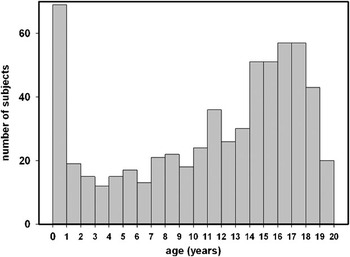
We obtained 24-hour Holter recordings from a total of 616 children and adolescents, 299 being males and 317 females, with palpitations or suspected paroxysmal supraventricular tachycardia, these suspected findings not being confirmed in any of the patients. The examinations were performed in two tertiary centres for paediatric cardiology. None of the subjects was receiving relevant medication, and none had undergone prior clinical intervention. In all cases, a full clinical and echocardiographic examination failed to detect evidence of structural congenital cardiac disease, significant arrhythmia, or of any other serious illness. Hyperthyroidism was excluded by thorough clinical investigation. Levels of thyroid hormone were not evaluated because of the absence of relevant clinical symptoms. The age of the subjects is displayed in Figure 1.
Age distribution.

Methods
All recordings were obtained on outpatient basis in the normal environment of the subjects. Analysis of the recordings was performed using Mars 8000 scanners from Marquette/GE Laboratories (Milwaukee, Wisconsin, USA), and by experienced Holter technicians applying careful review of template and heart rate profiles. All patients were in sinus rhythm during the recording, and all recordings were found to be normal by the supervising physicians. The software of the scanner used 5 consecutive RR intervals for automatic calculation of heart rate.
Statistics
Best-fit non-linear regressions were applied to correlate age and gender with minimal, mean, and maximal heart rate, and maximal RR-intervals. We then calculated 5th, 25th, 75th and 95th centiles for each variable. Maximal heart rate was not evaluated, since the degree of physical activity during the recording was not standardized. The data was analyzed using multivariate analysis of variance, taking as dependent variables the minimal and mean heart rate and maximal RR-interval, and as independent variables the gender, using a 2 step factor, and age as a covariate. This was followed by use of the Tukey HSD post-hoc test corrected for multiple measurements. The level of significance was set at a value for p of less than 0.05. The age-dependent changes were fitted to non-linear function using non-linear regression analysis as described above, followed by a post-hoc two-sample t-test with Bonferroni adjustment to evaluate the influence of gender in 5 different age groups. SigmaStat 3.10 and Systat 11 (Systat Software Inc., Richmond, California, USA) were used for analysis.
Results
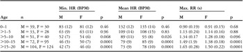
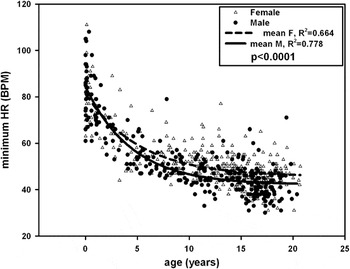
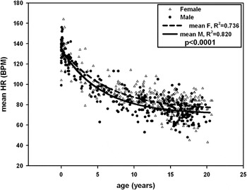
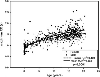
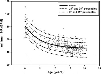
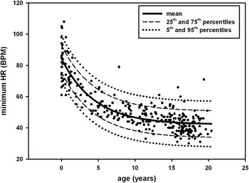
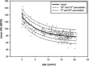
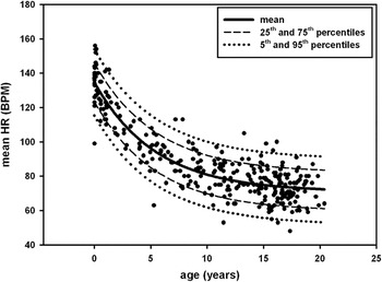
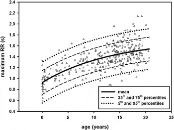
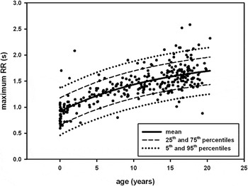
Correlation of heart rate with age could be established with sufficient accuracy using non-linear regression. For minimal and mean heart rate, a single exponential decay was found with 3 parameters, and for the maximal RR-interval, a double exponential rise to maximum with 4 parameters provided the best fit. In addition, we tried to elucidate the influence of gender on the age-dependent adaptation of the parameters. A more detailed post-hoc t-test analysis disclosed significant differences in females as opposed to males starting from the age of 10 years (Table 1). The male patients had significant lower minimal and mean heart rates, and correspondingly higher maximal RR-intervals, than did the female patients (p less than 0.0001 for the difference between the female and male regression curves, Figs 2–4). The corresponding gender specific regression equations with the 5% and 95% centiles are summarized in Table 2, and the regression curves along with the 5th, 25th, 75th and 95th centiles are displayed in Figures 5–10.
Average minimal and mean HR, maximal RR-interval and the corresponding p-values in the different age and gender groups with the standard deviation (SD) given in brackets.

Min. HR = minimum heart rate; Mean HR = mean heart rate; Max. RR = maximum RR-interval; M = male; F = female; n = number of patients in the male/female group.
Gender-dependent effect of age on minimal heart rate. Statistical significance is given for the difference between female and male subjects.

Gender-dependent effect of age on mean heart rate. Statistical significance is given for the difference between female and male subjects.

Gender-dependent effect of age on maximal RR-interval. Statistical significance is given for the difference between female and male subjects.

Regression equations for minimal and mean HR and maximal RR-interval versus age (x) as derived from analysis of 299 male and 317 female subjects.

Min. HR = minimum heart rate; Mean HR = mean heart rate; Max. RR = maximum RR-interval; M = male; F = female.
Minimal heart rate versus age of 317 female subjects.

Minimal heart rate versus age of 299 male subjects.

Mean heart rate versus age of 317 female subjects.

Mean heart rate versus age of 299 male subjects.

Maximal RR-interval versus age of 317 female subjects.

Maximal RR-interval versus age of 299 male subjects.

Discussion
The heart rate of healthy children changes markedly from birth to young adulthood. Although values for infants, children, and young adults have existed for quite some time, there is very limited data concerning age-related changes in a healthy population from birth up to the age of 20 years. Findings using Holter recordings at these different stages of life have also not yet been defined, preventing their correlation with normal values. Moreover, although it is widely accepted that there is a distinct difference in values of rate related to gender, to the best of our knowledge clear data is lacking on this issue. Hence, for the first time, we present a comprehensive study on a large cohort of normal subjects aged from birth to 20 years investigating the range of minimal and mean heart rate, as well as the maximal RR-interval, seeking to define limits of normal rate for use when evaluating Holter electrocardiograms. The age-dependent evaluation of the minimal and mean heart rate, as well as the maximal RR-interval, yielded a close and good correlation, and showed dependence on both age and gender. Previous studies with a large number of participants had also revealed a significant effect of age and gender on heart rate.Reference Rijnbeek, Witsenburg, Schrama, Hess and Kors7–Reference Davignon, Rautaharju, Boisselle, Soumis, Megdlas and Choquette9 In all these studies, nonetheless, heart rate had been determined using a standard resting electrocardiogram, and not a continuous 24-hour Holter recording. The latter technique has been used to examine cardiac rhythm in newborns,Reference Montague, Taylor, Stockton, Roy and Smith10 infants, childrenReference Porter, Gillette and MyNamara11, Reference von Bernuth, Toussaint, Mund, Rabe and Timbul12 and adolescents,Reference Dickinson and Scott13 confirming the influence of age on heart rate, no limits for normal rates have yet been defined.
Our study also revealed a gender-dependent splitting of the normal values after an age of about 10 years. A possible explanation could be that women have higher heart rates than men.Reference Abdel-Rahman, Merrill and Wooles14, Reference Liu, Kuo and Yang15 This has been related to a different regulation of heart rate due to gender differences in the autonomic nervous system. This gender dependent effect was absent when postmenopausal women were compared to men, yet became evident again when conjugated oestrogen was administered as replacement therapy. Taken together, these observations suggest that the divergence in heart rate between males and females after the age of about 10 years could be induced by puberty, and be related to the changes in levels of sex hormones. Due to the clinical situation, however, it was not possible to achieve equal distribution of age of our subjects. Thus, besides the pre-pubertal hormonal state, the lower number of patients ranging from 1 to 10 years could also contribute to the lack of statistically significant effect of gender within these younger subjects. Similarly, the number of patients below the age of 1 year might be too low appropriately to track the rapid changes in heart rate occurring in this period, thus limiting the validity of the normal limits established for the first year of life.
We could clearly define, nonetheless, the normal range of heart rate and RR-interval values from the neonatal period to adolescence, and in this way provide reliable information for the evaluation of heart rate as established using ambulatory Holter electrocardiograms. According to our data, it seems necessary to use gender-specific normal values. The displayed centile graphs and regression equations may allow the cardiologist to estimate visually, or to calculate, whether minimal and mean heart rates, and maximal RR-intervals, as derived from 24-hour electrocardiography are to be considered as normal or not in the populations of children and adults aged less than 20 years.
Acknowledgement
Roman A. Gebauer was supported by the Research project No. 64203 of the University Hospital Motol, Prague, Czech Republic.