Introduction
The application of synthetic biomaterials for the regeneration of bone tissue as an alternative to bone grafts, both in orthopedics and in orthodontics, has many advantages [Reference Gurtner, Callaghan and Longaker1]. These include a low risk of contamination by bacteria and viruses, providing a material according to the patient needs.
The most widely used biomaterials are polymers, ceramics, composites, and also metals. These materials can be used to replace some parts of the body, although its surface composition is tailored regarding the application and depending on the need and location that the device will be implanted. In this sense, metal biomaterials are commonly used in prostheses that need a greater mechanical resistance. In these classes of biomaterials, the investigations associated with titanium and its alloys are mainly motivated by some unique characteristics of this material, such as low compression, low corrosion [Reference Habazaki, Onodera, Fushimi, Konno and Toyotake2], low toxicity, and modulus of elasticity closer to human bone [Reference Holzapfel, Reichert, Schantz, Gbureck, Rackwitz, Nöth, Jakob, Rudert, Groll and Werner3].
However, to achieve this corrosion resistance, this metal is most often coated with a thin film of titanium dioxide (TiO2) [Reference Murugan and Ramakrishna4, Reference Macak, Tsuchiya, Taveira, Ghicov and Schmuki5, Reference Gong, Grimes, Varghese, Hu, Singh, Chen and Dickey6]. Finally, several studies have shown that the nanotexturization of titanium and its alloys can increase the biocompatibility of the implant and facilitate the process of osseointegration [Reference Kubies, Himmlová, Riedel, Chánová, Balík, Rová, Bártová and Pešáková7, Reference Le Guehennec, Enkel, Weiss, Amouriq and Layrolle8].
Processes of film deposition or surface modification can be employed for the purpose of improving the characteristics of a biomaterial. In the case of nanotexturization of titanium and its alloys, it is possible to create morphologies that mimic the human bone or even promote chemical modifications in the material that facilitate the process of osseointegration. In this regard, the anodic process is a surface modifying technique that can be used with a great success for such titanium-based biomaterial improvements [Reference Murugan and Ramakrishna4, Reference Ehrenfest, Coelho, Kang, Sul and Albrektsson9].
Nanotexturizated titanium implants obtained from electrochemical techniques show high interaction with osteoblastic cells [Reference Rieger, Dupret-bories, Salou, Layrolle, Debry, Lavalle and Vrana10]. Wei et al. [Reference Wei, Igarashi, Okumori, Igarashi, Maetani, Liu and Yoshinari11] have shown that the wettability increases the protein adsorption and also the initial attachment of osteoblastic cells. For other cell types, such as bacterial cells, surface hydrophilicity or hydrophobicity is also crucial in adsorption [Reference Hori and Matsumoto12]. Staphylococcus aureus is a bacterium that presents a hydrophilic surface [Reference Neoh, Hu, Zheng and Kang13]; thus, the adhesion of this bacterium to a substrate increases with the hydrophilicity of the surface material. The proliferation of these microorganisms might affect the process of osseointegration, causing the rejection of the implant. An alternative to enhance the adhesion of bone cells and to hinder the interaction of microorganisms is to incorporate antimicrobial drugs into the nanotexturized surface; for this purpose, nanostructured titanium (TiO2NT) may serve as drug reservoir to locally deliver these substances after implantation into the human body. These drugs, when administered locally around the implant, offer several advantages, minimizing the amount of drug required reducing the chances of systemic toxicity [Reference Yao and Webster14]. In addition, if a low drug release rate is achieved, it is possible to create a microorganism inhibition zone around the implant. This slow release process can be achieved by using a physical or chemical barrier.
Some studies [Reference Unagolla and Jayasuriya15, Reference Wei, Zhou, Feng, Li and Wang16, Reference Mohan, Anandan and Rajendran17] have shown the addition of chitosan as a diffusional barrier to control the drug releasing rate. In spite of its proven biocompatibility, chitosan is an exogenous material to the human body. Thus, an attractive alternative of a diffusional barrier might be a macromolecule present in human organisms, such as the collagen protein. This protein is found in the body as a connective tissue and it is related directly with the process of osseointegration [Reference Shoulders and Raines18, Reference Roehlecke, Witt, Kasper, Schulze, Wolf, Hofer and Funk19, Reference Landis and Silver20]. Besides these advantages, in the present study, the collagen was used as a retarding agent, thus allowing the control of the releasing rate of the drug. In this work, titanium dioxide coatings obtained from titanium and its Ti6Al4V alloy were prepared by anodization method. The synthesized coatings were used as a reservoir for a model antimicrobial drug. A set of drug that incorporated coatings was prepared. In order to delay the drug releasing rate, another set of samples were coated with a collagen drug-free film. The third set of samples with a collagen-drug film was also prepared. The drug-releasing kinetics, as well as the antimicrobial activity and the hemotoxicity of the obtained biomaterials, were also investigated.
Results and discussion
Coating characterization
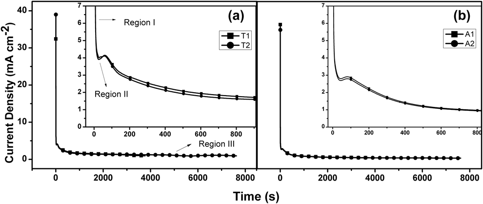
TiO2NT coatings are produced using anodization technique. The anodization curves obtained for both substrates (T and A) by the anodization process can be observed in Fig. 1.
Current density (mA/cm2) as a function of anodization time (s) for anodized samples, where (a) T1 and T2 are the commercial pure Ti synthesized at 1 and 2 h, and (b) A1 and A2 are the alloy Ti6Al4V anodized at 1 and 2 h. All anodization carried out applying an anodized potential of 25 V and at room temperature.

Anodization curves for both substrates (T and A) exhibited a similar behavior and can be divided into three regions. In the first region, there is an abrupt drop in current density, reaching a minimum value [Reference Macak, Tsuchiya and Schmuki21, Reference Macak, Tsuchiya, Ghicov, Yasuda, Hahn, Bauer and Schmuki22]. This behavior is a consequence of gradual resistance increase during a compact oxide film formation, which acts as a barrier to the passage of electrons [Reference Regonini, Bowen, Jaroenworaluck and Stevens23]. In region 2, since the oxide growth by ionic transport becomes difficult, nucleation of the nanotubes (NTs) (opening of channels) takes place. Nucleation provides less resistive pathways for the current and increases the surface area of the oxide. The presence of fluoride and simulated body fluid (SBF) ions in the electrolytic medium promotes the dissolution of the oxide through the formation of the [TiF6]−2 and other soluble complexes. In addition, the ionic species present in the solution can be incorporated into the oxide structure. The formation and dissolution reactions attained a steady state (Region III) that corresponds to the vertical growth of the nanotubular structure of TiO2NTs [Reference Ghicov and Schmuki24].
The insets of Figs. 1(a) and 1(b) show that nucleation of NTs occurs at higher current values for titanium substrate than for alloy, which can be related to the composition of the alloy that modify the anodic oxide resistivity. Nevertheless, both substrates presented a similar anodic profile in the electrolyte used.
The coating surface may play an important role in the osseointegration mechanism between the implant and the human body. In this sense, the study of its structural properties such as morphology, microstructure, and contact angle is extremely important to evaluate its performance as biomaterial.
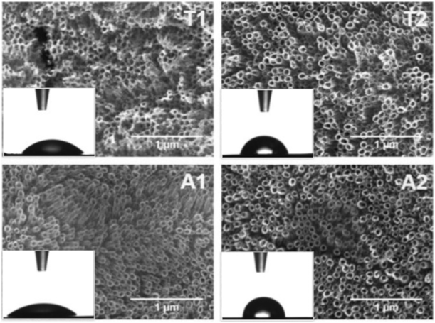
Scanning electron microscopy (SEM) analysis of the samples revealed a typical nanotubular structure, with homogenous NTs perpendicularly aligned to the substrate for both substrates (Fig. 2). The morphology of the coatings obtained from A presents a higher NT organization when compared to the morphology of the films obtained from T. This result shows that the heterogenous composition of the alloy favors a better growth of the nanostructures [Reference Mohammed, Khan and Siddiquee25].
SEM and contact angle images (detail in the figures). T1 and T2 are the commercial pure Ti synthesized at 1 and 2 h, and A1 and A2 are the alloy Ti6Al4V anodized at 1 and 2 h.

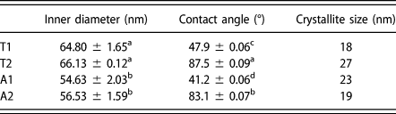
The coatings prepared from different substrates presented changes in their morphology regarding the length of the inner diameter of NTs. This parameter was determined from SEM image analysis and the data are presented in Table I. Analysis of variance (ANOVA) of NT inner diameter shows that there is no significant variation in this parameter as the anodization time increases from 1 to 2 h, for both substrates. However, a significant variation of the inner diameter was observed regarding the substrate type (A or T). The NTs grown from Ti alloy (A) are about 10 nm smaller in diameter than the NTs grown from titanium (T).
Parameters extracted from data processing. NT inner diameter obtained by ImageJ analysis, contact angle values, and crystallite size obtained by XRD analysis using the Scherrer equation.

From inspection of Table I, all Ti substrates produced NTs with diameters smaller than 100 nm, which is in accordance to Kulkarni [Reference Kulkarni, Mazare, Schmuki and Iglič26], who observed an effective cell adhesion and greater osseointegration of NTs coatings with internal diameters smaller than this magnitude.
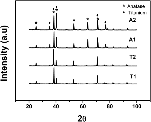
Figure 3 shows the X-ray diffraction (XRD) patterns of samples after heat treatment for 1 and 2 h. The analysis showed that all the synthesis conditions generate a well-defined XRD pattern characterized by sharp and narrow diffraction peaks, all matching with similar inorganic crystal structure database (ICSD). XRD patterns showed hexagonal titanium ICSD #00-005-0682 and tetragonal TiO2 (anatase) ICSD #01-071-1167. This later was easily identified by the peak (101) at 24.2°.
XRD patterns for prepared samples. T1 and T2 are the commercial pure Ti synthesized at 1 and 2 h, and A1 and A2 are the alloy Ti6Al4V anodized at 1 and 2 h. The phases are indexed as (♦) titanium (ICSD #00-005-0682) and (*) anatase (ICSD #01-071-1167).

From Scherrer equation [Eq. (2)] [Reference Scherrer27] applied in anatase (101) reflection peaks, crystallite sizes in the range of 18–27 nm were calculated. For coatings grown on titanium substrate, the increase in anodizing time promoted an increase in crystallite size. However, for coatings grown on alloy substrate, the increase in the anodization time promoted a small decrease in crystallite size.
The determined structural parameters, such as the internal diameter of the NTs and the crystallite size, may reflect directly on the surface characteristics of the coating. The contact angle measurements of each sample show these characteristics. The results are shown in Table I. Contact angle can be related to the surface properties, mainly the surface roughness, which could affect the initial contact angle of a water droplet on each coating [Reference Shin, Shokuhfar, Choi, Lee and Friedrich28]. Sun and Wang [Reference Sun and Wang29] showed that the increase in wettability could be associated not only to the roughness but also to the increase in the amount of chemisorbed water molecules and to the Ti–OH functional groups on the surface of the coating.
The results presented significant changes in the wettability of the material with increasing anodization time. For anodic oxide films formed on the alloy, the contact angle practically doubles with increasing anodizing time, transforming an initially hydrophilic surface material to a hydrophobic surface material. For coatings formed on titanium, the material also changes from a hydrophilic to hydrophobic character. According to Shin et al. [Reference Shin, Shokuhfar, Choi, Lee and Friedrich28], at the beginning of the anodizing process, there is the formation of Ti(OH)4 instead of TiO2. Titanium hydroxide presents a more hydrophilic character than the oxide. In this sense, it can be inferred that increasing the anodization time promotes the decrease of the amount of adsorbed water molecules, which can be related to the conversion of Ti–OH species into TiO2 in both metal substrates investigated.
Drug release
The presence of nanostructures significantly increases the surface area of the material [Reference Macak, Tsuchiya, Ghicov, Yasuda, Hahn, Bauer and Schmuki22]. For this reason, the incorporation of drugs into the nanostructure for targeted delivery of the therapeutic agent to the specific site has been extensively studied [Reference Unagolla and Jayasuriya15].
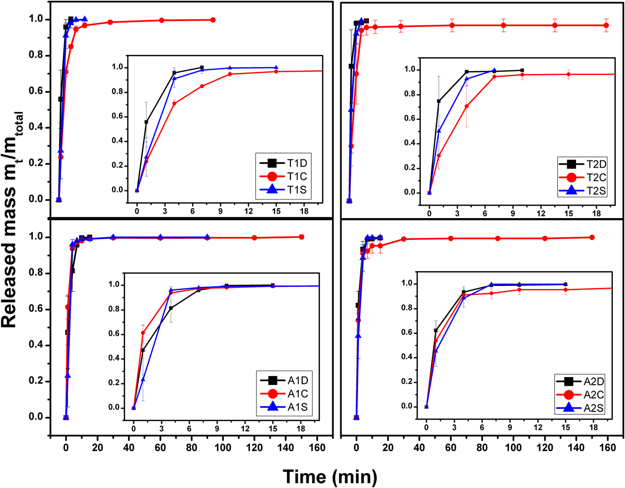
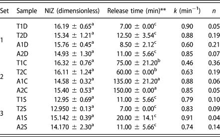
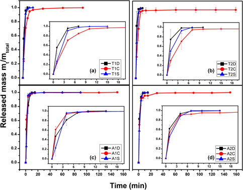
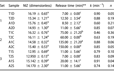
Figure 4 shows the cumulative ciprofloxacin hydrochloride (CIPRO) mass released from the TiO2NTs samples. As can be observed, the samples that showed longer release time were those coated with collagen (samples of set 2, Table II). However, statistical analysis using a post hoc Tukey’s HSD test showed that the drug release time of the set 3 samples is equivalent to the release time of set 1, where only the drug was added. As a consequence, the use of a mixed solution containing drug and collagen was not favorable for any of the synthesis conditions. This result may indicate that some physicochemical characteristics of this solution (collagen + drug), such as the high viscosity when compared with the aqueous solution of the drug, may hinder the permeation of the drug and collagen into nanotubular structure provided by the coating. Thus, the incorporated material was deposited only in the more superficial portion of the coatings.
Cumulative mass of CIPRO released as a function of time from samples synthesized according to Table III.
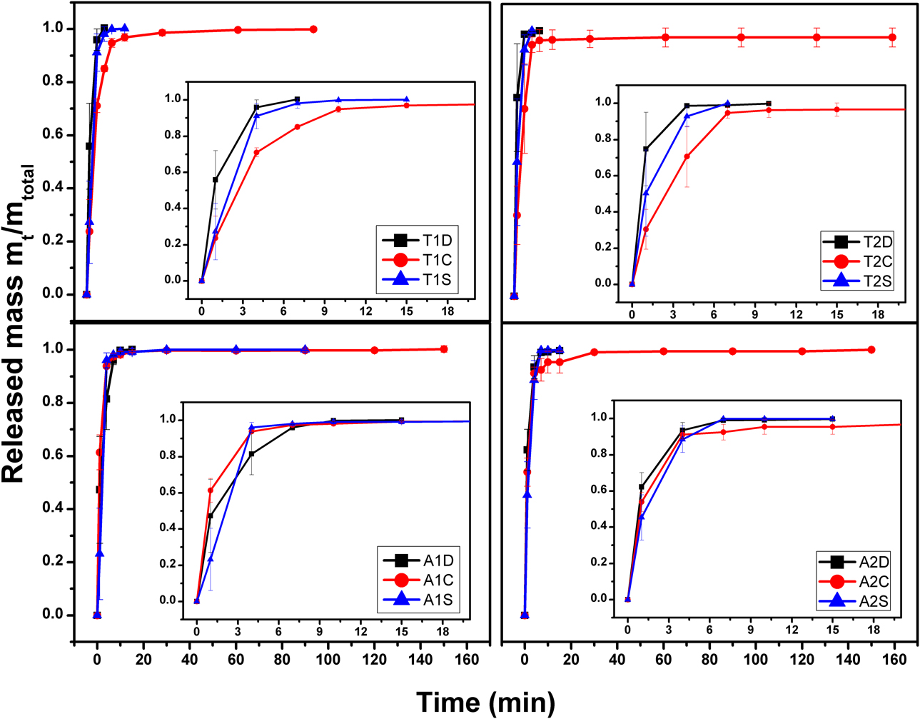
NIZ against S. aureus and kinetic parameters. According to Korsmeyer–Peppas model k is kinetic constant and n is the exponential factor. Both parameters were extracted from release curves (Fig. 4).
** The release time was defined as the time required for the release of 1 mg of drug.
The post hoc Tukey’s HSD test showed that samples that exhibited the longest release times were the samples prepared from the alloy. Analyzing the structural parameters obtained in Table I and the results from drug release (Fig. 4), it can be observed that the internal diameter of the NTs can present a significant influence in drug release. The internal diameter of the NTs grown on the alloy was smaller than the NTs grown on the titanium. Thus, there is a significant change in the surface area when the two substrates are compared. Coatings grown on the alloy presented a larger surface area, which is an interesting feature for materials to be used as drug nanoreservoirs.
The results of Korsmeyer–Peppas fitting are presented in Table II. According to Ritger and Peppas [Reference Ritger and Peppas30], when the value of diffusional exponent (n) is between 0.02 and 0.30, the mechanism is considered as Fickian diffusion; values between 0.43 < n < 0.85 are considered as non-Fickian; and values of n = 0.85 is considered as case II transport [Reference Unagolla and Jayasuriya15, Reference Ritger and Peppas30]. By analysis of the data presented in Table II, it can be observed that for all the conditions studied, drug release occurs following Fick’s model.
The value of k indicates the release rate of drugs within the matrices of TiO2NTs, where higher k values indicate a faster release, and lower values a slower release [Reference Holzapfel, Reichert, Schantz, Gbureck, Rackwitz, Nöth, Jakob, Rudert, Groll and Werner3]. The analysis of the release time and kinetic parameters (k and n) suggests that the release of the drug is likely to occur by a mixed mechanism. The linear analysis of the curve, where the rapid release of the drug occurs (burst), follows a Fickian kinetics. After this stage, the material continues to release but at a much lower rate.
A large quantity of the drug is released from the coatings in the burst, resulting a higher value of kinetic constant. When the internal diameter of the NTs is smaller, an amount of the drug can be retained due to the interaction with the surface of the oxide and/or due to a synergistic effect between the oxide, collagen, and the drug.
Biological activity
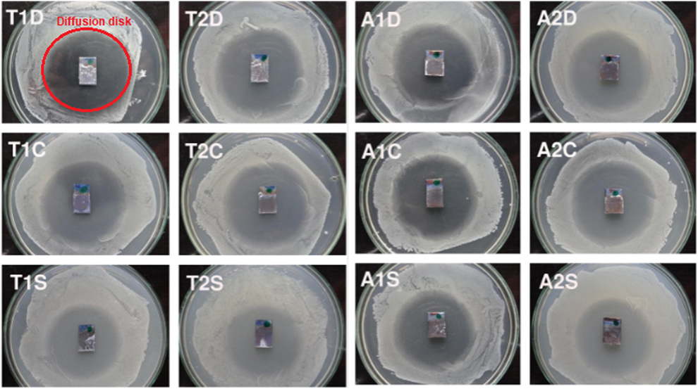
The antimicrobial activity of the samples was investigated from the inhibition halo against S. aureus. Figure 5 shows the inhibition halo of all samples. As can be seen, for all conditions there was a significant inhibition bacteria growth. The normalized inhibition zone (NIZ), presented in Table II, was calculated according to Eq. (3), where the inhibition area was determined by ImageJ software, National Institutes of Health (NIH), Bethesda, Maryland (represented by red circle in Fig. 5).
Petri dishes showing the inhibition halo of S. aureus promoted by the samples synthesized according to Table III.
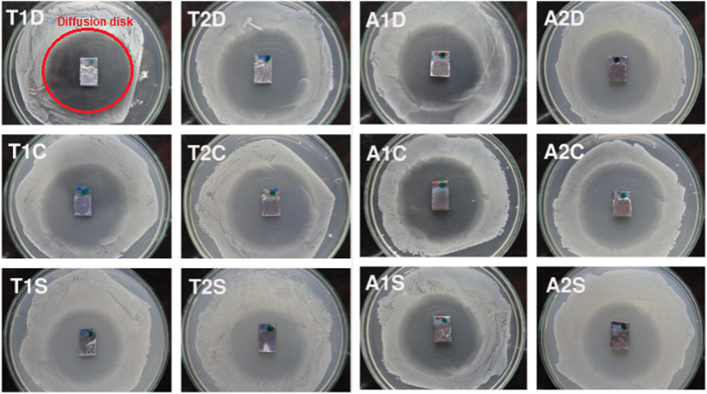
The results show that inhibition of bacterial growth in coatings occurs effectively for all the synthesis conditions. The NIZ values in Table II show that for all samples, there was an area of inhibition of at least 13 times the implant area. This result shows that for all prepared coatings there is a local inhibition of bacterial growth. Such feature is important in the development of bone implants, since these materials can prevent the adhesion of bacteria, avoiding the formation of biofilms that lead to rejection of the implant [Reference Feng, Geng, Li, Cui, Zhu, Liang, Liu, Wang and Yang31].
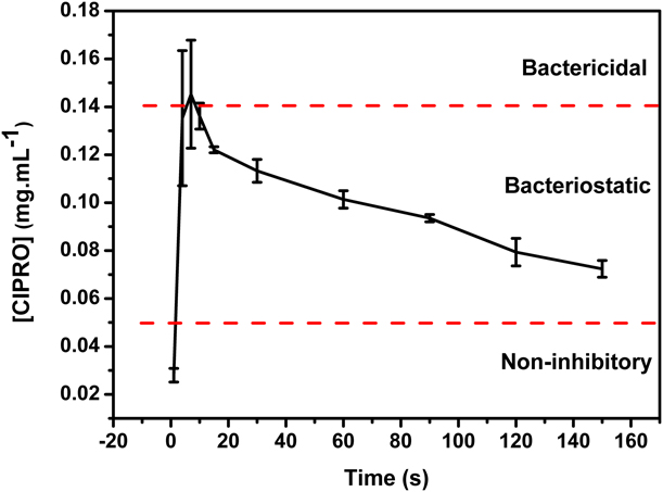
For slow release devices, two conditions must be considered. The first is that the released concentration of the drug at any time is nontoxic to the host organism. The second is that the concentration of the drug is effective for the inhibition of bacteria. The concentration inhibitory range is distinguished by bacteriostatic and bactericidal. The term bacteriostatic refers to a concentration range that prevents the growth of the bacterium but is not able to inactivate it. On the other hand, the term bactericide refers to a concentration capable of killing the microorganism.
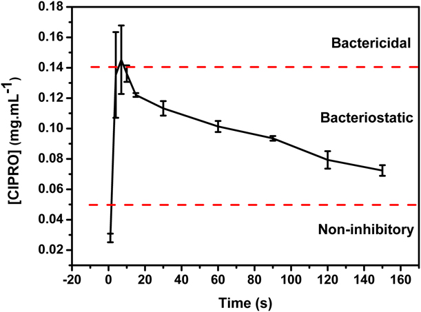
In order to verify the antimicrobial activity of the coatings, the minimum inhibitory concentration was obtained. For all conditions, the antimicrobial activity against S. aureus was in the bacteriostatic band, as can be seen in Fig. 6. Note that during the burst releasing stage, a high amount of the drug is released, although its concentration is within the bacteriostatic band. At the end of the process, when the entire amount of the drug was released, the local concentration remained in the bacteriostatic range not toxic to the human body.
Concentration of ciprofloxacin released from sample T1C as a function of time. The horizontal lines delimit the noninhibitory and inhibitory zones (bacteriostatic and bactericidal).
The hemolytic activity was studied to verify the toxicity of both bare substrates (T and A) used as references controls, as well as the samples synthesized in this study. The results are presented in Fig. 7.
Hemolytic activity of the samples obtained according to Table III (only for 1 h of anodiztion), compared with the metal substrates, with the negative control (PB) and the positive control (Triton-X). The letters at the top of the bars refer to ANOVA analysis. Samples that share the same letter do not present significant statistical difference.

According to Table II, no statistical difference between the samples synthesized in 1 and 2 h was observed. Therefore, for the hemolysis test, only the samples obtained in 1 h of anodization were studied. As can be seen, all samples (including reference metals) did not promote lysis of erythrocytes. This is an interesting result because the anatase phase found in the synthesized materials might not be inert to blood cells [Reference Zitting and Skyttä32]. All samples presented a low percentage of hemolysis, being statistically similar to the negative phosphate buffer (PB) control. The only exception observed was the sample T1S that showed a small hemolytic activity.
Conclusion
The results showed that the anodizing time influenced the structural properties of the obtained NTs. There was significant change in the wettability of the coatings, where the samples changed their surface character from hydrophilic to hydrophobic when the anodization time was increased. It was observed that the anodized coatings grown over the commercial pure Ti substrate presented NTs with an internal diameter of 10 nm larger than those prepared over the Ti6Al4V substrate. The drug release assays showed no statistical difference in drug releasing time between coating anodized at different anodization times. However, the coating coverage with a thin layer of collagen decreased the drug releasing rate. In the biological assays, the diffusion disc confirmed that there was no growth of S. aureus around the coatings. This result indicates that drug loaded inside the coatings inhibited the biofilm formation. Finally, the hemolytic activity did not show toxicity to the blood cells, which indicates that this methodology may be applied to the development of new active biomaterials.
Methods
Coatings preparation
The synthesis of TiO2NT coatings was performed using two types of substrates, commercially pure Ti (T) and Ti6Al4V alloy (A). The metal sheets were limited to 15 × 10 mm and polished with silicon carbide sandpaper (1200 mesh), then washed with distilled water, and dried at room temperature. The anodizing area was carefully held constant (1 cm2 each face), controlling the metal exposure (T and A) with PTFE tape. The oxidation of the metal was accomplished by applying 25 V using a DC voltage source (MPC303D) between the Ti electrodes (T and A) and a pair of platinum counter electrodes, all arranged in parallel. The electrolytic solution was adapted from the methodology developed by Simon [Reference Simon and Sikora33] and consists of 0.75% w/w of ammonium fluoride (NH4F), 9% v/v water, and 1% v/v SBF [Reference Kokubo, Kim and Kawashita34] in ethylene glycol. The anodizing process was carried out for 1 and 2 h and several samples were produced, according to Table I. After the synthesis, the coatings were submitted to the annealing in a muffle at 450 °C for 2 h, using a heating ramp of 2 °C/min, to obtain crystalline oxide (anatase).
Characterization of TiO2 coatings
Samples morphology was evaluated by High Resolution FE-SEM (FEI Quanta 650 FEG, Hillsboro, Oregon) and the micrographs were evaluated for the quantification of the inner diameter NTs using ImageJ software [Reference Schneider, Rasband, Eliceiri and Instrumentation35].
The microstructure of the TiO2 coatings was evaluated by X-ray diffraction (XRD) (Rigaku D/max 2500 PC diffractometer, Tokyo, Japan), in a Bragg–Brentano mode, operating between 20 and 80° (2θ) with a continuous scan speed of 2°/min with Cu Kα = 1.5406 Å and radiation at 40 kV and 150 mA. The grain size was evaluated by Scherrer equation [Reference Scherrer27] [Eq. (1)],
 $$\tau = {{K \cdot \lambda } \over {B \cdot \cos \;\theta }}\quad ,$$
where τ is the mean crystallite diameter, K is a constant that depends on the shape of the particles, in this study it was used the usual value (0.9), λ is the wavelength of electromagnetic radiation, B is the width of half height of the diffraction peak, and θ is the Bragg diffraction angle.
Water contact angle measurements were performed to examine the samples surface wettability. Experiments were carried out using a Hamé-hart Goniometer/Tensiometer Model 250 connected with a video camera (Randolph, New Jersey). The contact angle was measured with respect to deionized water. Droplets of 0.5 μL were spread over three different locations on each sample. The contact angle for each drop was recorded by calculating the mean of left and right angles. The average of three readings has been reported and analyzed statistically using ANOVA.
Drug loading
CIPRO was used as model drug. For the incorporation of the drug, the top filling technique was used, a physical adsorption method adapted from the methodology of Feng [Reference Feng, Geng, Li, Cui, Zhu, Liang, Liu, Wang and Yang31]. A 10 g/L solution of CIPRO was added and gently spread onto the anodized area (1 cm2 each face) and then dried at 25 °C in an incubator with air circulation. This process was repeated 5 times on each face, loading the coatings with 1 mg of CIPRO.
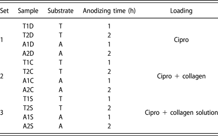
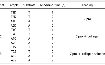
After loading, samples were subdivided into three sets. In the set 1, samples were loaded just with the drug. In the set 2, samples were loaded with the drug and then coated with collagen film. A solution of 2 g/L of collagen in 2% w/v acetic acid was used. In the set 3, the samples were loaded with a mixed solution of collagen and CIPRO at the same concentrations of the set 2 [Reference Mohan, Anandan and Rajendran17]. The conditions of sample preparation are shown in Table III.
Planning matrix of the samples preparation conditions. Sample denominations are related to substrate, anodizing time, and loading methodology.
Drug release assay
The CIPRO release study was carried out by immersing the samples in 5 mL of ultrapure water (Mili-Q) maintained at 37 °C in a thermostatic bath according to the methodology adapted from Gulati [Reference Gulati, Kogawa, Prideaux, Findlay, Atkins and Losic36]. Aliquots of 0.5 mL were collected at times of 1, 4, 7, 10, 15, 30, 60, 90, 120, 150, 180, 240, and 300 min. Each aliquot removed was replaced by 0.5 mL of fresh ultrapure water to maintain the sink condition [Reference Abdou37]. The amount of drug released was acquired using ultraviolet-visible (Shimadzu, UV-1800, Kyoto, Japan) spectroscopy at 241 nm and a calibration curve. The drug release profiles were constructed from the mean value of the cumulative mass released as a function of time.
To study the release mechanism, it was used the Korsmeyer–Peppas model [described by Eq. (2) [Reference Sinn Aw, Kurian and Losic38]]. The equation kinetic parameters were extracted from the average of releasing data,
 $${{{M_{\rm{t}}}} \over {{M_{{\rm{tm\acute{a}x}}}}}} = k{t^n}\quad ,$$
where  ${{{M_{\rm{t}}}} \!\left/{\vphantom{{{M_{\rm{t}}}} {{M_{{\rm{tm\'a x}}}}}}}\right.{{M_{{\rm{tm\acute{a}x}}}}}}$ is the cumulative release of the drug over the maximum mass released, k is the release kinetic constant, and n is the release exponent used to characterize different release patterns for thin films.
Bacterial susceptibility assay
Samples were investigated for their antibacterial potential against Staphylococcus aureus (ATCC 25923). An inoculum was prepared in BHI broth and incubated for 18 h. The initial amount of cells was standardized using the Mc Farland 0.5 scale (108 CFU/mL). After standardization, the initial amount was set at 106 CFU/mL by dilution in liquid BHI medium. 100 µL of this bacterial solution was dispersed on the surface of Petri dishes containing nutrient agar. The coatings were placed in the center of the plate for the inhibition halo investigation. The Petri dishes were incubated for 18 h at 36–37 °C and after this time measurements of the inhibition halos were performed, using ImageJ software. The NIZ was calculated according to Eq. (3). All analyzes were done in duplicate and the results are presented with mean ± standard deviation,
 $${\rm{NIZ}} = {{{\rm{Inhibition}}\;{\rm{zone}}\;\left( {{\rm{m}}{{\rm{m}}^{\rm{2}}}} \right)} \over {{\rm{Anodized}}\;{\rm{area}}\;\left( {{\rm{m}}{{\rm{m}}^{\rm{2}}}} \right)}}\quad .$$
In addition, a minimum inhibitory concentration (MIC) assay was performed to assess the antimicrobial potential of time-released drug amounts. The determination of MIC was carried out by the Mueller-Hinton broth dilution technique according to CLSI [39, Reference Krichen, Karoud, Sila, Abdelmalek, Ghorbel, Ellouz-Chaabouni and Bougatef40].
Hemolytic activity assay
For the hemolytic activity assay, healthy volunteer adult blood was collected in EDTA-containing tubes. The collected blood was centrifuged at 6 °C at 4000 RPM for 10 min for the separation of erythrocytes (precipitate) and plasma, the latter being discarded. For the hemolysis assay, the methodology of Bulmus [Reference Bulmus, Woodward, Lin, Murthy, Stayton and Hoffman41] was adapted. The hemolytic activity can be calculated by the Eq. (4) [Reference Palanivelu and Ruban Kumar42],
 $$\% {\rm{Hemolysis}} = {{{\rm{Absorbance}}\;{\rm{of}}\;{\rm{sample}}} \over {{\rm{Absorbance}}\;{\rm{of}}\;{\rm{positive}}\;{\rm{control}}}} \times 100\% \quad .$$
Statistical analysis
Statistical analysis was performed using one-way ANOVA with post hoc Tukey HSD test in order to verify the statistical relevance between coating synthesis conditions. Values of P < 0.05 were considered to indicate statistical significance.
Acknowledgment
The authors thank the UTFPR, Central Analysis of UTFPR-PB, UFSCar, Biocenter Clinical Analysis Laboratory, and to Brazilian Nanotechnology National Laboratory (LNNano) in National Center of Energy and Materials Research (CNPEM) and Analysis Center of UTFPR-PB. This work was supported by UTFPR [PAPCDT 06/2016 and 07/2017]. This study was financed in part by the Coordenação de Aperfeiçoamento de Pessoal de Nível Superior—Brasil (CAPES)—Finance Code 001.