INTRODUCTION
Eimeria is a genus of at least 1200 species of obligate intracellular protozoan parasites within, in hierarchical order, the Coccidiasina, Conoidasida, Apicomplexa and Alveolata (Adl et al. Reference Adl, Simpson, Farmer, Andersen, Anderson, Barta, Bowser, Brugerolle, Fensome, Fredericq, James, Karpov, Kugrens, Krug, Lane, Lewis, Lodge, Lynn, Mann, McCourt, Mendoza, Moestrup, Mozley-Standridge, Nerad, Shearer, Smirnov, Spiegel and Taylor2005). The genus is relatively closely related to other coccidian genera such as Cyclospora, Toxoplasma, Neospora, Hammondia, Isospora and Sarcocystis, less closely to Cryptosporidium, and more distantly to haemosporidians such as Plasmodium (Adl et al. Reference Adl, Simpson, Farmer, Andersen, Anderson, Barta, Bowser, Brugerolle, Fensome, Fredericq, James, Karpov, Kugrens, Krug, Lane, Lewis, Lodge, Lynn, Mann, McCourt, Mendoza, Moestrup, Mozley-Standridge, Nerad, Shearer, Smirnov, Spiegel and Taylor2005).
Eimeria infect fish, reptiles, birds and mammals, with most species being highly host-specific. Most are parasites of the intestine but some do infect other gastric organs. The Eimeria that infect chickens are the best known and studied because they cause losses as high as US$2 billion per year to poultry industries around the world (Wallach et al. Reference Wallach, Ashash, Michael and Smith2008); species that infect chickens include Eimeria tenella, Eimeria maxima, Eimeria acervulina, Eimeria brunetti, Eimeria mitis, Eimeria necatrix and Eimeria praecox. Genome sequencing projects are underway for E. tenella (www.genedb.org/Homepage/Etenella) and E. maxima (Blake et al. Reference Blake, Alias, Billington, Clark, Mat-Isa, Mohamad, Mohd-Amin, Tay, Smith, Tomley and Wan2012).
The life cycles of poultry Eimeria are, arguably, the most straightforward of any apicomplexan parasites. Transmission is via the fecal–oral route and infection begins with the ingestion of sporulated oocysts, each of which contains eight haploid sporozoites, housed within four sporocysts (Fig. 1). These sporozoites invade intestinal cells, immediately enter sequential phases of asexual reproduction (referred to as merogony or schizogony) resulting in the production of merozoites, which are also haploid and reinvade the intestinal epithelia. The main purpose of this asexual phase of reproduction seems to be to amplify the sheer numbers of merozoites within the host in preparation for sexual reproduction, which is an important feature of all apicomplexan life cycles. Furthermore, as these parasites are haploid for much of their life cycle, there is an important implication for their genetics, namely that the progeny of asexual proliferation will be clonal. In addition, their life cycle is such that there is a real possibility of self-fertilization, which will also result in clonal offspring.
Diagram illustrating the life cycle of Eimeria species in the chicken. Asexual development (merogony) of the poultry Eimeria species occurs in the intestine, giving rise to microgametes and macrogametes, the sexual stages of the life cycle. Fertilization of macrogametes by the flagellated, motile microgametes results in diploid zygotes, around which form oocyst walls. Unsporulated oocysts are excreted in the feces and sporulate under appropriate conditions of temperature and humidity. These sporulated oocysts, each containing eight infectious sporozoites, within four sporocysts, contaminate the external environment and infect new hosts when ingested with food or water. (Image supplied by D. J. P. Ferguson.)

That sex exists in Eimeria, and apicomplexan parasites, generally, is beyond dispute. Direct evidence for it includes the development of sexually dimorphic stages, genetic recombination events including meiosis, and the production of spores/cysts as part of the sexual phase of the life cycle. Morphological analyses of the dimorphic sexual stages and evidence of fertilization events have been documented to varying degrees for Eimeria (Klimes et al. Reference Klimes, Rootes and Tanielian1972; Long, Reference Long1972; Fernando, Reference Fernando1973; Joyner and Norton, Reference Joyner and Norton1975; Speer and Danforth, Reference Speer and Danforth1976; Ferguson et al. Reference Ferguson, Birch-Andersen, Hutchison and Siim1977; Madden and Vetterling, Reference Madden and Vetterling1977; Shirley, Reference Shirley1978; Pittilo and Ball, Reference Pittilo and Ball1979, Reference Pittilo and Ball1980; Ball et al. Reference Ball, Pittilo, Joyner and Norton1981; Elwasila, Reference Elwasila1983; Sutton et al. Reference Sutton, Shirley and McDonald1986; Laxer et al. Reference Laxer, Healey and Youssef1987; Shirley and Harvey, Reference Shirley and Harvey1996; Blake et al. Reference Blake, Hesketh, Archer, Carroll, Smith and Shirley2004), Toxoplasma gondii (Pelster and Piekarski, Reference Pelster and Piekarski1971, Reference Pelster and Piekarski1972; Ferguson et al. Reference Ferguson, Hutchison and Siim1975; Grigg et al. Reference Grigg, Bonnefoy, Hehl, Suzuki and Boothroyd2001; Speer and Dubey, Reference Speer and Dubey2005; Sibley and Ajioka, Reference Sibley and Ajioka2008), Cryptosporidium (Beier and Sidorenko, Reference Beier and Sidorenko1990; Cheadle et al. Reference Cheadle, Toivio-Kinnucan and Blagburn1999; Feng et al. Reference Feng, Rich, Tzipori and Widmer2002; Hijjawi et al. Reference Hijjawi, Meloni, Ng'anzo, Ryan, Olson, Cox, Monis and Thompson2004), Theileria (Mehlhorn et al. Reference Mehlhorn, Weber, Schein and Buscher1975; Mehlhorn and Schein, Reference Mehlhorn and Schein1976, Reference Mehlhorn and Schein1977; Katzer et al. Reference Katzer, Ngugi, Oura, Bishop, Taracha, Walker and McKeever2006) and Plasmodium (see, for example, MacCallum, Reference MacCallum1897; Walliker et al. Reference Walliker, Carter and Morgan1971, Reference Walliker, Carter and Sanderson1975, Reference Walliker, Quakyi, Wellems, McCutchan, Szarfman, London, Corcoran, Burkot and Carter1987; Sinden and Croll, Reference Sinden and Croll1975; Sinden et al. Reference Sinden, Canning and Spain1976; Ranford-Cartwright et al. Reference Ranford-Cartwright, Balfe, Carter and Wallijer1991; Day et al. Reference Day, Koella, Nee, Gupta and Read1992; Babiker et al. Reference Babiker, Ranford-Cartwright, Currie, Charlwood, Billingsley, Teuscher and Walliker1994; Ramasamy et al. Reference Ramasamy, Kulasekera, Wanniarachchi, Srikrishnaraj and Ramasamy1997; Conway et al. Reference Conway, Roper, Oduola, Arnot, Kremsner, Grobusch, Curtis and Greenwood1999; Moreira et al. Reference Moreira, Marrelli and Jacobs-Lorena2004; Hastings and Watkins, Reference Hastings and Watkins2005).
The apicomplexan sexual phase can be broadly divided into three main events: gametocytogenesis, producing gametocytes from merozoites; gametogenesis, producing haploid microgametes and macrogametes from gametocytes; and fertilization of macrogametes by microgametes, producing diploid zygotes with ensuing meiosis marking the end of the sexual phase (Sinden, Reference Sinden1983; Smith et al. Reference Smith, Walliker and Ranford-Cartwright2002). However, variations in apicomplexan sexual reproduction patterns are apparent. Thus, in Eimeria and other Coccidia, there is no transfer of gametocytes between hosts; gametocytogenesis and gametogenesis represent a continuous process within one host cell. In contrast, in Plasmodium, gametocytogenesis and gametogenesis are distinct. In Plasmodium gametocytogenesis, merozoites produce gametocytes whose development is ‘frozen’, preventing gamete formation within mammalian hosts; gametogenesis only occurs in insects, producing haploid microgametes and macrogametes from gametocytes. An additional difference is that, in Eimeria and other Coccidia, there is a single, synchronized phase of gametocyte production whilst in Plasmodium there can be multiple, staggered phases of gametocyte formation, the result being that gametocytes can be observed circulating in the blood for many months, presumably to ensure increased opportunities for infecting insect vectors.
The genetic determination of the sexually dichotomous microgametes and macrogametes in Eimeria is distinct from the development of male and female gametes in, for example, mammals, in which it is the presence or absence of a single gene, on the Y chromosome, that dictates gender assignment in diploid males and females, respectively (Tilmann and Capel, Reference Tilmann and Capel2002). Thus, the production of either of the dimorphic haploid mammalian gametes, the sperm or ovum, is restricted to the sexually mature male or female, respectively. In contrast, Eimeria, Toxoplasma and Plasmodium are known to develop into either micro- or macrogametocytes from cloned lines of haploid merozoites (reviewed by Cornelissen, Reference Cornelissen1988). Hence, unlike mammalian gender types, micro- and macrogametes of any clonal strain of an apicomplexan species are genetically identical, yet still develop into morphologically distinct cells (Ferguson, Reference Ferguson2002). Thus, apicomplexan sexual differentiation is a purely phenotypic phenomenon. This indicates that sexual differentiation and development is dependent on the differential expression of sexual stage-specific genes, rather than presence or absence of sex chromosomes.
In this review, we aim to piece together the molecular events that underpin sexual reproduction in Eimeria and use any additional detail that can be gleaned from analogous events in Plasmodium to speculatively fill current gaps in our knowledge. Such information is intrinsically fascinating from a biological standpoint and, potentially, extremely valuable in the design of novel control strategies.
GAMETOCYTOGENESIS
In Eimeria, a set number of rounds of asexual reproduction (merogony) precedes gametocytogenesis (McDonald and Rose, Reference McDonald and Rose1987). The exact number of rounds of merogony that takes place before gametocytogenesis varies for different species (Klimes et al. Reference Klimes, Rootes and Tanielian1972) but the norm may be three, at least in poultry Eimeria (McDonald and Rose, Reference McDonald and Rose1987). The commitment of final-generation merozoites to sexual differentiation is often considered to be genetically programmed and not reliant on environmental cues (Smith et al. Reference Smith, Walliker and Ranford-Cartwright2002). This is less tightly controlled in T. gondii in which a mixture of asexual and sexual stages is the norm. There is also at least one exception amongst Eimeria species, namely Eimeria vermiformis in immunodeficient mice, in which the parasite continues into additional rounds of asexual reproduction in the absence of immune pressure from the host (Rose, Reference Rose and Soulsby1987).
The morphologically distinct micro- and macrogametocytes are generally regarded as the sexual stages in Eimeria (and other Apicomplexa) but it can be inferred that sex determination occurs in the generation of merozoites immediately preceding gametocytogenesis. Thus, despite lack of sexual dimorphism, the final-generation merozoites must also be considered sexually committed (reviewed by Cornelissen, Reference Cornelissen1988). Klimes et al. (Reference Klimes, Rootes and Tanielian1972) concluded that third-generation E. tenella merozoites can be differentiated by periodic acid Schiff's (PAS) staining, with PAS-positive merozoites developing into PAS-positive macrogametocytes whereas PAS-negative merozoites develop into PAS-negative microgametocytes (though this has proved difficult to confirm; D. J. P. Ferguson, unpublished observations). It is possible, therefore, that sexual stage genes may be expressed in merozoites, well before the biological development of gametocytes. The identity of these genes is unknown in Eimeria but some clues may be found in studies on Plasmodium falciparum. Although gametocytogenesis takes only a few hours in Eimeria, it is a complex, time-consuming process in P. falciparum, taking 9–12 days, and involving five stages of morphogenesis (see Dixon et al. Reference Dixon, Dearnley, Hanssen, Gilberger and Tilley2012). This extended period of gametocytogenesis has been a boon for the study of this process and, as a result, far more information has been gathered for P. falciparum than for any other apicomplexan. However, any extrapolations from Plasmodium to Eimeria, although potentially very useful, must be done with caution.
Analogous to Eimeria, schizonts of P. falciparum, in the generation preceding gametocytogenesis, are committed to producing daughter cells of a single gender only (Silvestrini et al. Reference Silvestrini, Alano and Williams2000; Smith et al. Reference Smith, Lourenco, Carter, Walliker and Ranford-Cartwright2000). There is, likewise, some controversy regarding the role of parasite genetics vs environmental cues in Plasmodium gametocytogenesis within the mammalian host (Alano, Reference Alano2007), with observations that the rate of asexual multiplication (Smalley et al. Reference Smalley, Brown and Bassett1981) and drug treatments (Buckling et al. Reference Buckling, Crooks and Read1999a, Reference Buckling, Ranford-Cartwright, Miles and Readb), amongst a wide variety of stressors (Baker, Reference Baker2010), can dictate the onset of gametogenesis. Nevertheless, there is also clearly a parasite genetic component governing gametocytogenesis in Plasmodium (Day et al. Reference Day, Karamalis, Thompson, Barnes, Peterson, Brown, Brown and Kemp1993; Kuehn and Pradel, Reference Kuehn and Pradel2010; Sinden et al. Reference Sinden, Carter, Drakeley and Leroy2012).
Not surprisingly, given their functional and morphological differences from asexual parasites, the gametocytes of malaria parasites possess unique molecular signatures (reviewed expertly by Sinden, Reference Sinden2009; Baker, Reference Baker2010). Transcriptomic analyses have detected specific upregulation of expression of over 200 genes during gametocyte development (Silvestrini et al. Reference Silvestrini, Bozdech, Lanfrancotti, Giulio, Bultrini, Picci, Derisi, Pizzi and Alano2005). Similarly, P. falciparum DNA microarrays have been used to observe expression patterns of gametocyte stages both in comparison to asexual stages (Le Roch et al. Reference Le Roch, Zhou, Blair, Grainger, Moch, Haynes, de la Vega, Holder, Batalov, Carucci and Winzeler2003) and within different developmental stages of the gametocyte (Young et al. Reference Young, Fivelman, Blair, de la Vega, Le Roch, Zhou, Carucci, Baker and Winzeler2005), identifying clusters of at least 218 and 246 genes, respectively, expressed in gametocytes only. What regulates the expression of such clusters is, as yet, not known but a role for the evolutionarily conserved, eukaryotic translational repressor, Puf2, is conceivable since it has been implicated in gametocytogenesis in P. falciparum (Miao et al. Reference Miao, Li, Fan, Li and Cui2010), though a similar role was not confirmed in Plasmodium berghei (Hughes et al. Reference Hughes, Philip, Starnes, Taylor and Waters2010).
At the protein level, pioneering analyses (Lasonder et al. Reference Lasonder, Ishihama, Andersen, Vermunt, Pain, Sauerwein, Eling, Hall, Waters, Stunnenberg and Mann2002) documented over 900 proteins in gametocytes, 315 of which were found exclusively in gametocytes. Subsequently, in what is proving to be a landmark study, Khan et al. (Reference Khan, Franke-Fayard, Mair, Lasonder, Janse, Mann and Waters2005) were able to separate highly pure micro- and macrogametocytes prior to proteomic analysis. Six hundred and fifty proteins were identified in the microgametocyte samples, with 236 (36%) of these determined to be exclusive to this parasite stage, while 101 of the 541 proteins (19%) in the macrogametocyte samples showed stage-exclusive expression. Furthermore, 69 proteins were present in the pure samples of both micro- and macrogametocytes but not in asexual parasite samples. Moreover, aside from the high percentage of hypothetical proteins identified in the microgametocyte- (68%) and macrogametocyte-exclusive (54%) proteomes, the respective proteomes mirror the known biology of these distinct parasite stages (Khan et al. Reference Khan, Franke-Fayard, Mair, Lasonder, Janse, Mann and Waters2005). For example, macrogametocytes contain more ribosomal and mitochondrial proteins, in accordance with the higher abundance of ribosomes observed in this cell type (Janse and Waters, Reference Janse, Waters, Janse and Waters2004). In contrast, microgametocytes contain more axoneme-related and DNA replication proteins (Janse and Waters, Reference Janse, Waters, Janse and Waters2004) and, again, this is reflected in their biology, specifically, the generation of flagella and the genome replication for a possible eight motile gametes. Proteomic analyses of purified microgametes (Sinden et al. Reference Sinden, Talman, Marques, Wass and Sternberg2010; Wass et al. Reference Wass, Stanway, Blagborough, Lal, Prieto, Raine, Sternberg, Talman, Tomley, Yates and Sinden2012) confirmed these observations, identifying several dynein components (dyneins are conserved flagellar proteins; Sinden et al. Reference Sinden, Talman, Marques, Wass and Sternberg2010) and the armadillo repeat protein, PF16, which was demonstrated to be a key flagellar structural element of microgametes (Straschil et al. Reference Straschil, Talman, Ferguson, Bunting, Xu, Bailes, Sinden, Holder, Smith, Coates and Tewari2010). Given the conserved nature of many of these flagellar proteins, it is reasonable to expect that similar proteins will be uncovered in coccidian parasites; indeed, the genomes for both E. tenella and T. gondii contain sequences corresponding to PF16 (Briggs et al. Reference Briggs, Davidge, Wickstead, Ginger and Gull2004).
GAMETOGENESIS
Gametogenesis – the formation of microgametes and macrogametes – has been well studied in Eimeria (Klimes et al. Reference Klimes, Rootes and Tanielian1972; Fernando, Reference Fernando1973; Speer and Danforth, Reference Speer and Danforth1976; Ferguson et al. Reference Ferguson, Birch-Andersen, Hutchison and Siim1977, Reference Ferguson, Belli, Smith and Wallach2003; Madden and Vetterling, Reference Madden and Vetterling1977; Pittilo and Ball, Reference Pittilo and Ball1979, Reference Pittilo and Ball1980; Ball et al. Reference Ball, Pittilo, Joyner and Norton1981; Elwasila, Reference Elwasila1983; Laxer et al. Reference Laxer, Healey and Youssef1987) and it is recognized that these processes are similar in T. gondii (Ferguson et al. Reference Ferguson, Hutchison, Dunachie and Siim1974, Reference Ferguson, Hutchison and Siim1975; Ferguson and Dubremetz, Reference Ferguson, Dubremetz, Weiss and Kim2007). However, the molecular basis for gametogenesis in Eimeria is not well understood; some clues may, again, be available from studies on malaria parasites.
Microgametogenesis
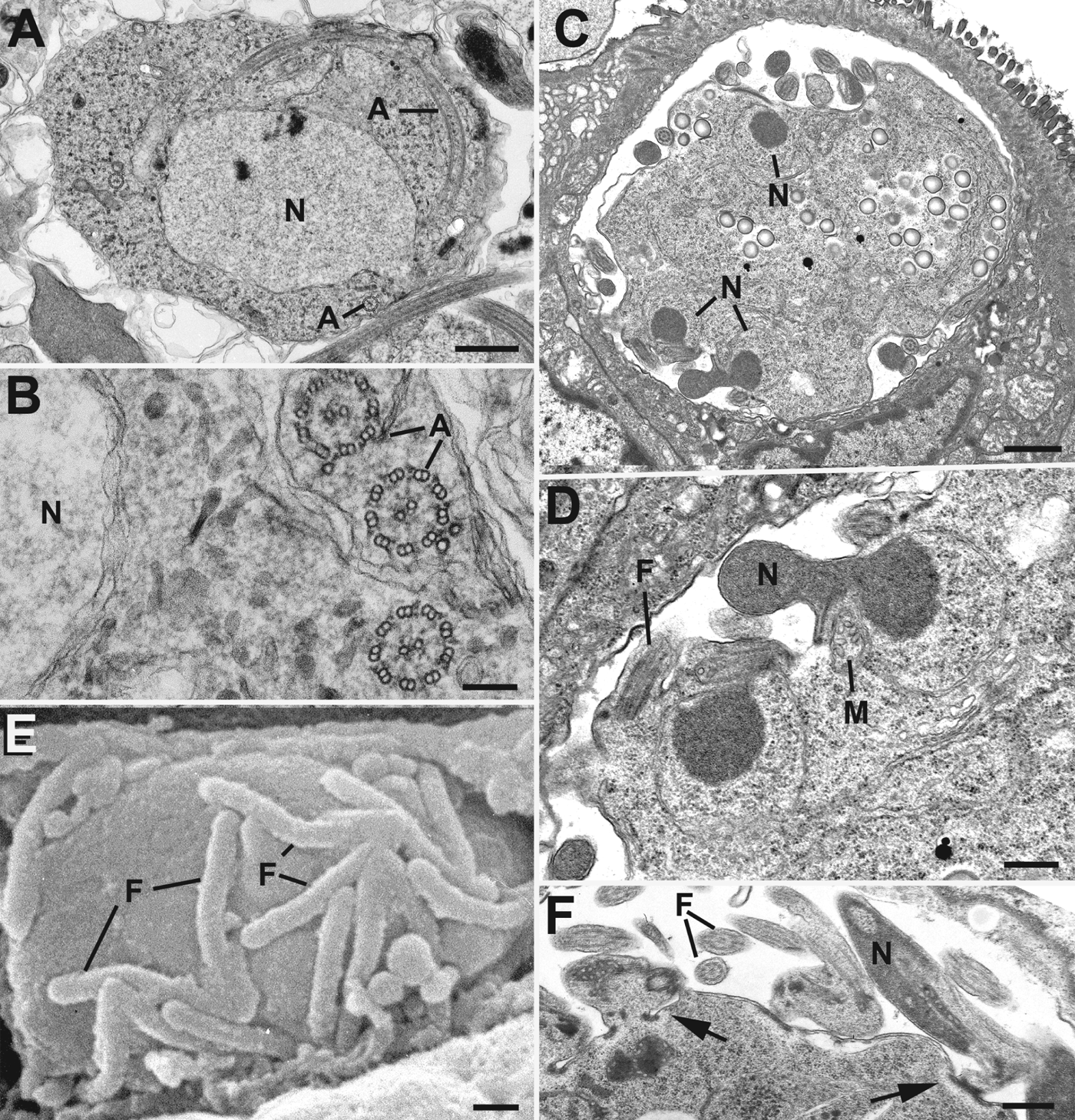
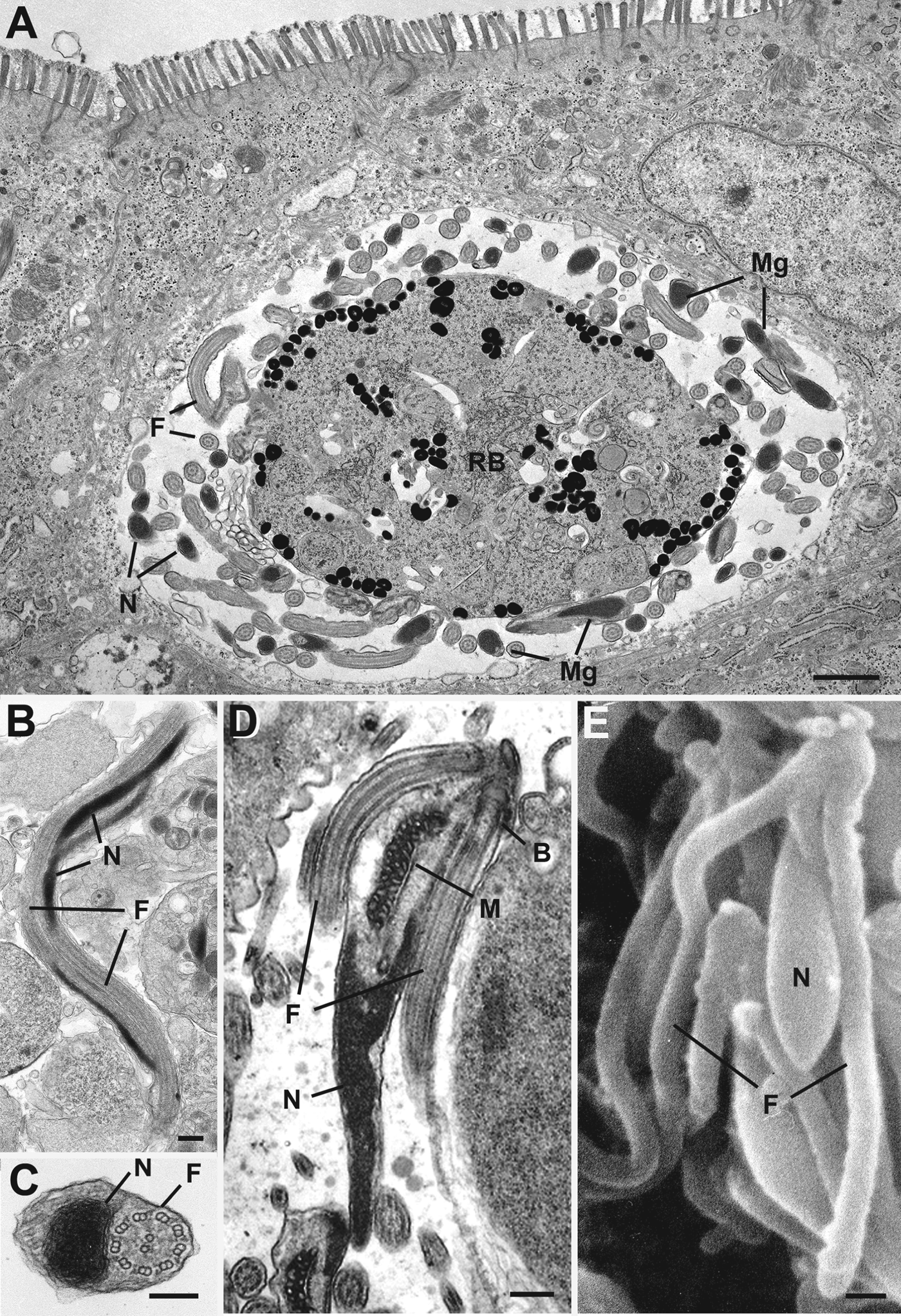
Microgametogenesis (Figs 2 and 3) in Eimeria (and T. gondii) takes place within a parasitophorous vacuole inside host gut epithelial cells and can be observed as early as 130 h post infection for E. maxima (Ball et al. Reference Ball, Pittilo, Joyner and Norton1981) or 120 h post infection for E. tenella (Laxer et al. Reference Laxer, Healey and Youssef1987). The process of microgamete formation involves a growth phase, in which there is repeated nuclear division, followed by a differentiation phase in which the nuclei move to the gametocyte periphery (Fig. 2C). The two centrioles become the basal bodies from which the axonemes develop, resulting in flagellar growth into the parasitophorous vacuole (Fig. 2D and F). The axonemes of Eimeria (and T. gondii) have the typical 9+2 microtubular structure also characteristic of Plasmodium microgametes (Sinden et al. Reference Sinden, Canning and Spain1976) (Fig. 2A and B; Fig. 3B and C). The emergence of the developed flagella generally precedes that of the microgamete body in E. maxima (Ball et al. Reference Ball, Pittilo, Joyner and Norton1981), Eimeria magna (Speer and Danforth, Reference Speer and Danforth1976), E. brunetti (Ferguson et al. Reference Ferguson, Birch-Andersen, Hutchison and Siim1977) and E. tenella (Madden and Vetterling, Reference Madden and Vetterling1977). This is followed by budding of the basal bodies, the electron-dense portion of the nucleus and an associated mitochondrion to form the microgamete (Fig. 2D). The mature microgametes are released into the parasitophorous vacuole, and consist of a nucleus, a mitochondrion, a number of longitudinally running microtubules, and two or three flagella (Scholtyseck et al. Reference Scholtyseck, Mehlhorn and Hammond1972; Fernando, Reference Fernando1973; Ferguson et al. Reference Ferguson, Birch-Andersen, Hutchison and Siim1977; Madden and Vetterling, Reference Madden and Vetterling1977; Elwasila, Reference Elwasila1983) (Figs 2E and 3A). The final number of microgametes formed from a single microgametocyte varies depending on the species, ranging from 20–30 for E. acervulina to 100 or more for E. maxima (Scholtyseck et al. Reference Scholtyseck, Mehlhorn and Hammond1972; West et al. Reference West, Smith and Read2000). A similar process operates in T. gondii (Fig. 2D and F, Fig. 3D and E), though each microgametocyte only produces 15–30 biflagellated microgametes (Ferguson and Dubremetz, Reference Ferguson, Dubremetz, Weiss and Kim2007).
Electron micrographs of microgametocytes and developing microgametes of Plasmodium berghei (A, B) Eimeria tenella (C, E) and Toxoplasma gondii (D, F). (A) Mid stage P. berghei microgametocyte showing the large nucleus (N) and a number of axonemes (A) located within the cytoplasm. Bar is 1 μm; (B) Detail of the P. berghei microgametocyte cytoplasm showing axonemes (A) with the classical 9+2 arrangement of microtubules. N – nucleus. Bar is 100 nm; (C). Mid-stage E. tenella microgametocyte showing a number of nuclei (N) located around the periphery. Bar is 1 μm; (D) Detail showing the electron dense portion of the nucleus (N) with associated mitochondrion (M) and flagella (F) protruding from the T. gondii microgametocyte surface into the parasitophorous vacuole. Bar is 200 nm; (E) Scanning electron micrograph of the surface of an E. tenella microgametocyte showing early stages of microgamete formation each with the two developing flagella (F). Bar is 100 nm; (F) Section through the periphery of a late-stage T. gondii microgametocyte showing microgametes within the parasitophorous vacuole but still attached by their anterior end to the gametocyte (arrows). N – nucleus, F – flagellum. Bar is 200 nm. (Images supplied by D. J. P. Ferguson.)
Electron micrographs of mature microgametes of Eimeria tenella (A), Plasmodium berghei (B, C) and Toxoplasma gondii (D, E). (A) Low power micrograph showing a mature E. tenella microgametocyte surrounded by fully formed microgametes (Mg) within a parasitophorous vacuole in the apical cytoplasm of a gut epithelial cell. F – flagellum, N – nucleus, RB –residual body. Bar is 1 μm; (B, C) Longitudinal (B) and cross (C) section through a P. berghei microgamete showing the undulating flagellum (F) and the electron dense nucleus (N) enclosed by the plasmalemma. Bar is 100 nm; (D) Longitudinal section through a T. gondii microgamete showing the electron-dense nucleus (N) anterior to which is the mitochondrion (M) and the basal bodies (B) giving rise to two flagella (F). Bar is 100 nm; (E) Scanning electron micrograph of a mature T. gondii microgamete showing the body of the microgamete containing the nucleus (N) and the two long posteriorly running flagella (F). Bar is 100 nm. (Images supplied by D. J. P. Ferguson.)
Despite the wealth of ultrastructural data on microgametogenesis in Eimeria, little is known about the molecular mechanisms that might dictate this development. However, it is known that the protein, MORN1, is associated with a ring-like structure from which microgametes bud from microgametocytes in both Eimeria and T. gondii (Ferguson et al. Reference Ferguson, Sahoo, Pinches, Bumstead, Tomley and Gubbels2008) (Fig. 4C). There is also genomic evidence that E. tenella and T. gondii have intraflagellar transport proteins that are absent in Plasmodium but which are associated with axoneme/flagellar formation (Briggs et al. Reference Briggs, Davidge, Wickstead, Ginger and Gull2004). Nevertheless, we can again speculate that similarities between Plasmodium and Eimeria may be found in other aspects of microgamete development, particularly in the molecular mechanisms relating to the signal cascades that accompany transformation of gametocytes into microgametes, the egress of microgametes from host cells and fertilization.
Light micrographs of plastic (A, B) and wax (C) sections stained with Azure A (A) or by immunofluorescence (B, C) of gut tissue infected with Eimeria maxima. (A) Part of a villus showing a mid stage microgametocyte (Mi) with numerous peripherally located dark blue nuclei (N), mature macrogamete (Ma) with large wall forming bodies (W) and a developing oocyst (O) with formation of the oocyst wall (OW). Bar is 10 μm; (B) Similar section to A showing a number of macrogametes characterized by the positive staining (green) of the WFB1 and 2 (W1, W2) labelled with antibodies to affinity purified gametocyte antigens (see Ferguson et al. Reference Ferguson, Belli, Smith and Wallach2003) and a developing oocyst (O) characterized by the positive staining of the developing oocyst wall. Bar is 10 μm; (C) Similar section to A showing a macrogamete (Ma) identified by positive staining (green) with antibodies to affinity purified gametocyte antigens (see Ferguson et al. Reference Ferguson, Belli, Smith and Wallach2003), while two mid-stage microgametocytes (M) show positive staining (red) with anti-MORN1. Bar is 10 μm. (Images supplied by D. J. P. Ferguson.)
The development of Plasmodium microgametocytes into microgametes is the result of a sophisticated molecular response of the parasite to its changing environment as it transits from mammalian to insect host. One of the earliest apparent responses is activation of the phosphoinositol pathway and release of calcium from cytoplasmic storage (Martin et al. Reference Martin, Jett and Schneider1994; Raabe et al. Reference Raabe, Wengelnik, Billker and Vial2011); this activates various protein kinases (Billker et al. Reference Billker, Dechamps, Tewari, Wenig, Franke-Fayard and Brinkmann2004; Khan et al. Reference Khan, Franke-Fayard, Mair, Lasonder, Janse, Mann and Waters2005; Rangarajan et al. Reference Rangarajan, Bei, Jethwaney, Maldonado, Dorin, Sultan and Doerig2005; Reininger et al. Reference Reininger, Billker, Tewari, Mukhopadhyay, Fennell, Dorin-Semblat, Doerig, Goldring, Harmse, Ranford-Cartwright and Packer2005, Reference Reininger, Tewari, Fennell, Holland, Goldring, Ranford-Cartwright, Billker and Doerig2009; Tewari et al. Reference Tewari, Dorin, Moon, Doerig and Billker2005; Ishino et al. Reference Ishino, Orito, Chinzei and Yuda2006; Siden-Kiamos et al. Reference Siden-Kiamos, Ecker, Nyback, Louis, Sinden and Billker2006; Dorin-Semblat et al. Reference Dorin-Semblat, Quashie, Halbert, Sicard, Doerig, Peat and Ranford-Cartwright2007; McRobert et al. Reference McRobert, Taylor, Deng, Fivelman, Cummings, Polley, Billker and Baker2008; Ranjan et al. Reference Ranjan, Ahmed, Gourinath and Sharma2009). These kinases regulate everything from parasite egress to DNA replication to nuclear division to flagella assembly (Sinden, Reference Sinden2009). Another kinase, the serine/arginine-rich protein kinase, is essential specifically for microgamete development; this kinase is highly conserved in eukaryotes, contributing to alternative splicing of mRNA and regulating mitosis, which may be underscoring its importance in microgamete development (Tewari et al. Reference Tewari, Straschil, Bateman, Bohme, Cherevach, Gong, Pain and Billker2010). There are orthologues of several of these kinases in T. gondii and Cryptosporidium parvum (Billker et al. Reference Billker, Lourido and Sibley2009) and it seems unthinkable that orthologues will not be found in Eimeria in due course. Similarly, conserved cell cycle proteins are crucial for microgamete formation in P. berghei (Guttery et al. Reference Guttery, Ferguson, Poulin, Xu, Straschil, Klop, Solyakov, Sandrini, Brady, Nieduszynski, Janse, Holder and Tewari2012a) and their involvement in coccidian microgametogenesis might also be anticipated. Likewise, coccidian microgametes can reasonably be expected to contain the evolutionarily conserved histone chaperone, FACT-L, a protein identified by proteomic analysis (Khan et al. Reference Khan, Franke-Fayard, Mair, Lasonder, Janse, Mann and Waters2005) and subsequently demonstrated to be important for microgamete development (Laurentino et al. Reference Laurentino, Taylor, Mair, Lasonder, Bartfai, Stunnenberg, Kroeze, Ramesar, Franke-fayard, Khan, Janse and Waters2011). Finally, transcripts of several proteases are expressed during Plasmodium gametogenesis (reviewed by Kuehn and Pradel, Reference Kuehn and Pradel2010) and two independent studies (Torres et al. Reference Torres, Rodriguez, Rodriguez and de la Cruz Hernandez-Hernandez2005; Rupp et al. Reference Rupp, Bosse, Schirmeister and Pradel2008) have shown that treatment of Plasmodium gametocytes with cysteine/serine protease inhibitors can affect microgamete formation; E. tenella possesses a number of protease genes, the expression of which is upregulated or specific for the sexual stages (Katrib et al. Reference Katrib, Ikin, Brossier, Robinson, Slapetova, Sharman, Walker, Belli, Tomley and Smith2012).
Macrogametogenesis
Macrogametogenesis in Eimeria occurs in a parasitophorous vacuole within the same type of host cell as the other stages. It occurs at specific post-infection time-points, which may vary depending on the species but, importantly, coincides with microgamete formation. Eimeria tenella macrogametogenesis can be observed as early as 120 h post infection (Klimes et al. Reference Klimes, Rootes and Tanielian1972) while, in E. maxima, the same process can be observed as early as 130 h post infection.
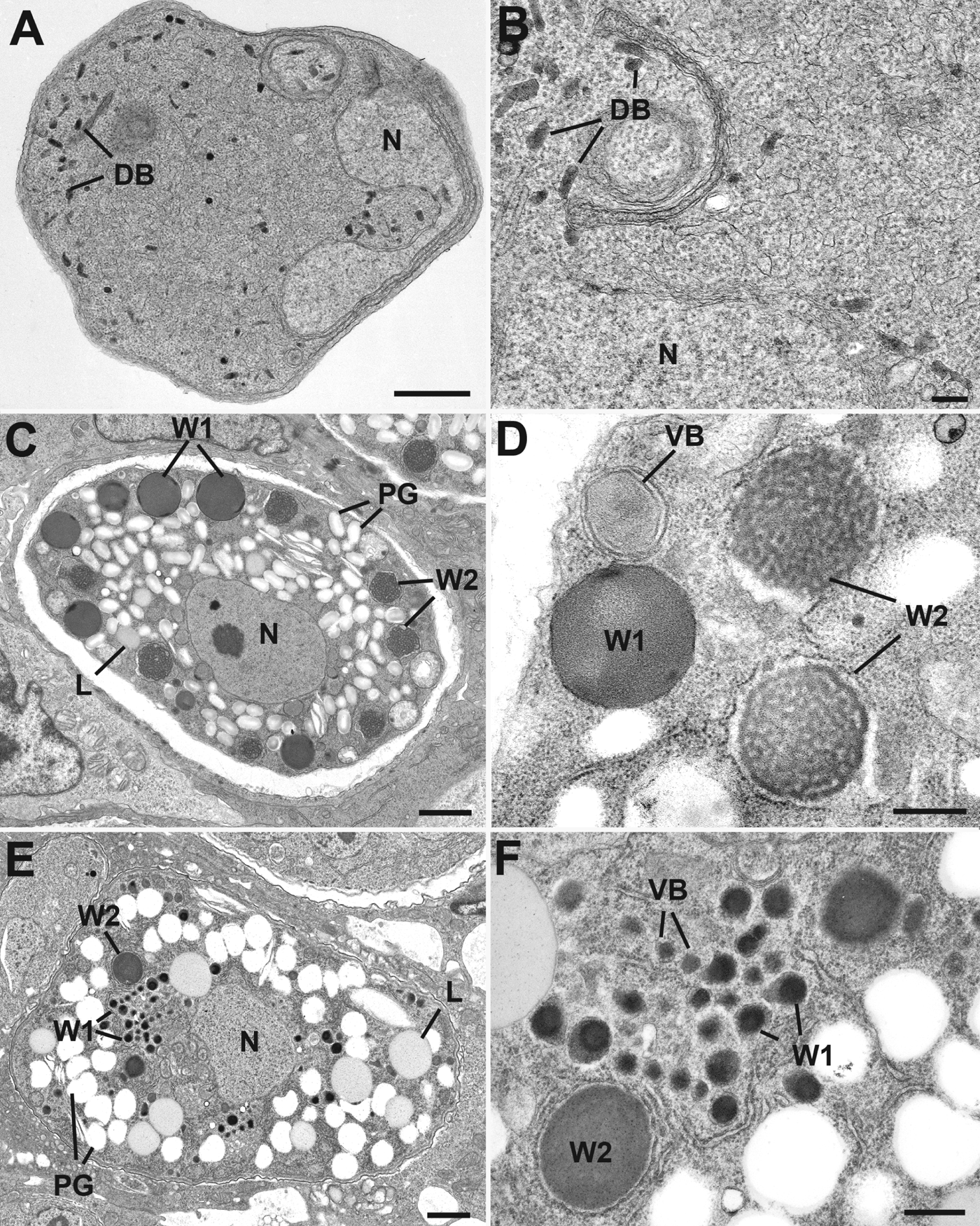
Development of the macrogametes is characterized, most notably, by the synthesis of a repertoire of unique organelles – the veil-forming bodies (VFB), the wall-forming bodies, type 1 (WFB1) and type 2 (WFB2) – as well as storehouses in the form of polysaccharide granules and lipid droplets (see, e.g. Pittilo and Ball, Reference Pittilo and Ball1979; Ferguson et al. Reference Ferguson, Belli, Smith and Wallach2003; reviewed by Belli et al. Reference Belli, Smith and Ferguson2006). The VFB and WFB2 start to form before the WFB1. In chicken Eimeria, the WFB1 are osmiophilic (i.e. possibly containing lipid and protein) and are larger than the WFB2 (Pittilo and Ball, Reference Pittilo and Ball1979; Ferguson et al. Reference Ferguson, Belli, Smith and Wallach2003) (Fig. 4A and B, Fig. 5C and D). However, in certain other Eimeria species and in T. gondii, the WFB1 are smaller than the WFB2 (Ferguson et al. Reference Ferguson, Hutchison, Dunachie and Siim1974; Scholtyseck et al. Reference Scholtyseck, Mehlhorn and Hammond1971) (Fig. 5E and F). This is in marked contrast to Plasmodium in which there are no VFB, WFB, polysaccharide granules or lipid droplets (Sinden, Reference Sinden1982) (Fig. 5A and B).
Electron micrographs of macrogametes of Plasmodium berghei (A, B), Eimeria tenella (C, D) and Toxoplasma gondii (E, F). (A, B) Low power (A) and detail (B) of the macrogamete of P. berghei showing the nucleus (N) and the cytoplasm containing a few cigar-shaped dense bodies (DB). Bars are 1 μm and 100 nm, respectively; (C, D) Low power (C) and detail (D) of the macrogamete of E. tenella showing the nucleus (N) while the cytoplasm contains a number of veil-forming bodies (VB), large electron-dense WFB1 (W1) and a number of WFB2 (W2), which are located within membranes of rough endoplasmic reticulum, plus large numbers of polysaccharide granules (PG) and lipid droplets (L). Bars are 1 μm and 500 nm, respectively; (E, F) Low power (E) and detail (F) of the macrogamete of T. gondii showing the nucleus (N) while the cytoplasm contains a number of veil-forming bodies (VB) (identified by immunoelectron microscopy), small electron dense WFB1 (W1), and a few larger WFB2 (W2), plus large numbers of polysaccharide granules (PG) and lipid droplets (L). Bars are 1 μm and 200 nm, respectively. (Images supplied by D. J. P. Ferguson.)
The VFB are released during macrogamete formation and, following presumptive fertilization, the WFB1 and WFB2 ultimately form the outer and inner layers of the oocyst wall, respectively (discussed below). A limited number of proteins have been identified during eimerian macrogamete development, almost all of which are stored in wall-forming bodies and are, ultimately, incorporated into the developing oocyst wall (discussed below). Additionally, eimerian macrogametes carry the non-nuclear genome of the apicoplast, which exhibits maternal inheritance (Ferguson et al. Reference Ferguson, Campbell, Henriquez, Phan, Mui, Richards, Muench, Allary, Lu, Prigge, Tomley, Shirley, Rice, McLeod and Roberts2007). However, information on other macrogamete-specific proteins that might be involved in other aspects of sexual stage biology is lacking and, again, clues may be sought from malaria parasites.
Plasmodium macrogametocytes respond to their uptake by mosquitoes in an almost identical manner to, and using similar molecular machinery as, microgametocytes, at least initially. They revert to a rounded morphology and egress from the red blood cell within a few minutes of arrival in their insect host's midgut (Sinden, Reference Sinden2009). Electron-dense, gametocyte-specific, osmiophilic bodies (Fig. 5A and B) (Sinden, Reference Sinden1982) are mobilized and release Pfg377 (Alano et al. Reference Alano, Read, Bruce, Aikawa, Kaido, Tegoshi, Bhatti, Smith, Luo, Hansra, Carter and Elliott1995; Severini et al. Reference Severini, Silvestrini, Sannella, Barca, Gradoni and Alano1999) and MDV-1/PEG-3 (Furuya et al. Reference Furuya, Mu, Hayton, Liu, Duan, Nkrumah, Joy, Fidock, Fujioka, Vaidya, Wellems and Su2005; Silvestrini et al. Reference Silvestrini, Bozdech, Lanfrancotti, Giulio, Bultrini, Picci, Derisi, Pizzi and Alano2005; Lanfrancotti et al. Reference Lanfrancotti, Bertuccini, Silvestrini and Alano2007; Lal et al. Reference Lal, Delves, Bromley, Wastling, Tomley and Sinden2009), proteins that, along with GEST (Talman et al. Reference Talman, Lacroix, Marques, Blagborough, Carzaniga, Menard and Sinden2011), play crucial roles in egress (de Koning-Ward et al. Reference de Koning-Ward, Olivieri, Bertuccini, Hood, Silvestrini, Charvalias, Berzosa Diaz, Camarda, McElwain, Papenfuss, Healer, Baldassarri, Crabb, Alano and Ranford-Cartwright2008; Ponzi et al. Reference Ponzi, Siden-Kiamos, Bertuccini, Curra, Kroeze, Camarda, Pace, Franke-Fayard, Laurentino, Louis, Waters, Janse and Alano2009).
Whilst microgametes are little more than a vehicle to deliver DNA, macrogametes are a molecular storehouse for the next generation of parasites. In the Coccidia, examples of this include polysaccharide granules and lipid droplets, which are abundant in macrogametes (Fig. 5C and E), and the storage, in the wall-forming bodies of coccidian parasites, of precursor proteins that are destined for processing and incorporation into the oocyst wall (see below and Belli et al. Reference Belli, Smith and Ferguson2006 for a review). Another example is the translational repression that is initially evident during macrogametocytogenesis in malaria parasites and carried through the macrogamete until after fertilization. This phenomenon is mediated by DOZI (development of zygote inhibited), a member of the DDX6 family of RNA helicases, and CITH (a homologue of Caenorhabditis elegans CAR-1 and Drosophila Trailer Hitch), both of which are essential for zygote development (Mair et al. Reference Mair, Braks, Garver, Wiegant, Hall, Dirks, Khan, Dimopoulos, Janse and Waters2006, Reference Mair, Lasonder, Garver, Franke-Fayard, Carret, Wiegant, Dirks, Dimopoulos, Janse and Waters2010). DOZI and CITH are core to the formation of Plasmodium P granules, within which around 20% (> 370) of macrogamete mRNA transcripts are stabilized, hidden from the cell's translational machinery and, hence, stored until after fertilization (Mair et al. Reference Mair, Braks, Garver, Wiegant, Hall, Dirks, Khan, Dimopoulos, Janse and Waters2006, Reference Mair, Lasonder, Garver, Franke-Fayard, Carret, Wiegant, Dirks, Dimopoulos, Janse and Waters2010). This process of translational repression is highly conserved in eukaryotic germ-line cells (Hughes et al. Reference Hughes, Philip, Starnes, Taylor and Waters2010) and, therefore, it may be anticipated that DOZI and CITH will be detected in the macrogametes of Eimeria in the not too distant future. However, it must be kept in mind that the needs of Eimeria and Plasmodium are very different. Within the Coccidia, there is a need to take up and store all the molecular requirements to fulfil subsequent development (sporulation), which occurs within the oocyst in the external environment and forms only a limited number of infectious sporozoites (eight in Eimeria and Toxoplasma), and also to maintain their viability for months. In contrast, the Plasmodium macrogamete, upon fertilization, converts into a motile ookinete before forming an oocyst at the basal lamina of the insect host's gut wall, where it obtains nutrients, undergoes marked growth and produces large numbers of sporozoites.
FERTILIZATION AND MEIOSIS
The observation of fertilization per se has proved elusive in Apicomplexa such as Toxoplasma or Eimeria but it is known that zygotes form and develop into oocysts, which are excreted into the outside world and, within which, the sexual phase continues as meiosis, followed by mitosis, leading to the generation of haploid sporozoites over several days. It has, however, been postulated, due to the lack of solid empirical observations of fertilization, that the development of oocysts in Coccidia could take place without the necessity of fertilization, for instance, via parthenogenesis (Ferguson, Reference Ferguson2002). However, in Eimeria there is some evidence that fertilization of macrogametes by microgametes is necessary for the production of a viable oocyst. First, monoclonal antibodies raised against purified E. tenella microgametes have been shown to reduce oocyst formation in vitro by greater than 50% (Laxer et al. Reference Laxer, Healey and Youssef1987) and, second, a correlation was noted between reduced microgamete numbers (following prolonged culturing at sub-standard temperatures) and poor sporulation rates in oocysts (Long, Reference Long1972). Together, these findings suggest that, while some oocysts may still be produced and excreted in the absence of fertilization, they may ultimately fail to sporulate (i.e. produce sporozoites) and are, therefore, non-infective. Furthermore, although fertilization may be empirically presumptive in Eimeria, genetic recombination has been shown convincingly in E. tenella (Shirley and Harvey, Reference Shirley and Harvey1996), E. maxima (Joyner and Norton, Reference Joyner and Norton1975), E. acervulina (Sutton et al. Reference Sutton, Shirley and McDonald1986) and E. mitis (Shirley, Reference Shirley1978), and also for T. gondii (Pfefferkorn and Pfefferkorn, Reference Pfefferkorn and Pfefferkorn1980). Furthermore, genetic recombination has been shown to be a potential mechanism for the development of drug resistance in E. maxima (Blake et al. Reference Blake, Hesketh, Archer, Carroll, Smith and Shirley2004).
Little is known of the physical process of fertilization in Coccidia with the exception of a few preliminary ultrastructural studies based on limited observations showing microgametes in contact with macrogametes (Scholtyseck et al. Reference Scholtyseck, Mehlhorn and Hammond1971; Madden and Vetterling, Reference Madden and Vetterling1977; Elwasila, Reference Elwasila1983) (Fig. 6A). It is not even clear when or where fertilization takes place, since microgametes have no cell-invading machinery, which could suggest it occurs extracellularly (in the gut lumen). However, oocyst wall formation is initiated while the macrogamete is still within the host cell (Fig. 4A and B, Fig. 6B). How the microgamete could penetrate the oocyst wall is unclear. Currently, no proteins or molecular events underpinning fertilization or meiosis in Eimeria have been discovered and, again, we must turn to Plasmodium for clues.
Images of macrogamete and oocyst formation in Toxoplasma gondii (A, G), Eimeria tenella (B, E, F) and Eimeria maxima (C, D). (A) Scanning electron micrograph showing a number of microgametes (Mg) characterized by their flagella in contact with a macrogamete (Ma) that could represent a stage in fertilization in T. gondii. Me – merozoite. Bar is 1 μm; (B) Light micrograph of a section through a chicken caecum infected with E. tenella showing a number of mature macrogametes (Ma), a few microgametocytes (M) and a developing oocyst (O) that is still located within a host cell. Bar is 5 μm; (C) Immunofluorescent images showing E. maxima WFB1, positively stained (green) with antibodies to affinity purified gametocyte antigens (see Ferguson et al. Reference Ferguson, Belli, Smith and Wallach2003), coalescing at the surface to initiate oocyst wall formation (arrowheads) in E. maxima. Bar is 5 μm; (D) Immunofluorescent image showing a developing E. maxima oocyst with the wall (OW) positively stained (green) with antibodies to affinity purified gametocyte antigens (see Ferguson et al. Reference Ferguson, Belli, Smith and Wallach2003) in E. maxima. Bar is 5 μm; (E) Low-power transmission electron micrograph of a fully formed oocyst of E. tenella in the gut lumen, showing the oocyst wall (OW) enclosing a cytoplasmic mass that, having lost its WFBs, consists of a central nucleus (N) surrounded by polysaccharide granules (PG) and lipid droplets (L). Bar is 1 μm; (F) Detail of the periphery of the E. tenella oocyst in E showing the loose outer veil (V) and the wall consisting of the thick electron dense outer layer (OL) and the more electron lucent inner layer (IL), while the cytoplasmic mass is limited by the plasmalemma. Bar is 100 nm; (G) Detail of the periphery of an oocyst of T. gondii showing the loose veil (V) and the wall consisting of a thin electron dense outer layer (OL) and the more electron lucent inner layer (IL). Bar is 100 nm. (Images supplied by D. J. P. Ferguson.)
Fertilization in Plasmodium, in contrast to other Apicomplexa, is well documented (Kuehn and Pradel, Reference Kuehn and Pradel2010); indeed, it was observed over 100 years ago (MacCallum, Reference MacCallum1897) and described in detail nearly 40 years ago (Sinden and Croll, Reference Sinden and Croll1975; Sinden et al. Reference Sinden, Canning and Spain1976). Interaction between microgametes and macrogametes in the lead-up to fertilization appears to be mediated by a number of surface proteins with adhesive properties (reviewed by Sinden, Reference Sinden2009; Baker, Reference Baker2010). These include P45/48, P47 and P230, which are all members of a family of proteins characterized by the possession of 6-cysteine domains. P47 is macrogamete-specific (van Schaijk et al. Reference van Schaijk, van Dijk, van de Vegte-Bolmer, van Gemert, van Dooren, Eksi, Roeffen, Janse, Waters and Sauerwein2006) whereas P45/48 and P230, though found in both micro- and macrogametes, seem to be more important in microgamete fertility (van Dijk et al. Reference van Dijk, Janse, Thompson, Waters, Braks, Dodemont, Stunnenberg, van Gemert, Sauerwein and Eling2001; Sinden, Reference Sinden2009). These proteins may form surface complexes as a prelude to fertilization (Sinden, Reference Sinden2009). Another group of adhesive or attachment proteins is the CCP family (Simon et al. Reference Simon, Scholz, Moreira, Templeton, Kuehn, Dude and Pradel2009), at least one of which is known to associate with P230 (Scholz et al. Reference Scholz, Simon, Lavazec, Dude, Templeton and Pradel2008), which is highly suggestive of an important role in microgamete to macrogamete binding (Baker, Reference Baker2010). What role recently discovered, nanotube-like filaments (Kuehn and Pradel, Reference Kuehn and Pradel2010) play in bringing microgametes and macrogametes together, and whether this involves any of these adhesive proteins, remains to be determined. Whether similar processes operate in Eimeria (or other apicomplexans) is unknown but, hopefully, will be more readily analysable with the completion of Eimeria genome-sequencing projects.
In Plasmodium, once contact between micro- and macrogametes has been made, fusion of the gamete membranes proceeds quickly, mediated by HAP2/GCS1 (Hirai et al. Reference Hirai, Arai, Mori, Miyagishima, Kawai, Kita, Kuroiwa, Terenius and Matsuoka2008; Liu et al. Reference Liu, Tewari, Ning, Blagborough, Garbom, Pei, Grishin, Steele, Sinden, Snell and Billker2008). Homologous sequences for hap2/gcs1 are also found in the genomes of T. gondii and E. tenella (Sinden et al. Reference Sinden, Carter, Drakeley and Leroy2012). Following membrane fusion, the microgamete moves into the cytoplasm of the macrogamete and, within hours, nuclear fusion occurs, resulting in a diploid zygote (Sinden et al. Reference Sinden, Hartley and Winger1985). The zygote undergoes meiosis without cell division, becomes tetraploid and transforms into an ookinete over the next 24 h, thus marking the end of the sexual phase of the Plasmodium life cycle (Sinden et al. Reference Sinden, Hartley and Winger1985; Janse et al. Reference Janse, van der Klooster, van der Kaay, van der Ploeg and Overdulve1986). At a molecular level, the genes held dormant during gametocytogenesis in the mammalian host are now expressed, control being mediated by the combined efforts of transcription factors like AP2-O (Yuda et al. Reference Yuda, Iwanaga, Shigenobu, Mair, Janse, Waters, Kato and Kaneko2009), kinases such as Nek-1, Nek-4, PK7, GAK and CDPK1 (Reininger et al. Reference Reininger, Billker, Tewari, Mukhopadhyay, Fennell, Dorin-Semblat, Doerig, Goldring, Harmse, Ranford-Cartwright and Packer2005, Reference Reininger, Tewari, Fennell, Holland, Goldring, Ranford-Cartwright, Billker and Doerig2009; Tewari et al. Reference Tewari, Straschil, Bateman, Bohme, Cherevach, Gong, Pain and Billker2010; Sebastian et al. Reference Sebastian, Brochet, Collins, Schwach, Jones, Goulding, Rayner, Choudhary and Billker2012), the translational repressors, DOZI and CITH (Mair et al. Reference Mair, Braks, Garver, Wiegant, Hall, Dirks, Khan, Dimopoulos, Janse and Waters2006, Reference Mair, Lasonder, Garver, Franke-Fayard, Carret, Wiegant, Dirks, Dimopoulos, Janse and Waters2010), and the unique protein phosphatase with Kelch-like domains, PPKL (Guttery et al. Reference Guttery, Poulin, Ferguson, Szoor, Wickstead, Carroll, Ramakrishnan, Brady, Patzewitz, Straschil, Solyakov, Green, Sinden, Tobin, Holder and Tewari2012b), all of which are critical for zygote and/or ookinete development. At least some of these factors (e.g. Nek-1, Nek-4, DOZI) play important roles in, or set the stage for, meiosis (Reininger et al. Reference Reininger, Billker, Tewari, Mukhopadhyay, Fennell, Dorin-Semblat, Doerig, Goldring, Harmse, Ranford-Cartwright and Packer2005, Reference Reininger, Tewari, Fennell, Holland, Goldring, Ranford-Cartwright, Billker and Doerig2009; Mair et al. Reference Mair, Lasonder, Garver, Franke-Fayard, Carret, Wiegant, Dirks, Dimopoulos, Janse and Waters2010) and, given their evolutionarily conservative natures, might be anticipated to be important in zygote development in parasites like Eimeria and Toxoplasma.
The result of fertilization and meiosis in Plasmodium is the ookinete, which traverses the insect midgut epithelium and forms an oocyst between the basal lamina and the gut epithelium. However, unlike the oocysts of Eimeria, Plasmodium oocysts are not excreted and are not a site for any further sexual development. Rather, this oocyst stage remains in the insect, a marked growth phase ensues and a series of endomitotic divisions occurs; the resulting sporozoites burst from the oocyst and invade the salivary glands, ready for subsequent transmission to the mammalian host (Sinden, Reference Sinden2009). In contrast, for Eimeria and other Coccidia, the oocyst is the stage in which sexual reproduction is completed; meiosis proceeds, inside the protective oocyst wall, followed by mitosis to produce the infectious sporozoites. Thus, assembly of the oocyst wall is a critical part of sexual reproduction for coccidian parasites.
OOCYST WALL DEVELOPMENT
The formation of an oocyst wall through the coalescence of veil- and wall-forming bodies in macrogametes is an event that is well documented, not only in various species of Eimeria (Scholtyseck et al. Reference Scholtyseck, Mehlhorn and Hammond1971; Mouafo et al. Reference Mouafo, Weck-Heimann, Dubremetz and Entzeroth2002; Ferguson et al. Reference Ferguson, Belli, Smith and Wallach2003; Belli et al. Reference Belli, Ferguson, Katrib, Slapetova, Mai, Slapeta, Flowers, Miska, Tomley, Shirley, Wallach and Smith2009), but also in other cyst-forming Apicomplexa such as Sarcocystis (Vetterling et al. Reference Vetterling, Pacheco and Fayer1973), T. gondii (Scholtyseck et al. Reference Scholtyseck, Mehlhorn and Hammond1971; Ferguson et al. Reference Ferguson, Hutchison and Siim1975, Reference Ferguson, Becht and Soldati2000; Speer and Dubey, Reference Speer and Dubey2005) and C. parvum (Spano et al. Reference Spano, Puri, Ranucci, Putignani and Crisanti1997). Many of the components of the oocyst wall are produced and stored during macrogamete development; the similarity in macrogamete development in different cyst-forming Apicomplexa is clearly related to the conserved biology of this remarkable structure. The oocyst wall acts as a protective barrier that shields the parasite from harsh, external pressures faced during transmission between hosts, including environmental stress and chemical disinfection (Belli et al. Reference Belli, Smith and Ferguson2006).
Ultrastructural morphology and biochemical composition
The oocyst wall of Eimeria is a bilayered structure and is composed of an outer layer and inner layer, which incorporate the contents of WFBs type 1 and 2, respectively (Stotish et al. Reference Stotish, Wang and Meyenhofer1978; Pittilo and Ball, Reference Pittilo and Ball1980; Elwasila, Reference Elwasila1984; Ferguson et al. Reference Ferguson, Belli, Smith and Wallach2003) (Figs 4B and 6C–F). Early studies indicated that the outer layer is made up of lipid material, whereas the inner layer is comprised mainly of disulphide-linked glycoprotein, evidenced mainly by the dissociation effect of thiol reagents (Stotish et al. Reference Stotish, Wang and Meyenhofer1978). Recent analyses of purified E. maxima and E. tenella unsporulated oocyst walls (containing both the inner and outer layer) demonstrated a protein content exceeding 90% in both instances (Mai et al. Reference Mai, Sharman, Walker, Katrib, De Souza, McConville, Wallach, Belli, Ferguson and Smith2009). The T. gondii oocyst wall has also been described as bilayered, although, in contrast to Eimeria species, the electron-dense outer layer is thinner than the inner layer (Ferguson et al. Reference Ferguson, Hutchison and Siim1975) (cf. Fig. 6F and G). The developing oocyst is enclosed by an outer veil, initially described for T. gondii, but also present in Eimeria species (Ferguson et al. Reference Ferguson, Becht and Soldati2000, Reference Ferguson, Belli, Smith and Wallach2003; Belli et al. Reference Belli, Ferguson, Katrib, Slapetova, Mai, Slapeta, Flowers, Miska, Tomley, Shirley, Wallach and Smith2009) (Fig. 6F and G). This veil is lost by the time that oocysts are excreted with the feces.
Molecular composition
A number of proteins, ranging in size from 8 to 33 kDa, found in purified wall samples of E. maxima oocysts, can be mapped to the amino acid sequence and/or have been shown to cross-react with antibodies raised against the macrogamete-specific proteins, GAM56 (Belli et al. Reference Belli, Wallach, Luxford, Davies and Smith2003a) and GAM82 (Belli et al. Reference Belli, Wallach and Smith2003b), respectively. This suggests that gametocyte-specific proteins are processed into smaller peptides prior to their incorporation into the oocyst wall. Similar observations have been made for other Eimeria species (Mouafo et al. Reference Mouafo, Weck-Heimann, Dubremetz and Entzeroth2002; Krucken et al. Reference Krucken, Hosse, Mouafo, Entzeroth, Bierbaum, Marinovski, Hain, Greif and Wunderlich2008; Belli et al. Reference Belli, Ferguson, Katrib, Slapetova, Mai, Slapeta, Flowers, Miska, Tomley, Shirley, Wallach and Smith2009). It is not completely clear what mechanism is governing the processing of these gametocyte-specific proteins prior to their incorporation into the oocyst wall, although Katrib et al. (Reference Katrib, Ikin, Brossier, Robinson, Slapetova, Sharman, Walker, Belli, Tomley and Smith2012) have proposed that parasite-specific proteases are involved.
GAM56 and GAM82 contain one and two tyrosine-rich domains, respectively, in their amino acid sequences. Following the predicted processing of these proteins, fragments of GAM56 and GAM82 are proposed to cross-link, via the oxidative catalysis of dityrosine bond formation, to form an extracellular matrix around the developing zygote or oocyst (Belli et al. Reference Belli, Wallach, Luxford, Davies and Smith2003a, Reference Belli, Smith and Ferguson2006; Mai et al. Reference Mai, Smith, Feng, Karib, Slapeta, Slapetova, Wallach, Luxford, Davies, Zhang, Norton and Belli2011). The role of tyrosine-rich proteins in the development of extracellular matrices has also been documented for other parasites, including Schistosoma mansoni (Chen et al. Reference Chen, Rekosh and LoVerde1992) and Fasciola hepatica (Waite and Rice-Ficht, Reference Waite and Rice-Ficht1987), specifically as major components of their eggshells. No direct homologues of GAM56 or GAM82 can be found in T. gondii but six tyrosine-rich proteins have been found in the oocysts of this parasite (Fritz et al. Reference Fritz, Bowyer, Bogyo, Conrad and Boothroyd2012a, Reference Fritz, Buchholz, Chen, Durbin-Johnson, Rocke, Conrad and Boothroydb). No homologues exist in any of the Plasmodium genomes, probably reflecting the fact that malaria parasites do not produce an excreted oocyst or develop a hardened oocyst wall.
A second class of oocyst wall protein has also been identified in E. tenella and also appears to represent a WFB2-specific protein. This protein, GAM22, is a histidine- and proline-rich glycoprotein that has been detected in mature gametocytes (at 168 h post infection) and unsporulated oocysts (Krucken et al. Reference Krucken, Hosse, Mouafo, Entzeroth, Bierbaum, Marinovski, Hain, Greif and Wunderlich2008). It is expressed much later than GAM56 (Mouafo et al. Reference Mouafo, Weck-Heimann, Dubremetz and Entzeroth2002). Unlike GAM56, there is no indication that GAM22 is processed into smaller peptides prior to wall formation. However, as proposed by Krucken et al. (Reference Krucken, Hosse, Mouafo, Entzeroth, Bierbaum, Marinovski, Hain, Greif and Wunderlich2008), it is tempting to speculate that it may play a role in stabilizing the oocyst wall via cross-links between histidine and catechols, as seen in insect cuticles.
We have also very recently identified (unpublished) a third class of oocyst wall protein in E. tenella and E. maxima, namely, homologues of the cysteine-rich Cryptosporidium oocyst wall proteins (COWPs). There are nine known COWPs in Cryptosporidium, all displaying varying degrees of conservation of Type I and Type II cysteine-rich repeats (Lally et al. Reference Lally, Baird, McQuay, Wright and Oliver1992; Ranucci et al. Reference Ranucci, Muller, La Rosa, Reckmann, Morales, Spano, Pozio and Crisanti1993; Spano et al. Reference Spano, Puri, Ranucci, Putignani and Crisanti1997; Templeton et al. Reference Templeton, Lancto, Vigdorovich, Liu, London, Hadsall and Abrahamsen2004). At least some of these appear to localize to the inner oocyst wall, the wall-forming bodies of macrogametes and to a single large vesicle within immature macrogametocytes, indicating this protein is potentially an early indicator of sexual stage development. Furthermore, semi-quantitative RT-PCR analyses revealed upregulated transcription of all nine genes in parasite stages associated with sexual stage development and oocyst formation in C. parvum, supporting the hypothesis that these COWP proteins are involved in the formation of the oocyst wall. The study by Templeton et al. (Reference Templeton, Lancto, Vigdorovich, Liu, London, Hadsall and Abrahamsen2004) also uncovered a homologue of COWP6 in the T. gondii genome sequence (referred to as TgOWP1) and, subsequently, seven further COWP homologues have been uncovered in T. gondii (Possenti et al. Reference Possenti, Cherchi, Bertuccini, Pozio, Dubey and Spano2010). As is the case for tyrosine-rich proteins, no plasmodial homologues to COWP have been found.
Various authors (Lally et al. Reference Lally, Baird, McQuay, Wright and Oliver1992; Ranucci et al. Reference Ranucci, Muller, La Rosa, Reckmann, Morales, Spano, Pozio and Crisanti1993; Spano et al. Reference Spano, Puri, Ranucci, Putignani and Crisanti1997; Templeton et al. Reference Templeton, Lancto, Vigdorovich, Liu, London, Hadsall and Abrahamsen2004, Possenti et al. Reference Possenti, Cherchi, Bertuccini, Pozio, Dubey and Spano2010) have speculated that disulphide bonding between cysteine residues of COWPs may play a role in stabilizing the structure of the oocyst wall. Interestingly, the cyst walls that form around the gamonts of another apicomplexan, Gregarina niphandrodes (Gregarinasina), also have a cysteine-rich protein that shows high similarity to COWP proteins (Omoto et al. Reference Omoto, Toso, Tang and Sibley2004). Like Cryptosporidium, Toxoplasma and Eimeria, this organism produces a cyst that is excreted via the feces and transmitted to a new, susceptible host (a mealworm beetle). Even in more divergent organisms, like the cnidarian, Hydra vulgaris, the development of the cyst wall (in this case, the nematocyst wall) appears to represent a conserved mechanism involving the cross-linking of highly-repetitive, cysteine-rich proteins (Engel et al. Reference Engel, Ozbek, Streitwolf-Engel, Petri, Lottspeich and Holstein2002; Ozbek et al. Reference Ozbek, Pokidysheva, Schwager, Schulthess, Tariq, Barth, Milbradt, Moroder, Engel and Holstein2004).
Carbohydrates may also play critical roles in oocyst wall construction. The primary enzyme in the glycosylation pathway, glucosamine : fructose-6-phosphate aminotransferase has been characterized in E. tenella and is expressed specifically in macrogametes, transcription occurring early in gametocyte development but protein expression being restricted to mature gametes prior to their conversion into oocysts (Walker et al. Reference Walker, Slapetova, Slapeta, Miller and Smith2010). Genes coding for three other enzymes needed for N-galactosamine synthesis are also transcribed in the course of E. tenella macrogamete development and both GAM56 and GAM82 contain high levels of N-galactosamine (Walker et al. Reference Walker, Slapetova, Slapeta, Miller and Smith2010). Thus, it is possible that O-linked glycosylation of these proteins plays a role in directing them to the developing oocyst wall at the appropriate time. Additionally, carbohydrates may be important oocyst wall constituents in their own right since β-1,3-Glucan has been proposed to form a structurally critical, trabecular scaffold in the inner layer of the oocyst wall of both T. gondii and E. tenella (Bushkin et al. Reference Bushkin, Motari, Magnelli, Gubbels, Dubey, Miska, Bullitt, Costello, Robbins and Samuelson2012).
CONCLUSIONS AND PERSPECTIVES
Understanding of the mechanics of sexual reproduction and transmission of apicomplexan parasites has advanced significantly in recent years, proving to be more than just an esoteric pursuit; it has resulted in the commercialization of a transmission-blocking subunit vaccine against Eimeria species, preventing coccidiosis in flocks of poultry (Wallach et al. Reference Wallach, Ashash, Michael and Smith2008). This vaccine contains a complex of immunogenic proteins, purified from the gametocytes of E. maxima, including GAM56 and GAM82 (Belli et al. Reference Belli, Witcombe, Wallach and Smith2002, Reference Belli, Wallach and Smith2003b), proteins that localize to the wall-forming bodies of the macrogamete, the fused wall-forming bodies in the developing zygote, and the inner layer of the bi-layered wall of the resulting oocyst (Ferguson et al. Reference Ferguson, Belli, Smith and Wallach2003). Vaccination with these proteins stimulates an immune response that is hypothesized to negate construction of the oocyst wall, a protective barrier necessary for the completion of the parasite's sexual phase, and for survival in the outside world prior to transmission to the next host (Sharman et al. Reference Sharman, Smith, Wallach and Katrib2010). This vaccine illustrates that transmission-blocking immunity can be implemented successfully to control apicomplexan parasites.
It is not far-fetched to believe that similar control can be achieved for other diseases caused by apicomplexans. Indeed, irrepressible momentum to develop and implement strategies to interrupt transmission of Plasmodium, as a critical element of a campaign to eradicate malaria, has shone an intense spotlight on the sexual phase of the apicomplexan life cycle (Alonso et al. Reference Alonso, Brown, Arevalo-Herrera, Binka, Chitnis, Collins, Doumbo, Greenwood, Hall, Levine, Mendis, Newman, Plowe, Rodriguez, Sinden, Slutsker and Tanner2011; Sinden et al. Reference Sinden, Carter, Drakeley and Leroy2012). A transmission-blocking vaccine against T. gondii, in which case the sexual stage-specific feline host represents a bottleneck in the parasite's life cycle, is also an attractive target.
Garnering a thorough understanding of sexual stage biology in the Apicomplexa will be fundamental to designing and implementing any effective transmission-blocking strategy; the successful design of transmission-blocking vaccines that target the sexual stages of Eimeria species has only been possible due to decades of research dedicated to this biological process. And, even in the case of this (limited) success story, less than absolute inhibition of transmission is a feature of vaccination (Wallach et al. Reference Wallach, Smith, Petracca, Miller, Eckert and Braun1995, Reference Wallach, Ashash, Michael and Smith2008). This may be adequate, even optimal, for protection against poultry coccidiosis in the integrated, self-contained environments used to raise commercial poultry (Wallach et al. Reference Wallach, Ashash, Michael and Smith2008) but will likely be less satisfactory for similar strategies to prevent transmission of, for example, malaria parasites or T. gondii. Perhaps the answer is multipronged vaccines that target, simultaneously: (a) the amplification of merozoites and/or their transformation into gametocytes; (b) the flagella of the microgametes, halting their ability to move to macrogametocytes; (c) proteins (presumably on the surface) of microgametes and/or macrogametocytes that mediate binding of the two in preparation for fertilization; and (d) in the case of coccidian parasites at least, proteins that give the oocyst wall its form and function.
Although analogous in part to Eimeria species, sexual stage development in other apicomplexans is nonetheless sufficiently divergent to warrant an entirely new assessment of possible transmission-blocking strategies. Historically, the analysis of apicomplexan sexual stages has proven a challenging task, due in no small part to the lack of reliable systems for culturing and purifying these specific cell types. In recent years, the limitations associated with analysing minute quantities of often impure parasite samples have become more surmountable, with advances in both the reliability and affordability of high-throughput transcriptome sequencing (Otto et al. Reference Otto, Wilinski, Assefa, Keane, Sarry, Bohme, Lemieux, Barrell, Pain, Berriman, Newbold and Llinas2010; Fritz et al. Reference Fritz, Buchholz, Chen, Durbin-Johnson, Rocke, Conrad and Boothroyd2012b). Although transcriptomics is unlikely to hold all the answers to sexual stage development – selective translational repression in Plasmodium species, for example, confounds transcriptome analysis somewhat – it has already provided important insights into the mechanisms of gene regulation and associated sexual stage determination. Thus, the discovery and investigation of AP2-O transcription factors (Balaji et al. Reference Balaji, Babu, Iyer and Aravind2005; Yuda et al. Reference Yuda, Iwanaga, Shigenobu, Mair, Janse, Waters, Kato and Kaneko2009), as well as translation regulators like DOZI (Mair et al. Reference Mair, Braks, Garver, Wiegant, Hall, Dirks, Khan, Dimopoulos, Janse and Waters2006), CITH (Mair et al. Reference Mair, Lasonder, Garver, Franke-Fayard, Carret, Wiegant, Dirks, Dimopoulos, Janse and Waters2010) and CDPK1 (Sebastian et al. Reference Sebastian, Brochet, Collins, Schwach, Jones, Goulding, Rayner, Choudhary and Billker2012) has already begun to yield insights into their roles in apicomplexan parasite sexual stage biology. Targeting such mechanisms may well be an effective approach to parasite control in the future via interference with the spectacular transformations that occur during sexual differentiation and development in apicomplexan parasites.
ACKNOWLEDGEMENTS
The electron micrographs were obtained in collaboration with Professor F. Tomley and Drs R. Tewari, M-J. Gubbers and S. Belli.
FINANCIAL SUPPORT
DJPF is supported by an equipment grant from the Wellcome Trust.