Interatrial septal anomalies include atrial septal defect, patent foramen ovale, and atrial septal aneurysm. Atrial septal defect represents one out of three cardiac congenital abnormalities, yet it remains a disease that is diagnosed late.Reference Rosas and Attie1 On the other hand, data from medical literature suggest that patent foramen ovale is present in about 25% of adults,Reference Meissner, Khandheria and Heit2–Reference Hara, Virmani and Ladich4 whereas the prevalence of atrial septal aneurysm varies from 2% to 10%.Reference Mügge, Daniel and Angermann5, Reference Olivares-Reyes, Chan, Lazar, Bandlamudi, Narla and Ong6 Earlier, patent foramen ovale and atrial septal aneurysm were considered innocent anomalies; however, lately these disorders are considered of relevance due to their association with systemic thromboembolic events, particularly in patients under the age of 55 years.Reference Kizer and Devereux3, Reference Mügge, Daniel and Angermann5, Reference Homma and Sacco7–Reference Pearson, Nagelhout, Castello, Gomez and Labovitz10
Early detection of interatrial septal anomalies is of major importance in order to prevent haemodynamic consequences and/or thromboembolic events. Hence, it is important to find indicators that will guide to early diagnosis of these disorders. Electrocardiogram offers some clues that should serve as hints for further, more detailed investigation of interatrial septum.
The objective of our study was to analyse the interatrial septum by transoesophageal echocardiography in adult patients with electrocardiogram signs of right bundle branch block and in those without right bundle branch block.
Materials and methods
We completed a prospective, cross-sectional, transesophageal study on 87 adult patients; 41 of these patients had electrocardiogram signs of right bundle branch block and formed the study group, whereas 46 patients were without signs of right bundle branch block and formed the control group. The study was accomplished in the University Clinical Center of Kosova during the period of February, 2006 until December, 2008. Patients for the study group were selected from the patients who were referred to our echocardiography laboratory for suspicion of atrial septal defect and had right bundle branch block on electrocardiogram, as well as from patients who visited our outpatient department with signs of right bundle branch block of unexplained aetiology and underwent the transoesophageal echocardiography solely for the study. The control group was formed from the list of patients referred for transoesophageal echocardiography for other reasons, mainly for assessment of aorta and for detection of thrombi, which concomitantly fulfilled the criteria to be included in our study. Patients with chronic obstructive pulmonary disease and patients with a documented history of interatrial septal anomalies were excluded from the study. Patients who could not tolerate transoesophageal echocardiography were also excluded.
Comprehensive histories, physical examination, laboratory tests, electrocardiograms, transthoracic echocardiography, and transoesophageal echocardiography were performed on all patients who were included in the study. Written consent was taken from all the patients included in the research. The study was approved by our institution's ethics committee.
Electrocardiogram was analysed carefully with respect to heart rhythm, heart rate, electric axis, eventual presence of “Q” waves, and ventricular repolarisation abnormalities. Special emphasis was given to analysing disorders of impulse conduction, particularly the right bundle branch block, which was defined as QRS duration of more than 120 milliseconds in right precordial leads, slurred S wave in leads I and V6, as well as RSR’ pattern with R’ taller than R in lead V1.
Echocardiographic studies
Transthoracic echocardiography was performed with Philips iE33 machine (Philips, Bothell, WA, USA) from parasternal, apical, and subcostal views. Color-Doppler and Pulse-Doppler were applied on interatrial septum. Echocardiographic measurements were obtained from longitudinal parasternal view according to the recommendations of the American Society of Echocardiography.Reference Lang, Biering and Devereux11 Pulmonary artery systolic pressure was calculated from the tricuspid regurgitation jet, by calculating the right ventricle to right atrial pressure gradient using the modified Bernuli equation and then adding an assumed right atrial pressure.
Transoesophageal echocardiography was performed with a multiplane probe connected to the same apparatus. All the patients were told to be in a fasted state on the day of the transoesophageal echocardiography procedure. Patients were put on conscious sedation by intravenous Midazolam injection, which was given in a range from 1.5 to 5 milligrams. Topical anaesthesia of the hypopharynx was applied by lidocaine spray.
Interatrial septum was analysed in several projections in order to verify its continuity. Color-Doppler and Pulse-Doppler were applied on the interatrial septum to examine possible interatrial shunting. In cases where discontinuation of interatrial septum was detected, a contrast of agitated saline was injected in the cubital vein, after which the movement of microbubbles was assessed. When discontinuation of interatrial septum was found, subsequently the type of septal anomaly was determined, as well as its size. Interatrial septum was also evaluated for the eventual presence of atrial septal aneurysm, which was diagnosed in case of protrusion of atrial septum for 10 millimetres or more from the midline. The aneurysm was analysed for the presence of excursions and their direction, in order to classify them into five types according to Olivares-Reyes et al.Reference Olivares-Reyes, Chan, Lazar, Bandlamudi, Narla and Ong6 Both transthoracic echocardiography and transoesophageal echocardiography were carried out in the presence of at least two skilled cardiologists.
Statistical analysis
All values were expressed as mean plus or minus standard deviation or fractions. Comparison between parametric variables was made using two-tailed unpaired t-test, and for categorical variables the chi square test was used. Multivariate analysis was used when more than two variables were compared. For all tests, a p value less than 0.05 was considered statistically significant. All statistical analyses were performed using statistical software (SSP, version 2.80, 2005).
Results
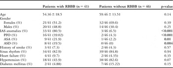
The two groups were similar with regard to age and gender. Interatrial septal anomalies were present in as many as 80.5% of the patients with electrocardiogram signs of right bundle branch block, with patent foramen ovale being the most prevalent disorder, followed by atrial septal aneurysm and atrial septal defect (Table 1). All interatrial septal disorders were diagnosed for the first time. Interatrial septal abnormalities were in total, as well as independently, significantly more frequent in the first group compared with the second group (Table 1). The rest of the baseline clinical features of the two patient groups are presented in Table 1.
Patient clinical characteristics of the two groups.Footnote *

ASA = atrial septal aneurysm; ASD = atrial septal defect; IAS = interatrial septum; PFO = patent foramen ovale; RBBB = right bundle branch block
* Data are presented as plus or minus standard deviation or fractions (%)
Bold values indicate that there is statistical significance between the groups
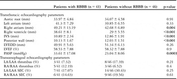
Right heart chambers were significantly larger in the study group (Table 2). Pulmonary artery systolic pressure was also significantly higher in the study group as compared to the control group, that is – 33.61 plus or minus 17.08 millimetres of mercury versus 23.04 plus or minus 8.06 millimetres of mercury, p value equal 0.0003. Interventricular and posterior wall thickness was significantly higher in the second group (Table 2), which might be explained by an almost twofold higher prevalence of arterial hypertension in the second group (Table 1). Other baseline transthoracic and transoesophageal echocardiographic parameters were similar between the two groups (Table 2).
Basic transthoracic echocardiography and transoesophageal echocardiography features.Footnote *

IVS = interventricular septum; LA = left atrium; LAA = left atrial appendage; LVEDD = left ventricular end-diastolic diameter; LVEF = left ventricular ejection fraction; PASP = pulmonary artery systolic pressure; RA = right atrium; RAA = right atrial appendage; RBBB = right bundle branch block; SEC = spontaneous echo contrast
* Data are presented as plus or minus standard deviation or fractions (%)
Clinical and echocardiographic aspects of patients with right bundle branch block and atrial septal anomalies
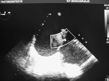
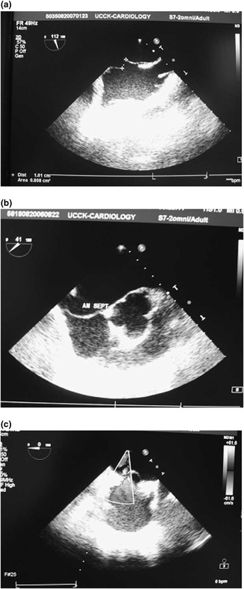
We diagnosed eight patients with atrial septal defect, five of whom had atrial septal defect type ostium secundum (Fig 1), one with atrial septal defect type sinusus venosus, one with atrial septal defect type ostium primum, and one patient had both atrial septal defect type ostium primum and secundum. Isolated atrial septal aneurysm was identified in nine patients, in combination with patent foramen ovale in six additional patients and with atrial septal defect in two cases. From the 17 patients with atrial septal aneurysm, seven were with atrial septal aneurysm type 3RL (Fig 2b), five with type 1R, three with type 4LR, and two patients with atrial septal aneurysm type 2L.
Transoesophageal echocardiography image of atrial septal defect associated with ventricular septal defect. The figure shows a large atrial septal defect type ostium secundum, measuring 29 millimetres and it also depicts a small size ventricular septal defect demonstrated by Color-Doppler.

Transoesophageal echocardiography images of patent foramen ovale and atrial septal aneurysm (a–c). (a) The presence of patent foramen ovale, measuring 10 millimetres at its maximal opening, in an 18-year-old male patient with right bundle branch block on electrocardiogram. (b) Atrial septal aneurysm type 3RL in a patient with right bundle branch block. (c) Atrial septal aneurysm type 4LR associated with patent foramen ovale, demonstrated in this image by Color-Doppler, in a 50-year-old patient with signs of right bundle branch block on electrocardiogram.

In the first group, 21 patients belonged to the 18- to 50-year age group, whereas 20 patients belonged to the 51- to 80-year age group. In the younger group, 20 patients (95.24%) had interatrial septal anomalies compared with 13 (65%) from the older group (p = 0.42). Atrial septal defect was present in seven (33.33%) younger patients and only in one (5%) older patient (p = 0.07). There was a slight difference between the age groups with regard to the prevalence of patent foramen ovale and atrial septal aneurysm (42.86% versus 35%, p = 0.73 and 19.05% versus 25%, p = 0.71, respectively).
In the first group, three of the patients had a history of stroke, with mean age of 60.33 plus or minus 15.95 years, including two female patients and one male patient. Of these three patients, two had patent foramen ovale, whereas the other patient did not have signs of interatrial disorders, but had chronic atrial fibrillation. The two patients with a coexistence of patent foramen ovale and right bundle branch block had 56.5 plus or minus 20.51 years and suffered a stroke 15.5 plus or minus 6.36 years earlier, thus being 41.5 plus or minus 27.58 years old at the time when they had the central cerebrovascular event. None of the patients suffered recurrent cerebral ischaemic attack.
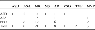
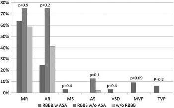
Interatrial septal anomalies were associated with one another and with other cardiac anomalies in our study group (Fig 1 and 2c,). Of the sixteen patients with patent foramen ovale, six had comorbidity of patent foramen ovale and atrial septal aneurysm (Fig 2c), making this the highest combination of interatrial septal disorders. Mitral regurgitation, on the other hand, was the most frequent cardiac pathology found with atrial septal disorders (Table 3). The remaining cardiac disorders associated with interatrial septal abnormalities are shown in Table 3. However, when compared with the patients with right bundle branch block and no atrial septal abnormalities and with the patients from the control group, statistical significance was not found with regard to the associated cardiac pathologies (Fig 3).
Interatrial and cardiac comorbidities in patients with interatrial septal anomalies.

AR = aortic regurgitation; ASA = atrial septal aneurysm; ASD = atrial septal defect; MR = mitral regurgitation; MS = mitral stenosis; MVP = mitral valve prolapsed; PFO = patent foramen ovale; TVP = tricuspid valve prolapse; VSD = ventricular septal defect
Cardiac comorbidities in the study population. The figure shows the prevalence of comorbidities in patients with right bundle branch block and atrial septal abnormalities, patients with right bundle branch block without atrial septal abnormalities, and patients without right bundle branch block. RBBB w ASA: patients with right bundle branch block and atrial septal abnormalities; RBBB w/o ASA: patients with right bundle branch block without atrial septal abnormalities; w/o RBBB: patients without right bundle branch block. MR = mitral regurgitation; AR = aortic regurgitation; MS = mitral stenosis; AS = aortic stenosis; VSD = ventricular septal defect; MVP = mitral valve prolaps; TVP = tricuspid valve prolaps.

As expected, patients with atrial septal defect had a significantly higher pulmonary artery systolic pressure, larger right ventricle and atrial size compared with patients with patent foramen ovale or atrial septal aneurysm (Table 4).
Anatomical and haemodynamic consequences of patients with interatrial septal disorders.

ASA = atrial septal aneurysm; ASD = atrial septal defect; PASP = pulmonary artery systolic pressure; PFO = patent foramen ovale; RA = right atrium; RV = right ventricle
*Data are presented as plus or minus standard deviation
We were able to diagnose by transthoracic echocardiography five (62.5%) out of eight patients with atrial septal defect. Transoesophageal echocardiography was helpful in these cases to precisely detect the size of the defect, as well as to diagnose an additional atrial septal defect in one case. Of the 16 patients with patent foramen ovale (Fig 2a), only five (31.25%) were suspected of having interatrial shunting by transthoracic echocardiography, and five (55.56%) patients with isolated atrial septal aneurysm were diagnosed before transoesophageal echocardiography procedure.
Discussion
Patients with atrial septal defect type ostium secundum frequently present with electrocardiogram signs of right axis deviation, right ventricular hypertrophy, rSR’ or rsR’ pattern in right precordial leads with normal duration of the QRS complex. It is not clear whether the delay in right ventricular activation is an expression of right ventricular volume overload or true delay of impulse conduction through the right bundle branch.Reference Sung, Tamer, Agha, Castellanos, Myerburg and Gelband12–Reference Boineau, Spach and Ayers17 Despite the fact that right bundle branch block is known to be associated with atrial septal defect, there are controversies of correlation of patent foramen ovale with right bundle branch block.Reference Belvís, Leta and Martínez-Domeño18 However, there are data suggesting that the “crochetage” pattern in the inferior leads correlates with the presence of patent foramen ovale.Reference Heller, Hagege, Besse, Desnos, Marie and Guerot19, Reference Ay, Buonanno, Abraham, Kistler and Koroshetz20
Chan et alReference Chan, Yip, Tay and Rajan21 in a wide screening of 651,794 schoolchildren found that 0.18% of these children had congenital cardiac disease, with mitral valve prolapse being the most frequent anomaly (0.08%), whereas atrial septal defect was present in 0.02% of them. Right bundle branch block was found in 132 cases, and it was the second most frequent electrocardiogram pathology found in these children.Reference Chan, Yip, Tay and Rajan21
We presented earlier two cases of middle-aged women diagnosed for the first time with atrial septal aneurysm associated with additional cardiac anomalies – such as patent foramen ovale, mitral valve prolapse, atrial septal defect type ostium primum, and atrial septal defect type ostium secundum – that presented with signs of right bundle branch block on electrocardiogram.Reference Bakalli, Kamberi, Pllana and Gashi22 We highlighted the importance of transoesophageal echocardiography as a method to diagnose interatrial septal abnormalities, as well as the value of right bundle branch block as a possible clue that may lead to early diagnosis of these disorders.Reference Bakalli, Kamberi, Pllana and Gashi22 These cases encouraged us to conduct the current study in order to evaluate the relationship of atrial septal abnormalities with right bundle branch block in a larger group of patients.
Data from this study demonstrated that all interatrial septal abnormalities were highly associated with right bundle branch block. Atrial septal abnormalities were present in 95% of patients with right bundle branch block from the younger age group. We were unable to explain precisely the relationship between right bundle branch block and atrial septal anomalies, particularly for patent foramen ovale and atrial septal aneurysm. However, possible mechanisms responsible for this relationship may be: abnormal delay in right ventricular activation as a result of enlarged right ventricular cavity size; progressive, chronic endocardial fibrosis, caused by turbulence in the region of the aneurysm and/or patent foramen ovale that could directly damage the conduction system; right ventricular overload and stretch of the conduction system. Other causes of right bundle branch – such as myocardial ischaemia, infarction, inflammation, cardiomyopathies, hypertension, fibrosis of the heart skeleton (Lev's disease or Lenegre's disease), advancing age or mechanical damage – could not be ruled out completely in some cases, but seemed less likely to be the cause. Moreover, we believe that further studies should investigate this relationship, especially in patients less than 50 years of age, keeping in mind that right bundle branch block is age dependent, although we found a very high association of right bundle branch block and atrial septal abnormalities in patients aged 18–50 years.
Clinical relevance of atrial septal abnormalities
Despite being well known that atrial septal defect causes anatomical and haemodynamic burden of the right heart chambers, patent foramen ovale and atrial septal aneurysm were considered innocent cardiac disorders. However, lately many authors consider patent foramen ovale as being responsible for the occurrence of ischaemic strokes in younger patients. Lechat et alReference Lechat, Mas and Lascault23 compared echocardiography findings in 60 patients under the age of 55 years with a history of ischaemic stroke with 100 subjects, and the prevalence of patent foramen ovale was significantly higher in the first group (40% versus 10%, p value less than 0.001). Mas et alReference Mas, Arquizan and Lamy24 in a prospective study with 598 patients, in the age group of 18–55 years, with a history of cryptogenic stroke found that 36% of these patients had patent foramen ovale, 1.7% had atrial septal aneurysm, and 8.5% of patients had both of these anomalies. This study also concluded that patients with a history of stroke who had patent foramen ovale combined with atrial septal aneurysm had a higher risk for recurrence of stroke. In our study, two patients in the first group with patent foramen ovale had a history of stroke, with mean age of 41.5 plus or minus 27.58 years. Among other clinical consequences of patent foramen ovale are also the decompression illnesses,Reference Torti, Billinger and Schwerzmann25 migraine and vascular headaches,Reference Anzola, Magoni, Guindani, Rozzini and Dalla Volta26 as well as platypnoea-orthodeoxia syndrome.Reference Cheng27
There are several studies suggesting the potential role of atrial septal aneurysm on cardioembolic stroke. The relationship between atrial septal aneurysm and cerebral ischaemic events was first reported by Belkin et alReference Belkin, Hurwitz and Kisslo28 in 1987 in a retrospective study, while subsequent multicentric studies confirmed this association.Reference Mügge, Daniel and Angermann5, Reference Marazanof, Roudaut and Cohen29 Nighoghossian et alReference Nighoghossian, Perinetti, Barthelet, Adeleine and Trouillas30 found atrial septal aneurysm in 34.5% of 79 patients who experienced stroke of unexplained aetiology. Right-to-left shunting through patent foramen ovale associated with atrial septal aneurysm has been suggested by many authors as the responsible mechanism for the occurrence of ischaemic stroke.Reference Mügge, Daniel and Angermann5, Reference Agmon, Khandheria and Meissner31, Reference Meissner, Whisnant and Khandheria32 Numerous studies have documented comorbidity of atrial septal aneurysm with patent foramen ovale,Reference Belkin, Hurwitz and Kisslo28, Reference Zabalgoitia-Reyes, Herrera, Gandhi, Mehlman, McPherson and Talano33–Reference Lechat, Lascault and Mas36 which was demonstrated in our study as well.
Transoesophageal echocardiography is a reliable method to detect interatrial septal abnormalities. Hanrath et alReference Hanrath, Schluter and Langenstein37 in their study, in which they compared transthoracic echocardiography with transoesophageal echocardiography for diagnosing atrial septal defect, reported that they were able to identify 10 out of 18 (56%) patients with atrial septal defect with transthoracic echocardiography from subcostal view, and 14 out of 18 (78%) when using contrast agents. Detection of atrial septal aneurysm and patent foramen ovale is more accurate with transoesophageal echocardiography as well. The prevalence of atrial septal aneurysm in earlier studies using transthoracic echocardiography was from 0.2% to 1%,Reference Zabalgoitia-Reyes, Herrera, Gandhi, Mehlman, McPherson and Talano33, Reference Hanley, Tajik and Hynes38 whereas the prevalence of this disorder is reported to be from 2% to 10% in transoesophageal echocardiography studies.Reference Mügge, Daniel and Angermann5, Reference Olivares-Reyes, Chan, Lazar, Bandlamudi, Narla and Ong6, Reference Pearson, Nagelhout, Castello, Gomez and Labovitz10, Reference Zabalgoitia-Reyes, Herrera, Gandhi, Mehlman, McPherson and Talano33, Reference Cabanes, Mas and Cohen35, Reference Comess, DeRook, Beach, Lytle, Golby and Albers39 Several studies using transoesophageal echocardiography, particularly with contrast agents, demonstrate that this is a more sensitive method for discovering patent foramen ovale than transthoracic echocardiography using contrast material.Reference Siostrzonek, Zangeneh and Gossinger40, Reference Di Tullio, Sacco, Venketasubramanian, Sherman, Mohr and Homma41
The limitation of this study is the small number of patients included in it. Another limitation is that we do not know whether right bundle branch block was present from birth in our patients, nor whether it had progressed from incomplete right bundle branch block. An additional limitation that has to be mentioned is that coronary angiography was not performed in patients with right bundle branch block in order to exclude ischaemia as a possible cause of this disorder.
In conclusion, right bundle branch block should serve as a valuable electrocardiogram marker to motivate a comprehensive search for interatrial septal abnormalities. In patients with right bundle branch block of unknown aetiology, even if no interatrial septal anomalies are suspected by transthoracic echocardiography, transesophageal echocardiography should be considered since it presents a reliable method for the detection of interatrial septal disorders. Early identification of these disorders is important in order to prevent their numerous consequences. Larger trials are needed to confirm our results.
Acknowledgement
We thank the staff of Echocardiography laboratory in University Clinical Center of Kosova for their assistance.