

INTRODUCTION
The marine noctilucoid dinoflagellates Abedinium dasypus (Cachon & Cachon-Enjumet) Loeblich & Loeblich III, Reference Loeblich and Loeblich1966 and Scaphodinium mirabile Margalef, Reference Margalef1963 are rarely reported worldwide and scarcely documented in the literature due to difficulties with their recognition from preserved samples. Despite their large size and characteristic shapes, these dinoflagellates with non-typical modified morphologies are often underestimated or absent from most of phytoplankton analysis (Larsen & Sournia, Reference Larsen, Sournia, Larsen and Patterson1991; Gómez & Furuya, Reference Gómez and Furuya2004).
Kuwait's waters have been monitored for phytoplankton community composition and density for a long time, and the phytoplankton diversity has been recently well documented based on Lugol's preserved material (Al-Yamani et al., Reference Al-Yamani, Bishop, Ramadhan, Al-Husaini and Al-Ghadban2004; Al-Kandari et al., Reference Al-Kandari, Al-Yamani and Al-Rifaie2009). Among Noctilucales, Noctiluca scintillans (Macartney) Kofoid & Swezy, 1921 has been reported from Kuwait (Al-Kandari et al., Reference Al-Kandari, Al-Yamani and Al-Rifaie2009). A recent phytoplankton survey was conducted on the basis of unpreserved samples examination with a focus on the poorly-preserved athecate dinoflagellates in Kuwait's waters. Careful analysis of the freshly collected samples revealed two morphologically modified dinoflagellates belonging to the family Leptodiscaceae Taylor, 1987 (order Noctilucales Haeckel, 1894)—Abedinium dasypus and Scaphodinium mirabile. The occurrence of these species has not been previously noted for Kuwait, or for the Arabian Gulf and the Indian Ocean as a whole. In this paper, previous records of S. mirabile and A. dasypus are summarized and new data on the distribution of these dinoflagellates within Kuwait's waters are provided.
MATERIALS AND METHODS
Sampling site
Kuwait's marine area occupies the north-western edge of the Mesopotamian shallow shelf of the northern part of the Arabian (Persian) Gulf, which is a marginal sea connected to the Indian Ocean via the Strait of Hormuz. Kuwait's waters are shallow, rarely deeper than 30 m, and generally well-mixed year-round, with mean salinity of 41.6. The mean annual seawater temperature is 23.8°C and ranges from 30.5°C (up to 36°C) in July and August to 14°C (down to 11.9°C) in January–February (Al-Yamani et al., Reference Al-Yamani, Bishop, Ramadhan, Al-Husaini and Al-Ghadban2004).
Sampling
A regular sampling programme was carried out from 2010 to 2012 throughout Kuwait's waters using speedboats. Systematic monthly monitoring of the poorly-preserved phytoplankton fraction was performed by sampling at seven sites, whereas some additional localities were sampled occasionally. Water samples from the surface layer (0–1 m) were collected with a 5 l Niskin bottle (General Oceanics, USA), stored in 1 l plastic bottles and brought directly to the laboratory in a cooler with ice.
Sample processing
Samples were either settled during 2–3 hours or gently concentrated by centrifugation (200 revolutions per minute, 3 minutes). Concentrated samples were scanned using an inverted Leica DMIL microscope (Leica, Wetzlar, Germany) at 50–200× magnification to estimate the phytoplankton composition. For detailed examination, cells were isolated by micropipetting and transferred to a glass slide in preparation for high-magnification photomicroscopy. Cells were examined and photographed using an upright Leica DMLM microscope (Leica, Wetzlar, Germany) equipped with Nomarski differential interference contrast optics and DFC 320 colour digital camera at 400–1000× magnifications. Cell dimensions were measured from images using Leica Application Suite v. 3.7 (Leica Microsystems (Switzerland) Ltd) software. Two specimens of Scaphodinium mirabile and five specimens of Abedinium dasypus were measured. All observations presented here are based on light microscopy examination of live cells that were isolated from freshly collected samples.
RESULTS
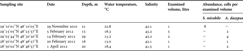
During a three-year course of regular sampling in Kuwait's waters, over which time approximately 180 samples were collected, cells of Scaphodinium mirabile and Abedinium dasypus occurred extremely rarely, they were only found on several sampling occasions in 2010 and 2012, mostly in the offshore area. Eight specimens of S. mirabile were found once (Table 1) in the surface layer sampled on 30 November 2010 in near-shore waters with temperature of 22.8°C and salinity of 42.1. The occurrence of A. dasypus was recorded on several occasions in winter and spring 2012 throughout the offshore waters with temperatures ranging from 15°C to 18.4°C and salinity range from 42.1 to 43.2 (Table 1).
Occurrence of Scaphodinium mirabile and Abedinium dasypus in Kuwait waters during 2010–2012 including geographical coordinates, dates, depth of the sampling site, water temperature of the surface layer, salinity, volume of sample and cell concentrations for each record of the species.

Live cells of these athecate dinoflagellates were extremely delicate, and they disintegrated easily during manipulation, but two specimens of Scaphodinium mirabile and five specimens of Abedinium dasypus were successfully isolated and their morphology was examined using high-magnification light microscopy (LM) under bright field (BF) and differential interference contrast (DIC) illumination.
SYSTEMATICS
Class DINOPHYCEAE West & Fritch, 1927
Order NOCTILUCALES Haeckel, 1894
Family LEPTODISCACEAE Taylor, 1987
Genus Scaphodinium Margalef, Reference Margalef1963
Scaphodinium mirabile Margalef, Reference Margalef1963
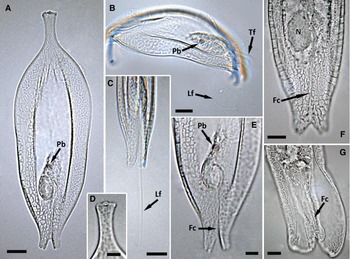
(Figure 1)
Light micrographs of Scaphodinium mirabile recorded from Kuwait waters in November 2010: (A) cell in non-folded view showing general appearance; (B) folding cell; (C, E, F) detail of the posterior cell end in folded view during swimming (C), and in non-folded view (E, F); (D) detail of the anterior cell end; (G) detail of the bilobate extension. Arrows point to the pigmented body (Pb); longitudinal (Lf) and transverse (Tf) flagella, flagellar canal (Fc) and nucleus (N). A–G, LM, BF. Scale bars: A–C, 20 µm; D, 5 µm; E–G, 10 µm.

SYNONYM:
Leptospathium navicula Cachon & Cachon-Enjumet, Reference Cachon and Cachon-Enjumet1964.
REFERENCES:
Margalef, Reference Margalef1963; Cachon & Cachon-Enjumet, Reference Cachon and Cachon-Enjumet1964; Gómez & Furuya, Reference Gómez and Furuya2004.
DESCRIPTION
In non-folded view cells were extremely flattened, elongated, 239–251 µm long and 77–80 µm wide (N = 2), strongly bilaterally symmetrical with slightly folded lateral margins (Figure 1A). Each cell end was terminated by distinct extension. One extension (here considered as anterior) was narrow, approximately 34–35 µm long, slightly widened to the end and arranged with three small processes (one of which was centrally located, while the other two were peripheral) (Figure 1A, D). Reverse extension (here considered as posterior) was wider and shorter than the anterior one, approximately 30–32 µm long, bilobate (Figure 1A, C, E–G). There was the cytoplasm swelling below the median, where large elongate encapsulated nucleus was located (Figure 1A, E, F). The cytoplasm was reticulated (Figure 1A, B, E) due to numerous myofibrils forming a uniform network. One small pigmented body (food particle?) was observed near the nucleus in both specimens examined (Figure 1A, B, E). Longitudinal flagellum was well visible, long (but shorter than the cell length) (Figure 1C), originated from the cytoplasm swelling, initially curving along one side of the nucleus and then extended straight along longitudinal cell axis within flagellar canal to the bilobate extension (Figure 1E–G). Transverse flagellum was observed only in folded cells (Figure 1B). Chloroplasts and trichocysts were not seen.
Live cells observed were more or less expanded, sliding slowly with narrow extension pointing forward due to longitudinal flagellum oscillations. The lateral margins of the cells were strongly folded during the active movement. Being disturbed, the cells folded rapidly into a ring on one of its sides, extension against extension.
Genus Abedinium Loeblich & Loeblich III, Reference Loeblich and Loeblich1966
Abedinium dasypus (Cachon & Cachon-Enjumet) Loeblich & Loeblich III, Reference Loeblich and Loeblich1966
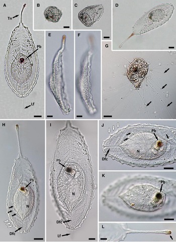
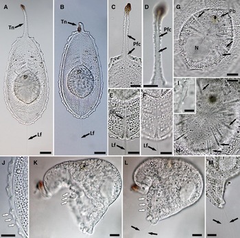
(Figures 2 & 3)
Light micrographs of Abedinium dasypus recorded from the north-western offshore Kuwait waters on 20 February 2012: (A) cell in non-folded view showing general appearance; (B–D) the same cell in folded (B, C) and non-folded (D) view; (E, F) the same cell in lateral view at the different focal planes; (G) folded cell just before lysis showing numerous long filaments (indicated by arrows) from discharged trichocysts; (H, I–K) other cell in non-folded view showing general appearance (H), and detail of the posterior cell end (I–K), numerous trichocysts (black arrows) and undulated cell margin (white arrows) are visible; (L) detail of the anterior cell end showing the tentacle with pigmented distal end (indicated by arrow). Pigmented body (Pb), distal flagellar canal (Dfc), longitudinal flagellum (Lf), nucleus (N), and tentacle (Tn) are indicated by arrows. A–D, G, LM, BF; E, F, H–L, LM, DIC. Scale bars: A–H, 20 µm; I–L, 10 µm.

Light micrographs of Abedinium dasypus recorded from the southern offshore Kuwait waters on 14 February 2012: (A, B) the same cell in non-folded view showing the different position of the tentacle; (C, D) detail of the anterior cell end showing the tentacle, the proximal flagellar canal and intracytoplasmic flagellum within flagellar canal (arrow); (E, F) detail of the distal flagellar canal; (G, H) detail of the fusiforme cytoplasm swelling showing numerous trichocysts (indicated by arrows); (I) detail of the trichocyst; (J) detail of the marginal area in non-folded cell showing the undulated cell margin (white arrow); (K, L) the same cell in folded view showing contracted cell margins (white arrows) and both flagella (black arrows) protruding from the posterior cell end; (M) detail of the posterior end in folded cell showing both flagella (black arrows) protruding from the distal flagellar canal. Pigmented body (Pb), distal flagellar canal (Dfc), proximal flagellar canal (Pfc), longitudinal flagellum (Lf), nucleus (N), and tentacle (Tn) are indicated by arrows. A–I, LM, BF; J–M, LM, DIC. Scale bars: A, B, 20 µm; C–H, G–M, 10 µm; I, 5 µm.

SYNONYM:
Leptophyllus dasypus Cachon & Cachon-Enjumet, Reference Cachon and Cachon-Enjumet1964.
REFERENCES:
Cachon & Cachon-Enjumet, Reference Cachon and Cachon-Enjumet1964; Gómez et al., Reference Gómez, Moreira and López-García2010.
DESCRIPTION
Cells were feather-shaped, extremely flattened, 163–251 µm long and 76–94 µm wide (N = 5), strongly bilaterally symmetrical (Figures 2A, D, H & 3A). Anterior part of the cell that points in the direction of the cell locomotion was terminated by long (52–57 µm), narrow (5–6 µm wide), and mobile tentacle with an orange-pigmented distal part (Figures 2A, E, H, L & 3A–D). Opposite cell end (here considered as posterior) was broadly rounded. Cell edge was bordered by a narrow undulated margin, 4–5 µm wide (Figures 2A, F, H, J & 3J). There was almost centrally located fusiforme cytoplasm swelling, 63–84 µm long and 47–56 µm wide, containing the nucleus, food vacuoles and trichocysts (Figures 2A, E, D, H–K & 3A, B, G). Nucleus was large, encapsulated, and elongate to roundish, 23 × 25–26 µm (Figures 2H–J & 3G). Single large pigmented body (food particle) was often located near nucleus (Figures 2A, D, H–K & 3G). Numerous elongate, clavate trichocysts were radiating out from the centre of the cytoplasm swelling (Figures 2A, H, J & 3A, B, G–I). The bigger ones were up to 15–24 µm long, whereas smallest trichocysts were 7–9 µm long (Figure 3H, I). Cytoplasm was transparent and densely granulated (Figures 2A, H–J & 3A, B, E, F, J). Cells were biflagellate. One of the flagella was included in the cytoplasm along its entire length, arising from the proximal part of the cytoplasm swelling, and lay throughout the cytoplasm along longitudinal axis up to end of tentacle within tube-like flagellar canal (Figures 2H, L & 3A, B, E, F). The other flagellum (longitudinal) was partially internal, originated from the distal part of the cytoplasm swelling and continued along longitudinal cell axis; its proximal part lay throughout tube-like flagellar canal, protruding with its distal part from the posterior cell end (Figures 2H–J & 3C, D). On one occasion both flagella were seen protruding from the posterior cell end just before cell lysis (Figure 3L, M). Myofibrils were arranged to form two zones of oblique striation above and below the cytoplasm swelling, radiating from the longitudinal cell axis to the cell margins (Figure 2A, H, I). Chloroplasts were not seen.
Freshly collected cells swam slowly, moving forward along the longitudinal axis due to longitudinal flagellum oscillations and feeling the space ahead with the tentacle. Cells were able to contract suddenly when the surrounding water was disturbed mainly due to contraction of the cell margins (Figure 3J–L). Being disturbed, cells rolled up into a ball immediately and folded tentacle inside (Figure 2B, C). The discharged trichocysts ejecting numerous long filaments were observed in the rolled cells just before lysis (Figure 2G).
DISCUSSION
The noctilucoid dinoflagellates (order Noctilucales) constitute an important component of the marine heterotrophic plankton. Among them, Noctiluca scintillans is well investigated due to its worldwide distribution and capacity to form red tides (e.g. Elbrächter & Qi, Reference Elbrächter, Qi, Anderson, Cembella and Hallegraeff1998). In contrast to N. scintillans, a number of the noctilucoid genera are rarely encountered, but on the basis of their descriptions they should be readily recognizable (Sournia, Reference Sournia1986; Larsen & Sournia, Reference Larsen, Sournia, Larsen and Patterson1991), including Scaphodinium and Abedinium.
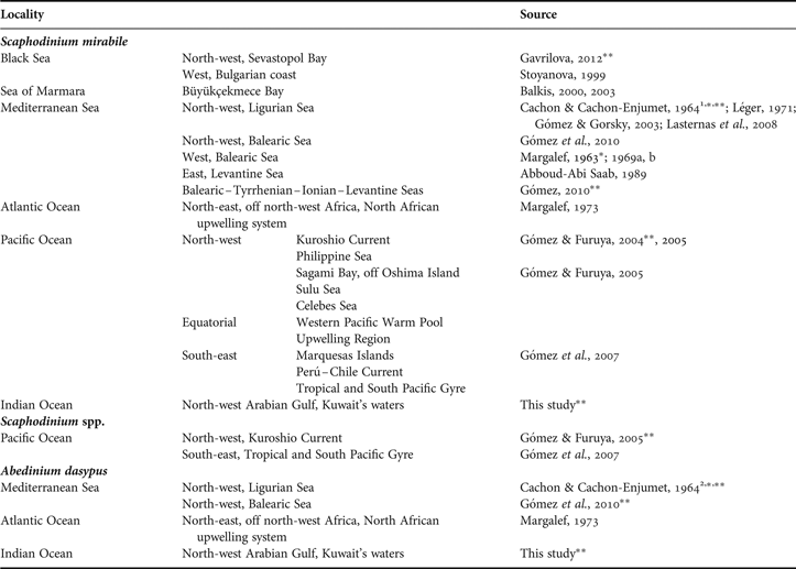
Worldwide records for Scaphodinium mirabile are scarce. Over half of the documented records for this species are restricted to the Mediterranean Sea and adjacent waters of the Marmara and Black Seas, and the eastern part of the North Atlantic Ocean (Table 2). In the Mediterranean Sea, the occurrence of S. mirabile is variable among sub-basins. While the species has been reported mostly from the coastal waters of the Ligurian and Balearic Seas in the western part of the basin (Table 2), it has been recorded as commonly occurred throughout the open Mediterranean waters (Gómez, Reference Gómez2010). Recently, the occurrence of S. mirabile in the Pacific Ocean has been documented, and its spatial distribution in various Pacific localities has been mapped (Gómez & Furuya, Reference Gómez and Furuya2004, Reference Gómez and Furuya2005; Gómez et al., Reference Gómez, Claustre, Raimbault and Souissi2007). Moreover, a few specimens that morphologically differed from the type species of Scaphodinium were found among Pacific population (Gómez & Furuya, Reference Gómez and Furuya2005) that casts doubt on the monospecific status of this genus. Despite the limited number of the worldwide records, S. mirabile can be considered as cosmopolitan species, being found in three oceans and in both northern and southern hemispheres (Table 2), with the highest occurrence in the Mediterranean Sea that could perhaps be attributed to the historical tradition of taxonomic studies and hydrographical peculiarity of the Mediterranean waters (e.g. Gómez, Reference Gómez2006).
Worldwide distribution of Scaphodinium spp. and Abedinium dasypus, as found in this and previous studies.

1, recorded as Leptospathium navicula; 2, recorded as Leptophyllus dasypus; *, record was illustrated by line drawings; **, record was illustrated by microphotographs.
Known records of Abedinium dasypus are much rarer and more geographically restricted (Table 2). This dinoflagellate has been originally described from the Villefranche Bay (north-west Mediterranean Sea) as Leptophyllus dasypus (Cachon & Cachon-Enjumet, Reference Cachon and Cachon-Enjumet1964). Due to the illegitimate generic name which is preoccupied under the International Code of Zoological Nomenclature, the genus was further renamed as Abedinium (Loeblich & Loeblich III, Reference Loeblich and Loeblich1966; Fensome et al., Reference Fensome, Taylor, Norris, Sarjeant, Wharton and Williams1993). After the initial record, Margalef (Reference Margalef1973) reported this species from the eastern part of the North Atlantic Ocean, and several specimens of A. dasypus were found in French coastal waters of the north-west Mediterranean Sea (Marseille and Banyuls-sur-Mer) much later (Gómez et al., Reference Gómez, Moreira and López-García2010).
Despite the intensive sampling throughout Kuwait's waters during three years combined with the thorough examination of freshly collected unpreserved samples, the observations of Scaphodinium mirabile and Abedinium dasypus were extremely rare and cells of these species occurred in very low concentrations, sometimes with a density of less than 1 cell per litre (Table 1). Records of A. dasypus specimens were numerous in comparison with those of S. mirabile, whereas the latter was more abundant per sample (Table 1). While the identification of these dinoflagellates is relatively easy due to their distinct morphology, these species could be easily overlooked during sample analysis because of their rarity and extreme delicacy.
Most of the Noctilucales have a predominantly tropical to warm-temperate distribution (Cachon & Cachon, Reference Cachon and Cachon1967, Reference Cachon and Cachon1969; Gomez & Furuya 2005, 2007). The northernmost records of Scaphodinium mirabile from temperate waters of the Black Sea (Stoyanova, Reference Stoyanova1999; Gavrilova, Reference Gavrilova2012) and in the vicinity of the Kuroshio Current in the north-west Pacific Ocean (Gomez & Furuya, 2004, 2005) coincided with warm seasons.
Our observation of Scaphodinium mirabile and Abedinium dasypus in subtropical Kuwait waters supported previous studies that have considered these species as thermophilic. Because of the extremely rare occurrence of S. mirabile and A. dasypus in Kuwait waters, there are not enough data to make a conclusion about the spatial distribution and seasonal prevalence pattern of these dinoflagellates, however, records of these species coincided with period of moderate water temperatures (15–22.8°C: Table 1) and were associated mainly with the offshore area.
Our observations of cells morphology in Scaphodinium mirabile and Abedinium dasypus were similar to those previously described and illustrated for these species (Margalef, Reference Margalef1963; Cachon & Cachon-Enjumet, Reference Cachon and Cachon-Enjumet1964; Sournia, Reference Sournia1986; Larsen & Sournia, Reference Larsen, Sournia, Larsen and Patterson1991; Fensome et al., Reference Fensome, Taylor, Norris, Sarjeant, Wharton and Williams1993; Gómez & Furuya, Reference Gómez and Furuya2004; Gómez et al., Reference Gómez, Moreira and López-García2010). Although not previously reported for Abedinium dasypus, we were able to document the specimen with two flagella protruding from the posterior cell end, which indicates that the intracytoplasmic character of one flagellum in A. dasypus could be considered as a variable feature. This study presents photographic records of both species from the Indian Ocean region for the first time.
The observations of Scaphodinium mirabile and Abedinium dasypus from this study can be considered as new records for Kuwait. To our knowledge, this is the first time that S. mirabile and A. dasypus have been observed in the Indian Ocean, and this indicates an extension of the spatial distribution of these species from that previously known.
ACKNOWLEDGEMENTS
We acknowledge the valuable help of Mr Alan Lennox (Kuwait Institute for Scientific Research, Kuwait) during the extensive field sampling. The authors thank Mr Alok Nigam (Behbehani United General Trading Co, Microscopy Products, Kuwait) for providing the light microscopy service. The authors are grateful to the Marine Biodiversity Records editorial team, especially Dr Cynthia Trowbridge, for their kind help and to two anonymous referees for their detailed and constructive comments. Deep appreciation is due to the Kuwait Institute for Scientific Research for funding this research study.