




No CrossRef data available.
Article contents
What is the role of p53 during the cyst formation of Trichinella spiralis? A comparable study between knockout mice and wild type mice
Published online by Cambridge University Press: 11 July 2005
Abstract
During the cyst formation of Trichinella spiralis, the infected muscle cell undergoes basophilic change and apoptosis, which results in nurse cell formation. This study revealed expression kinetics of some apoptosis genes such as p53 and its closely related genes (tumor suppressor genes p53, p53; mouse double minute 2, MDM2; cyclin-dependent kinase inhibitor p21, p21waf). RT-PCR (reverse transcription polymerase chain reaction) results showed that these genes were temporarily expressed in the infected muscles during the cyst formation period, but not in normal muscles (or very low if any), which suggested the involvement of these apoptosis genes in the nurse cell formation. Cysts and neighbouring muscle cells were separately collected and RT-PCR was performed, which suggested that p53 was expressed in the cysts. An immunocytochemical study showed that p53 was expressed in the nucleoplasm of basophilic cell in the cyst and Trichinella larvae, which suggested involvement of these apoptosis genes in the nurse cell formation. The same p53 expression kinetic study was performed on p53 knockout mice. The knockout mice did not express p53 genes, but expressed the other apoptosis genes in the same kinetics with only minor exceptions, suggesting that the expressions of these genes during the cyst formation were more or less p53-independent. There were no differences in the number and morphology of the cysts between the knockout mice and wild type mice. Thus apoptosis seen during the Trichinella cyst formation can be operated in the presence or absence of p53.
Keywords
Information
- Type
- Research Article
- Information
- Copyright
- © 2005 Cambridge University Press
INTRODUCTION
The larvae of Trichinella spiralis are released from muscles by host gastric juice after oral infection and grow to adults in the intestine. The second generation is produced from 5 days post-infection by the gravid female and migrates to the whole body of the host through the circulatory vessels. The newborn larvae invade the skeletal muscle and complete cyst formation by about 18 days post-infection (p.i.). To complete the cyst, the nurse cell has to be formed. The nurse cell formation is a result of muscle cell transformation and satellite cell misdifferentiation, where apoptosis by mitochondria-mediated factors is likely to be involved (Matsuo et al. 2000; Wu et al. 2001; Boonmars et al. 2004a). Additionally, a wide variety of apoptosis-related genes are possibly involved in Trichinella cyst formation, and the p53 gene and its related genes should be included in the list for precise analysis.
It is well established that the p53 gene plays important roles in many physiological functions including DNA repair, inhibition of angiogenesis, re-entry into the cell cycle, oxidative stress, and determination of cell fate and the oncogenic apoptosis pathway (Alarcon-Vargas and Ronai, 2002; Nakamura, 2004). In fact many genes are activated upon p53 gene expression including p21waf, MDM2, BAX, caspase 9 and caspase 3. Reportedly, possible functions of these factors are as follows. (1) p21waf is a cyclin-dependent kinase inhibitor that plays a role in the cell cycle, DNA synthesis, stress response and apoptosis (Kagawa et al. 1997; Dotto, 2000). (2) MDM2 is the abbreviation of mouse double minute 2 which acts in an auto-regulated feedback loop to limit the action of p53 (Alarcon-Vargas and Ronai, 2002; Wang et al. 2002). (3) BAX is a member of the Bcl-2 family, and can promote apoptosis through induction of cytochrome c release from mitochondria (Desagher and Martinou, 2000; Tsujimoto and Shimizu, 2000; Parone, James and Martinou, 2002). (4) Caspase 9 and caspase 3 are initiator caspases, both of which likely play roles in cascades that lead to cell death (Saleh et al. 1999; Desagher and Martinou, 2000; Parone, James and Martinou, 2002).
In spite of the possible importance of p53, there have been no reports so far about the kinetics of p53 expression in mice after Trichinella infection. The present study was designed to shed light on the role of p53 in the Trichinella cyst formation by correlating the expression of p53 gene with those of genes (p21waf, MDM2, BAX, caspase 9 and caspase 3) that may be influenced by the p53 gene. The p53 knockout (p53−/−) mouse was used as an experimental model to elucidate the role of p53.
MATERIALS AND METHODS
Experimental animals
Three kinds of mice were used including nude mice (purchased from Chubu Kagaku Shizai Co., Ltd), p53 knockout mice (p53−/−) and its wild type (p53+/+).
C57BL/6J p53 knockout (p53−/−, B6.129S2-Trp53−/−tm1Tyj) mice were produced by mating between homozygous male p53 (p53−/−, B6.129S2-Trp53−/−tm1Tyj) and heterozygous female (p53+/−, B6·129S2-Trp53−/−tm1Tyj), which was performed according to the instruction of The Jackson Laboratory (Bar Harbor, ME, USA). Genotyping was performed on each individual F1 mouse by polymerase chain reaction (PCR) that allows specific identification of p53+/+, p53+/− and p53−/−. The primers used were as follows: OIMR0013 (5′CTTGGGTGGAGAGGCTATTC3′), OIMR0014 (5′AGGTGAGATGACAGGAGATC3′), OIMR00336 (5′ATAGGTCGGC GGTTCAT3′), OIMR00337 (5′CCCGAGTATCTGGAAGACAG3′).
Wild type (C57BL/6J, p53+/+) mice were purchased from Chubu Kagaku Shizai Co., Ltd (Nagoya, Japan) and used as the non-transgenic control.
Parasites and infection
T. spiralis (ISS413) was used for the infection agent. The mice were orally infected and sacrificed for further analysis at certain time-points including 0 (control), 8, 13, 18, 23, 28, 33, 38 and 53 days post-infection (p.i.).
Primers for quantitative reverse transcription polymerase chain reaction (RT-PCR)
The primer pairs for apoptosis factors (p53, MDM2, p21waf, BAX, caspase 9 and caspase 3) and endogenous controls (MG3PDH, mouse glyceraldehyde-3-phosphate dehydrogenase; and 18S rRNA) were designed based on the published sequence as summarized in Table 1.

Detection of apoptosis-related genes in T. spiralis infected muscle by RT-PCR
Muscles were obtained from the hind limbs, immediately placed on ice, minced into tiny pieces and vigorously mixed. Then 100 mg of each sample was used for RT-PCR analysis. Muscles at day 0 p.i. (before infection), were used as a control. Total RNA was isolated using TRIZOL (Invitrogen, Carlsbad, CA, USA) according to the manufacturers instructions. The isolated RNA was treated with DNase (5 units RQ1 RNase-Free DNase, Promega, Co., Madison, WI, USA) and 119 units of Ribonuclease Inhibitor (Takara Shuzo, Co., Ltd, Kyoto, Japan), phenol/chloroform and ethanol. The precipitated total RNA was dissolved in RNase-free water (100 μl) and reverse transcribed into cDNA using Oligo(dT)12-18 primers (Amersham Phamacia Biotech, Inc. Piscataway, NJ, USA) and a Ready-to-Go First-Strand beads kit (Amersham Phamacia Biotech). Five μl of the RNA sample, 1 μl of 0·5 μg/μl Oligo(dT)12-18 and 27 μl of RNase free water were added to a Ready-to-Go tube. The tube was incubated at 37 °C for 60 min and then at 95 °C for 10 min.
The PCR reaction mixture comprised 3 μl of reverse transcription products (1[ratio ]10 diluted), 3 μl of 10X PCR buffer, 3 μl of deoxynucleoside triphosphate (2·5 mM each), 6 μl of primer pairs (5 μM), 0·12 μl of Taq polymerase (5 U/μl, Takara Shuzo) and 15 μl of distilled water to give a final volume of 30 μl. PCR amplification was performed under the condition of 1 cycle at 95 °C for 3 min; 22–35 cycles of 95 °C for 30 sec, 56–59 °C (depending on primers) for 30 sec and 72 °C for 2 min; and a final extension cycle at 72 °C for 10 min.
Aliquots of PCR products were analysed by gel electrophoresis and photographed under UV light. The photographs were analysed for density using Scion Image (Scion Co., Frederick, MD, USA). All reactions for the standard control and the experimental sample were performed in triplicate. The amount of p53, p21waf and MDM2 genes was calculated as follows:

The ratio of density of the apoptosis-related genes and the standard gene were plotted in a graph.
Detection of apoptosis-related genes in the nurse cell by RT-PCR
Cryosections (8 μm in thickness) of muscles from 18 days p.i. were prepared and placed onto glass slides (pre-heated at 200 °C for 2 h to destroy RNase) and dried immediately with room air using a conventional dryer. The nurse cell complexes were micro-dissected using the laser micro-beam of the Robot-MicroBeam system (P.A.L.M. GmbH, Bernried, Germany) equipped with a 40× dry long-distance objective lens. The control cells were collected from the unaffected area of the same sample used for the experiment. The nurse cell complexes were cut together with their supporting optimal cutting temperature compound with a laser beam. One μl of mineral oil was dropped into a 0·5 ml tube cap. The test samples contained about 150–200 cysts (1×10⁶/μm2 of tissue section), and the control, having the same area in μm, was prepared from the unaffected muscles. Those samples were analysed for apoptosis-related genes by means of the same method described above except an RNeasy protect mini-kit (Qiagen GmbH, Hilden, Germany) was used instead of the TRIZOL protocol.
Immunohistochemical staining for p53
Muscles of 18, 28 and 48 days p.i. were first processed for cryosectioning using a well-established protocol and were then processed for immunostaining. The sections were blocked with 5% skim milk in phosphate-buffered saline (PBS), incubated with the rabbit polyclonal antibodies to 1[ratio ]100 diluted p53 (p53 (FL-393), Santa Cruz Biotechnology Inc., Santa Cruz, CA, USA) for 1·5 h at 37 °C and washed 3 times with PBS. The second antibody labelling and peroxidase (visual marker) development were performed using Histostain®SP (ZYMED Laboratories Inc. San Francisco, CA, USA) according to the manufacturer's instructions. A negative control was performed using normal serum instead of the primary antibody.
Kinetics of apoptosis related-genes during T. spiralis infection
To explore the possible relations of p53 and its correlated genes (MDM2, p21waf, BAX, caspase 9 and caspase 3) in cyst formation of T. spiralis, RT-PCR was performed to detect expression kinetics of these genes in p53 knockout mice and wild type mice. Muscle tissues were obtained from the hind limbs of 0, 13, 23 and 38 days p.i., and processed for RT-PCR analysis as described above.
Morphology of the cyst
Muscles of mice (p53 knockout mice and its wild type) were obtained at 13, 23 and 30 days p.i. and processed for light microscopical observation according to well-established methods (H&E staining).
Muscle larva recovery
In order to know the susceptibility to T. spiralis infection, the p53 knockout mice (n=5) and its wild type mice (n=5) were orally infected with 100 larvae, and muscle larvae were recovered at 30 days p.i. In brief, the whole body of mice without skin and viscera was minced and subjected to complete digestion with 1% pepsin-HCl solution. The number of worms was counted under the light microscope.
RESULTS
Semi-quantitative RT-PCR in T. spiralis-infected nude mice muscle
Fig. 1 shows the relative level of gene expression of p53, p21waf and MDM2 genes, which were determined by quantitative RT-PCR at various stages in infected nude muscles or non-infected muscles. The housekeeping gene MG3PDH was used as an internal control, and its mRNA levels remained similar in infected muscle and the non-infected muscle control. Specific bands of p53, MDM2 and p21waf genes were also observed in all stages of infection but the expression level of each stage was different. The expression level of p53 was increased from 13 days p.i., reached a peak at day 18 p.i., and then at day 28 p.i. the expression level gradually decreased. The kinetics of p21waf was similar to that of p53. However, the MDM2 expression level that was observed in normal muscle at high level slightly changed during cyst formation.

Fig. 1. RT-PCR results on expression levels of apoptosis-related genes (p53, MDM2, and p21waf) in the infected muscle tissue. Total RNA was isolated from infected muscle tissue at various stages (0, 8, 13, 18, 23, 28, 33, 38 and 53 days p.i.) and amplification products were analysed by agarose gel electrophoresis and shown in the left panel (A). Using Scion Image software, the relative density of each sample band against the control band (MG3PDH) was analysed and shown in the right panel (B).
p53 apoptosis-related genes in the nurse cell of T. spiralis
To determine the origin of p53, p21waf and MDM2 genes, from cysts and/or muscle cells, RT-PCR was performed to detect the mRNA expression level in each specimen. Fig. 2 shows the quantitative RT-PCR of the p53, p21waf and MDM2 genes and the internal control gene (18S rRNA) at day 18 p.i. in cysts and muscle cells, which were separately collected by the laser microscopic capturing. The mRNA level of the control gene was similar in the cyst specimen and muscle cell specimen (panel A is an electrophoresis result; panel B is an expression ratio). Specific bands of p21waf and MDM2 genes were also observed in both specimens, but the much higher expression was observed in the cyst specimen. On the other hand, the specific band of the p53 gene was observed in the cyst specimen but not in the muscle cell specimen.

Fig. 2. RT-PCR results on expression levels of apoptosis-related genes (p53, MDM2, and p21waf) in the cyst or non-infected muscle cells that were obtained from the Trichinella spiralis-infected mouse at day 18 p.i. Total RNA was isolated from laser micro-dissected cysts or muscle cells, and RT-PCR of the sample genes and the standard gene (18S rRNA) was performed. PCR products were analysed by agarose gel electrophoresis (A), and the relative density of each sample band against the control gene band was analysed using Scion Image software (B).
Immunohistochemical staining
Fig. 3 shows the immunohistochemical staining of p53 protein on cryosections of infected nude mice at 18 (panel A), 28 (panel B) and 48 (panel C) days p.i. The parasite nucleus was positive for p53 staining (panels A, B and C). Strong positive staining for p53 was also observed in the nucleus of the basophilic cells (surrounding the parasite) at 18 and 28 days p.i., but not in that of eosinophilic cells (situated beneath the cyst wall). The well-developed round nucleoli were negative for the staining. At 48 days p.i. the staining in the nucleus was very light. The control slides were negative for the staining which confirmed the staining specificity (panel D).

Fig. 3. Immunohistochemical staining for p53. Cryosections of nude mouse muscles infected with Trichinella spiralis for 18 (A), 28 (B) and 48 (C) days p.i. were reacted with anti-p53 antibody. A negative control was performed using normal rabbit serum (D). The brown colour indicates a positive reaction. L, larva.
Kinetics of p53-related genes in T. spiralis p53 knockout or wild type mice muscle
Fig. 4 shows the kinetics of gene expressions in muscles (panel A) with emphasis on the comparison of p53 and other genes in T. spiralis infected knockout mice and wild type mice (panel B). The MG3PDH mRNA level was the same in the all-experimental conditions which confirmed the validity of the RT-PCR technique. A specific band of p53 was observed in all stages of wild type mice muscle but not in any stage of p53 knockout mice muscle sample, which confirmed that the p53 knockout mice were really devoid of p53 gene expression. The expression of p53 in the wild type mice increased and decreased following similar kinetics as shown in the nude mouse system (Fig. 1). The specific band of p21waf, MDM2, BAX, caspase 9 and caspase 3 genes was also observed in all stages of infection in both mice type but the expression level of each stage was different. The p21waf expression level increased in both types of mice, with the peak being at 13 days p.i. observed in the knockout mice but at 23 days p.i. in the wild type mice, although the expression level at 38 days p.i. was the same. MDM2 expression levels remained unchanged in both types of mice. Caspase 9 and BAX expression levels slightly increased in both type of mice. There was a sharp increase of caspase 9 in the knockout mice at 13 days p.i. The caspase 3 expression level in both types of mice increased from day 13 p.i. and remained high.

Fig. 4. RT-PCR results on expression levels of apoptosis-related genes (p53, MDM2, and p21waf, BAX, caspase 9 and caspase 3) in wild type and knockout mice infected muscle and uninfected muscle. Total RNA was isolated from infected muscle tissue at various stages (0, 13, 23 and 38 days p.i.) and amplification products were analysed by agarose gel electrophoresis and shown in the upper panel (A). Using Scion Image software, the relative density of each sample band against the control gene (MG3PDH) band was analysed and shown in the lower panel (B).
Histopathology
The myopathy of Trichinella infection in wild type and knockout mice (Fig. 5) was quite similar to that which was published previously (Matsuo et al. 2000; Wu et al. 2001). Basophilic change was observed from day 13 p.i. in both strains of mice (Fig. 5A and D). The cyst morphology of T. spiralis in both strains was not different at any stage of infection examined.

Fig. 5. Haematoxylin and eosin staining of Trichinella spiralis-infected wild type mice (A, B, C) and knockout mice (D, E, F) mouse muscle at day13 p.i. (A, D), day 23 p.i. (B, E) and day 30 p.i.(C, F). L, larva. Original magnification, 200×.
Muscle larva recovery from knockout mice or its wild type
In order to observe whether the knockout of p53 has an effect on the muscle larva burden or not, the numbers of muscle larvae recovered from the knockout mice (n=5) and wild type mice (n=5) were compared. The averaged numbers of larvae recovered from knockout and wild type mouse are 19345·6±2033·475 and 18610·8±3984·348 worms, respectively (Table 2), no significant difference was observed (P>0·01).

DISCUSSION
We have shown ultrastructural, cytochemical and molecular-biological evidence that mitochondria-mediated apoptosis may play some role in cystogenesis of T. spiralis (Boonmars et al. 2004a). The mitochondria-mediated apoptosis genes (BAX, caspase 9 and Apaf-1) were elevated at 13–28 days p.i., which corresponds to the time of the cyst formation (muscle cell transformation, nurse cell formation). Expression of the genes is restricted to the basophilic cytoplasm of the nurse cells. In fact the basophilic cytoplasm seems to be undergoing apoptosis because it has a high-level of activity of a marker enzyme of apoptosis (acidophosphatase; Boonmars et al. 2004b), and nuclear condensation and fragmentation (Matsuo et al. 2000). The basophilic cytoplasm disappears but the eosinophilic cytoplasm appears in turn and takes over the whole cyst and complete cyst formation (Boonmars et al. 2004a).
Reportedly, p53 plays a key role that may govern the mitochondria-mediated apoptosis pathway by activating the BAX gene (Wu and Deng, 2000; Schuler and Green, 2001; Luu et al. 2002), p21waf which leads to cell arrest and death (El-Deiry, 1998; Dotto, 2000), and MDM2 which acts in an auto-regulated feedback loop limiting the action of p53 (Alarcon-Vargas and Ronai, 2002; Wang et al. 2002). Such an important role of p53 led us to study the possible involvement of p53 in cyst formation. In the present study with nude mice, we showed that p53 was expressed in the nucleus of the basophilic cytoplasm, and the expression was up-regulated during cyst formation with the same kinetics as p21. Reportedly, BAX, caspase 9 and Apaf-1 (Boonmars et al. 2004a) and caspase 3 (Wu et al. 2005) are also expressed with the same kinetics as p53 in Trichinella infected nude mice.
Considering the importance of this p53 gene function and our previous and present data in nude mice, we repeated the same experiments using the p53 knockout mice. We anticipated that there would be some defects of the T. spiralis cysts because the apoptosis is interfered in p53-deficient mice and such experiments may elucidate the role of apoptosis in the cyst formation (including basophilic change and nurse cell formation).
The results turned out to be opposite of what we had predicted. The p53 knockout mice did not express the p53 gene, which confirms the validity of our experimental system. But, even without p53 expression, the expression of MDM2, BAX, caspase 9 and caspase 3 were totally not affected. Only p21 was affected in a minor way in that the increase of expression was somewhat delayed. More importantly, the absence of p53 did not interfere with cyst formation in terms of its morphology and its number. As far as judging from H&E sections, the cysts were quite the same as those obtained in comparable experimental systems, and the recovered numbers of parasites from the knockout mice and control mice were the same.
These results imply that p53 may indeed play some role in mitochondria-mediated apoptosis in the basophilic cytoplasm, but it is not indispensable for the induction of apoptosis in the basophilic cytoplasm. There must therefore be an alternative pathway that activates such genes as BAX, Apaf-1, caspase 9 and caspase 3 which are more directly involved in apoptosis. The basophilic cytoplasm undergoes apoptosis and its nucleus disappears with cyst formation (Boonmars et al. 2004a), which explains the decrease of p53 expression after the cyst formation. However, the expression level of p53 never returned to the level prior to infection, and this is probably because the infected larvae expressed p53.
Another gene, which directly induces apoptosis, is p21waf although the relationship between p53 and p21waf is controversial. Some have reported that the induction of p21waf is p53 dependent (El-Deiry, 1998; Gartel and Tyner, 1999), whereas others have reported it is independent (Kobayashi et al. 2000; Datto, Yu and Wang, 1995. Our experiment with p53 knockout mice supports the latter hypothesis. Although we have no ready information as to which pathway(s) caused apoptosis in the basophilic cytoplasm, it is plausible to conclude that BAX, caspase 9, caspase 3 and p21waf can be expressed without influence of p53 genes.
MDM2 gene is another example that is shown to be independent of p53 in the present system. This is in good agreement with some reports (Zhang et al. 2004; Daujat, Neel and Piette, 2001), but not with other reports that showed that the two genes (MDM2 and p53) are mobilized in both a parallel and unparallel manner (Almog et al. 2001; Demidenko and Blagosklonny, 2004; Halaschek-Wiener et al. 2004).
Previous reports suggested that MDM2 has a function in some important cell activities such as cell cycle control, differentiation, cell fate determination and DNA repair (Ganguli and Wasylyk, 2003). Our result is not in favour of such a hypothesis because the MDM2 expression was unchanged after Trichinella infection, during which time extensive and important alterations in muscle cells are supposed to occur, including re-entry into the cell cycle (Jasmer, 1993; Yao and Jasmer, 2001) and redifferentiation (Matsuo et al. 2000; Wu et al. 2001).
Based on the present data the following conclusions may be drawn. (1) p53 may play a role in apoptosis of the basophilic cytoplasm, but is not indispensable in that process. (2) The absence of p53 can be compensated with other genes in this apoptotic pathway. (3) The cyst can be formed without any defects, suggesting the presence of the complementary pathway. (4) The absence of p53 is not disadvantageous for Trichinella to establish parasitism in mice.
This study was partly supported by a Grant-in-Aid for Scientific Research (15590366) from the Ministry of Education, Culture, Sports, Science and Technology of Japan.
References
REFERENCES
Alarcon-Vargas, D. and
Ronai, Z. (
2002).
p53-Mdm2 – the affair that never ends.
Carcinogenesis
23,
541–547.CrossRefGoogle Scholar
Almog, N.,
Milyavsky, M.,
Stambolsky, P.,
Falcovitz, A.,
Goldfinger, N. and
Rotter, V. (
2001).
The role of the C' terminus of murine p53 in the p53/mdm-2 regulatory loop.
Carcinogenesis
22,
779–785.CrossRefGoogle Scholar
Boonmars, T.,
Wu, Z.,
Nagano, I. and
Takahashi, Y. (
2004 a).
Expression of apoptosis related factors in muscles infected with Trichinella spiralis.
Parasitology
128,
323–332. DOI: 10.1017/S0031182003004530.CrossRefGoogle Scholar
Boonmars, T.,
Wu, Z.,
Nagano, I.,
Nakada, T. and
Takahashi, Y. (
2004 b).
Differences and similarities of nurse cells in cysts of Trichinella spiralis and T. pseudospiralis.
Journal of helminthology
78,
7–16. DOI: 10.1079/JOH2003203.CrossRefGoogle Scholar
Datto, M. B.,
Yu, Y. and
Wang, X. F. (
1995).
Functional analysis of the transforming growth factor beta responsive elements in the WAF1/Cip1/p21 promoter.
The Journal of Biological Chemistry
270,
28623–28628.CrossRefGoogle Scholar
Daujat, S.,
Neel, H. and
Piette, J. (
2001).
MDM2: life without p53.
Trends in Genetics
17,
459–464. DOI: 10.1016/SO168-9525(01)02369-1.CrossRefGoogle Scholar
Demidenko, Z. N. and
Blagosklonny, M. V. (
2004).
Flavopiridol induces p53 via initial inhibition of Mdm2 and p21 and, independently of p53, sensitizes apoptosis-reluctant cells to tumor necrosis factor.
Cancer Research
64,
3653–3660.CrossRefGoogle Scholar
Desagher, S. and
Martinou, J. C. (
2000).
Mitochondria as the central control point of apoptosis.
Trends in Cell Biology
10,
369–377.CrossRefGoogle Scholar
Dotto, G. P. (
2000).
p21(WAF1/Cip1): more than a break to the cell cycle?
Biochimica et Biophysica Acta
1471,
43–56. DOI: 10.1016/S0304-419X(00)00019-6.CrossRefGoogle Scholar
El-Deiry, W. S. (
1998).
Regulation of p53 downstream genes.
Seminar in Cancer Biology
8,
345–357.CrossRefGoogle Scholar
Ganguli, G. and
Wasylyk, B. (
2003).
p53-independent functions of MDM2.
Molecular Cancer Research
1,
1027–1035.Google Scholar
Gartel, A. L. and
Tyner, A. L. (
1999).
Transcriptional regulation of the p21(WAF1/CIP1) gene.
Experimental Cell Research
246,
280–289. DOI: 10.1006/excr.1998.4319.CrossRefGoogle Scholar
Jasmer, D. P. (
1993).
Trichinella spiralis infected skeletal muscle cells arrest in G2/M and cease muscle gene expression.
The Journal of Cell Biology
121,
785–793.CrossRefGoogle Scholar
Halaschek-Wiener, J.,
Wacheck, V.,
Kloog, Y. and
Jansen, B. (
2004).
Ras inhibition leads to transcriptional activation of p53 and down-regulation of Mdm2: two mechanisms that cooperatively increase p53 function in colon cancer cells.
Cellular Signalling
16,
1319–1327. DOI: 10.1016/j. cellsig.2004.04.003.CrossRefGoogle Scholar
Kagawa, S.,
Fujiwara, T.,
Hizuta, A.,
Yasuda, T.,
Zhang, W. W.,
Roth, J. A. and
Tanaka, N. (
1997).
p53 expression overcomes p21WAF1/CIP1-mediated G1 arrest and induces apoptosis in human cancer cells.
Oncogene
16,
1903–1909.CrossRefGoogle Scholar
Kobayashi, N.,
Takada, Y.,
Hachiya, M.,
Ando, K.,
Nakajima, N. and
Akashi, M. (
2000).
TNF-alpha induced p21(WAF1) but not Bax in colon cancer cells WiDr with mutated p53: important role of protein stabilization.
Cytokine
12,
1745–1754. DOI: 10.1006/cyto.2000.0782.CrossRefGoogle Scholar
Luu, Y.,
Bush, J.,
Cheung, K. J. Jr and
Li, G. (
2002).
The p53 stabilizing compound CP-31398 induces apoptosis by activating the intrinsic Bax/mitochondrial/caspase-9 pathway.
Experimental Cell Research
276,
214–222. DOI: 10.1006/excr.2002.5526.CrossRefGoogle Scholar
Matsuo, A.,
Wu, Z.,
Nagano, I. and
Takahashi, Y. (
2000).
Five types of nuclei present in the cyst of Trichinella spiralis.
Parasitology
121,
203–210.CrossRefGoogle Scholar
Nakamura, Y. (
2004).
Isolation of p53-target genes and their functional analysis.
Cancer Science
95,
7–11.CrossRefGoogle Scholar
Parone, P. A.,
James, D. and
Martinou, J. C. (
2002).
Mitochondria: regulating the inevitable.
Biochimie
84,
105–111.CrossRefGoogle Scholar
Saleh, A.,
Srinivasula, S. M.,
Acharya, S.,
Fishel, R. and
Alnemri, E. S. (
1999).
Cytochrome c and dATP-mediated oligomerization of Apaf-1 is a prerequisite for procaspase-9 activation.
The Journal of Biological Chemistry
274,
17941–17945.CrossRefGoogle Scholar
Schuler, M. and
Green, D. R. (
2001).
Mechanisms of p53-dependent apoptosis.
Biochemical Society Transactions
29,
684–688.CrossRefGoogle Scholar
Tsujimoto, Y. and
Shimizu, S. (
2000).
VDAC regulation by the Bcl-2 family of proteins.
Cell Death and Differentiation
7,
1174–1181.CrossRefGoogle Scholar
Vaslet, C. A.,
Messier, N. J. and
Kane, A. B. (
2002).
Accelerated progression of asbestos-induced mesotheliomas in heterozygous p53+/− mice.
Toxicological Sciences
68,
331–338.CrossRefGoogle Scholar
Wang, X.,
Michael, D.,
de Murcia, G., and
Oren, M. (
2002).
p53 Activation by nitric oxide involves down-regulation of Mdm2.
Biological Chemistry
277,
15697–15702. DOI: 10.1074/jbc.M112068200.CrossRefGoogle Scholar
Wu, X. and
Deng, Y. (
2000).
Bax and BH3-domain-only proteins in p53-mediated apoptosis.
Frontiers in Bioscience
7,
151–156.Google Scholar
Wu, Z.,
Matsuo, A.,
Nakada, T.,
Nagano, I. and
Takahashi, Y. (
2001).
Different response of satellite cells in the kinetics of myogenic regulatory factors and ultrastructural pathology after Trichinella spiralis and T. pseudospiralis infection.
Parasitology
123,
85–94.CrossRefGoogle Scholar
Wu, Z.,
Nagano, I.,
Boonmars, T. and
Takahashi, Y. (
2005).
Tumor necrosis factor receptor-mediated apoptosis in Trichinella spiralis infected muscle cells.
Parasitology (in the Press).CrossRefGoogle Scholar
Yao, C. and
Jasmer, D. P. (
2001).
Trichinella spiralis-infected muscle cells: abundant RNA polymerase II in nuclear speckle domains colocalizes with nuclear antigens.
Infection and Immunity
69,
4065–4071. DOI: 10.1128/IAI.69.6.4065–4071.CrossRefGoogle Scholar
Zhang, Z.,
Wang, H.,
Li, M.,
Agrawal, S.,
Chen, X. and
Zhang R. (
2004).
MDM2 is a negative regulator of p21WAF1/CIP1, independent of p53.
The Journal of Biological Chemistry
279,
16000–16006. DOI: 10.1074/jbc.M312264200.CrossRefGoogle Scholar
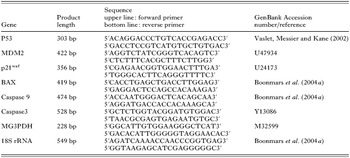
Table 1. Summary of the primer pairs for apoptosis-related genes (p53, MDM2, and p21waf, BAX, caspase 9 and caspase 3) and endogenous controls (MG3PDH and 18S rRNA)
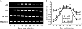
Fig. 1. RT-PCR results on expression levels of apoptosis-related genes (p53, MDM2, and p21waf) in the infected muscle tissue. Total RNA was isolated from infected muscle tissue at various stages (0, 8, 13, 18, 23, 28, 33, 38 and 53 days p.i.) and amplification products were analysed by agarose gel electrophoresis and shown in the left panel (A). Using Scion Image software, the relative density of each sample band against the control band (MG3PDH) was analysed and shown in the right panel (B).
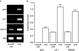
Fig. 2. RT-PCR results on expression levels of apoptosis-related genes (p53, MDM2, and p21waf) in the cyst or non-infected muscle cells that were obtained from the Trichinella spiralis-infected mouse at day 18 p.i. Total RNA was isolated from laser micro-dissected cysts or muscle cells, and RT-PCR of the sample genes and the standard gene (18S rRNA) was performed. PCR products were analysed by agarose gel electrophoresis (A), and the relative density of each sample band against the control gene band was analysed using Scion Image software (B).

Fig. 3. Immunohistochemical staining for p53. Cryosections of nude mouse muscles infected with Trichinella spiralis for 18 (A), 28 (B) and 48 (C) days p.i. were reacted with anti-p53 antibody. A negative control was performed using normal rabbit serum (D). The brown colour indicates a positive reaction. L, larva.
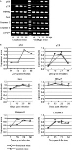
Fig. 4. RT-PCR results on expression levels of apoptosis-related genes (p53, MDM2, and p21waf, BAX, caspase 9 and caspase 3) in wild type and knockout mice infected muscle and uninfected muscle. Total RNA was isolated from infected muscle tissue at various stages (0, 13, 23 and 38 days p.i.) and amplification products were analysed by agarose gel electrophoresis and shown in the upper panel (A). Using Scion Image software, the relative density of each sample band against the control gene (MG3PDH) band was analysed and shown in the lower panel (B).
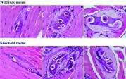
Fig. 5. Haematoxylin and eosin staining of Trichinella spiralis-infected wild type mice (A, B, C) and knockout mice (D, E, F) mouse muscle at day13 p.i. (A, D), day 23 p.i. (B, E) and day 30 p.i.(C, F). L, larva. Original magnification, 200×.

Table 2. Worm recovery of Trichinella spiralis-infected wild type and knockout mice
 You have
Access
- 22
- Cited by