Introduction
The cotton whitefly, Bemisia tabaci Gen. (Hem., Aleyrodidae), is an important pest of many crops, including vegetables and ornamentals (De Barro et al., Reference De Barro, Liu, Boykin and Dinsdale2011). This whitefly has more than 600 host plants causing economic damages in both greenhouse and field cucumber crops (Oliveira et al., Reference Oliveira, Henneberry and Anderson2001). The damage is caused through (i) sucking the plant sap leading to plant growth and yield reductions; (ii) excreting honeydew resulting in the invasion of the sooty molds, potentially decreasing photosynthesis and reducing the yield quality and quantity, and (iii) virus transmission (Berlinger, Reference Berlinger1986; Thompson, Reference Thompson2011).
The application of synthetic insecticides such as imidacloprid is the main strategy to control B. tabaci in many regions of the world (Palumbo et al., Reference Palumbo, Horowitz and Prabhaker2001). Pesticide resistance, side effects on non-target organisms, pest resurgence, and secondary pest outbreak are some limitations of the chemical control (Pedigo, Reference Pedigo2002; Safaei et al., Reference Safaei, Rajabpour and Seraj2016; Shahbi and Rajabpour, Reference Shahbi and Rajabpour2017).
Microbial control agents can serve as environmentally friendly components of integrated pest management (IPM) programs due to their selectivity, safety, and compatibility with other natural enemies (Lacey and Shapiro-Ilan, Reference Lacey and Shapiro-Ilan2008). Entomopathogenic fungi (EPF) are the most abundant group, ~60%, of insect pathogens (Liu et al., Reference Liu, Xie, Xue, Gao, Zhang, Zhang and Tan2009). EPF can directly penetrate through the arthropod cuticle, unlike bacteria and viruses which are ingested to induce pathogenicity (Ortiz-Urquiza and Keyhani, Reference Ortiz-Urquiza and Keyhani2013). Therefore, EPF can serve as efficient biocontrol agents against sap-feeding pests, e.g. the whiteflies.
There are several EPF that have been reported as efficient biocontrol agents against B. tabaci (Osborne and Landa, Reference Osborne and Landa1992; Faria and Wraight, Reference Faria and Wraight2001; Cuthbertson and Walters, Reference Cuthbertson and Walters2005; Cuthbertson et al., Reference Cuthbertson, Blackburn, Northing, Luo, Cannon and Walters2008; Cuthbertson et al., Reference Cuthbertson, Blackburn, Northing, Luo, Cannon and Walters2010). These agents have been systematically classified into phylum Ascomycota, order Hypocreales, and family Cordycipitaceae (Litwin et al., Reference Litwin, Nowak and Różalska2020). The important species within Cordycipitaceae include Beauveria bassiana (Balsamo-Crivelli) Vuillemin (Reference Vuillemin1912), Cordyceps fumosorosea (Wize) Kepler, B. Shrestha, and Spatafora (Reference Kepler, Luangsa-ard, Hywel-Jones, Quandt, Sung, Rehner, Aime, Henkel, Sanjuan, Zare, Chen, Li, Rossman, Spatafora and Shrestha2017) [Syn.: Isaria fumosoroseus Wize (Reference Wize1904)], A. lecanii (Zimmerman) Spatafora, Kepler and B. Shrestha (Reference Kepler, Luangsa-ard, Hywel-Jones, Quandt, Sung, Rehner, Aime, Henkel, Sanjuan, Zare, Chen, Li, Rossman, Spatafora and Shrestha2017) [Syn.: Lecanicillium lecanii (Zimmerman) Zare and Gams (Reference Zare and Gams2001)], and A. muscarius (Petch) Spatafora, Kepler and B. Shrestha (Reference Kepler, Luangsa-ard, Hywel-Jones, Quandt, Sung, Rehner, Aime, Henkel, Sanjuan, Zare, Chen, Li, Rossman, Spatafora and Shrestha2017) [Syn.: Lecanicillium muscarium Petch (Reference Petch1931)]. However, no study has been carried out to determine the pathogenicity of Iranian isolates of Akanthomyces spp. on whitefly. Iranian isolates can show significantly different potentials to control the pest due to the different climate of Iran in comparison with other climates in which the previous isolates were originated. Various entomopathogenic isolates with different geographic origins may indicate different pathogenicity to determine pests (Feng et al., Reference Feng, Poprawski and Khachatourians1994; Sani et al., Reference Sani, Ismail, Abdullah, Jalinas, Jamian and Saad2020). Furthermore, Lecanicillium isolates originating from a single geographical region were reported to exhibit different mortality rates against the target insect (Zhu and Kim, Reference Zhu and Kim2011; Manfrino et al., Reference Manfrino, Schuster, Saar, López Lastra and Leclerque2019; Xie et al., Reference Xie, Jiang, Li, Hong, Wang and Jia2019; Abdulle et al., Reference Abdulle, Nazir, Keerio, Ali, Zaman, Anwar, Nam and Qiu2020). Although it was mostly demonstrated that, in comparison to B. bassiana, L. lecanii isolates are more pathogenic to B. tabaci (Abdel-Raheem and Al-Keridis, Reference Abdel-Raheem and Al-Keridis2017; Espinosa et al., Reference Espinosa, da Silva, Duarte, Gonçalves and Polanczyk2019), a Russian isolate of L. lecanii was found to display relatively low virulence against the insect (Keerio et al., Reference Keerio, Nazir, Abdulle, Jatoi, Gadhi, Anwar, Sokea and Qiu2020). Therefore, during the current study, the potentials of four Iran-originated isolates of A. lecanii and A. muscarius to control B. tabaci on cucumber were studied under laboratory conditions. In Iran, cucumber is one of the important hosts of the whitefly causing significant quantitative and qualitative losses every year. Moreover, vegetative growth and conidiation of the isolates on various media were investigated to find the optimum medium for vegetative growth and conidiation of the EPF.
Materials and methods
Insect and host plant cultures
Seeds of greenhouse cucumber (Cucumis sativus cv. Negin) were sown in pots containing perlite-cocopeat mix (1:1, v: v) and were daily moistened with Hoagland's nutrient solution. The pots were placed in the rearing cages (1 × 0.6 × 1.2 m) in a growth chamber at 25 ± 1°C, 65 ± 5% relative humidity (RH), and 16:8 h (light:dark) photoperiod (Mohammadi et al., Reference Mohammadi, Seraj and Rajabpour2015).
Adults of the whitefly, B. tabaci biotype B, were collected using an aspirator from a commercial cucumber greenhouse in Shushtar district, Khuzestan province, southwest of Iran (32°05′30.5″N 48°45′25.2″E(. After their identification by Martin (Reference Martin1987) identification key, the whiteflies were introduced to the rearing cages. The cages were kept inside an air-conditioned room at a temperature of 25 ± 2°C, RH of 65 ± 5%, and a photoperiod of 14:10 h (light:dark). The plants were replaced biweekly. The colony was maintained until the end of the bioassay trials.
Isolation and identification of the fungi
Isolation
The EPF were isolated from the orange Pulvinaria scale, Pulvinaria aurantii Cock. (Hem., Coccidae), and cotton aphid, Aphis gossypii Glover (Hem., Aphididae), in citrus orchards of Citrus and Subtropical Fruits Research Center of Iran, Ramsar district, Mazandaran province, northern Iran (36°54′24.2″N 50°39′26.7″E). The fungus isolation was carried out using the method described by Kumar et al. (Reference Kumar, Jacob, Devasahayam, D'Silva and Kumar2015) with some modifications. Briefly, dead individual of each species was superficially sterilized using 10% sodium hypochlorite for 3 s and washed twice with distilled water. The specimens were then air-dried and transferred to Petri dishes lined with wet Whatman paper (No. 1) and incubated at 25°C for 48 h without light. Cadavers exhibiting fungal growth were selected and placed onto Sabouraud dextrose agar (SDA) medium (Merck, Germany) and incubated at 25°C. The single spore method was applied to obtain purified cultures of each fungal isolate.
Morphological identification
The fungal isolates were morphologically identified using classical taxonomy following general and specific identification keys (Petch, Reference Petch1925; Humber, Reference Humber and Lacey1997; Zare and Gams, Reference Zare and Gams2001; Zare and Gams, Reference Zare and Gams2004). Fungal stocks were prepared by sub-culturing the pure culture of the isolates into glass tubes containing potato dextrose agar (PDA) medium and incubating at 4°C for 72 h and then stored at room temperature. Microscope slides were prepared from each fungal isolate and morphological features including conidia, conidiophores, and phialides were photographed using a digital camera-equipped microscope (Olympus, Tokyo, Japan) at 400× magnification.
DNA extraction
The fungal DNA was extracted using cetyl-trimethyl-ammonium bromide (CTAB) solution as described by Gawel and Jarret. Fungal isolates were grown in potato sucrose broth (PS) for 7 days at room temperature. Then, the liquid medium was removed and mycelia were ground to a fine powder under liquid nitrogen. Pre-heated [60°C] extraction buffer (1.4 M NaCl, 20 mM EDTA, 2.5% CTAB, 2% 2-mercaptoethanol, 10 mM Tris-HCl, pH 8) was added and the mixture was incubated at 65°C for 30 min. The DNA was extracted with an equal volume of chloroform/isoamyl alcohol (24:1), precipitated with one volume of ice-cold isopropanol, washed for two times with 70% ethanol, and re-suspended in TE buffer (10 mM Tris-HCl, 0.1 mM EDTA, pH 8.0). The resulting DNA was stored at −20°C.
DNA amplification
PCR was performed using two primer pairs designed to amplify two regions of the fungal genome including internal transcribed spacer (ITS: ITS4/ITS5) and mitochondrial DNA (mtDNA: NMS1/NMS2) (White et al., Reference White, Bruns, Lee, Taylor, Innis, Gelfand, Sninsky and White1990). PCR was performed in a volume of 25 μl containing 10–20 ng of fungal DNA template, 1 μM of each primer, 12.5 μl of Taq DNA Polymerase Master Mix (Ampliqon, Denmark), and 9.5 μl of PCR-grade H2O. The mixture was heated for 3 min at 94°C and subjected to a 30 cycle-PCR program of 1 min at 96°C, 30 min at 55°C, and 1 min at 72°C. The final cycle was followed by 5 min incubation at 72°C. PCR products were excised from the gel and purified using a PCR clean-up kit (Denazist, Iran). Purified DNAs were then subjected to Sanger sequencing at BIONEER Corporation (South Korea).
Phylogenetic analysis
Base-calling was performed using the DNAStar Lasergene suite (DNAStar, Madison, WI, USA). Nucleotide sequences of the ITS region from the fungal isolates were deposited on the NCBI database under the accession numbers of MT130430, MT130431, MT130432, and MT130433. The resulting sequences were compared to those sequences whose information is available in the GenBank database. A total of 35 ITS sequences from EPF within Cordycipitaceae, including four sequences determined in this study and 31 sequences from different sources and diverse regions previously deposited on GenBank were used to carry out a phylogenetic analysis (Supplemental material, table S1). Multiple alignments of the sequences were performed using CLC Main Workbench software (ver. 7.6.2), and the maximum likelihood approach using General Time Reversible model test (Tavaré, 1986) with 100 bootstrap replicates was used to construct a phylogenetic tree. One corresponding sequence from Lecanicillium psalliotae was included in the analysis as an outgroup.
Fungal vegetative growth and conidiation
To find the optimum medium for vegetative growth and conidiation of the EPF, the fungal isolates (PAL6, PAL7, PAL8, and AGM5) were grown on different media including PDA (Himedia, India), SDA (Merck, Germany), SDA + yeast extract (SDA + Y) (Merck, Germany), PCA (Himedia, India), and malt agar (MA) (Merck, Germany). Three-mm discs were excised from the peripheral mycelium of 5-day-old cultures and were separately placed upside down in the center of 9-cm plates containing the aforementioned media. The cultured media were kept in a growth chamber under the conditions of 23 ± 1°C, 16/8 photoperiod (light/dark), and 70 ± 5% RH. The growth of fungal isolates was determined by measuring the colony diameter (in millimeter) using a ruler at 7-days post-incubation. Moreover, the conidial concentration (conidia ml−1) of each isolate cultured in different media was determined using a hemocytometer (HGB, Germany) after 7 days (see below). Each treatment was replicated three times.
Bioassay trials
Second instar nymphs of B. tabaci (2 days old) were used for the experiments due to their susceptibility to the EPF (Cuthbertson et al., Reference Cuthbertson, Walters and Northing2005). The developmental stage was obtained according to the method described by Banihashemi et al. (Reference Banihashemi, Seraj, Yarahmadi and Rajabpour2017). Males and females of the whitefly (about 50 pairs) were maintained together for mating for 48 h. After this period, the mated females were collected using an aspirator and introduced into the rearing cages. Nine days after introducing the mated adults to cucumber plants (at 25 ± 2°C, 60 ± 5% RH, and a photoperiod of 16 h light:8 h dark), the second instar nymphs were obtained for the experiments.
The protocol described by Wraight et al. (Reference Wraight, Carruthers, Bradley, Jaronski, Lacey, Wood and Galaini-Wraight1998) was used for the bioassay test. For each fungal isolate, five concentrations of conidial suspensions (104, 105, 106, 107, and 108 conidia ml−1) were prepared. Briefly, 12 ml of sterile distilled water + 0.05% Tween-80 (Merck, Germany) were added to the 15-day old PDA cultures and conidia and mycelium of each isolate was harvested using a sterile glass rod. To obtain the conidia, the mixture was filtered through four layers of cheesecloth. The conidia concentration (as conidia ml−1) was estimated using a hemocytometer (HGB, Germany). Three replications were considered for each counting. Two to three days before the bioassay, conidial viabilities of the EPF were checked using the method described by Castillo et al. (Reference Castillo, Moya, Hernández and Primo-Yufera2000). Briefly, 0.1 ml of 107 conidia ml−1 concentration was spread onto PDA media and incubated at 23 ± 1°C for 18 h. The germination of 50 conidia was randomly checked under a light microscope. A cucumber leaf infested by 10-s instar nymphs was sprayed once with each concentration using a handle sprayer from ~5 cm distance. In this situation, the preliminary test indicated that about 1 ml of the suspension was uniformly spread on the leaf surface in each spray. The treated leaf was immediately transferred to a Petri dish, 10 cm diameter, placed upside down on a 20 ml layer of water agar (5%) (Merck, Germany). The dishes were enclosed using plastic bags and incubated at 23 ± 1°C, 70 ± 5% RH, and a photoperiod of 16:8 h (light:dark) for 6 h. Subsequently, the dishes were removed from the bags, allowed to air dry, covered with ventilated lids (lids with an 8-cm diameter hole covered with a fine screen), and returned to the incubator. The treated leaves were incubated ventral surface up to prevent entrapment of humid air at the leaf surface. Mortalities of the nymphs were recorded at intervals of 24 h for 8 days. Mortality was defined by the presence of the fungal mycelia (Cuthbertson et al., Reference Cuthbertson, Walters and Northing2005). Seven replications, treated leaf, were used for each conidial concentration.
Data analysis
For vegetative growth and conidiation trials, the data were analyzed as a 5 (media) by 4 (EPF isolates) factorial experiment with three replications, based on a completely randomized design using the generalized linear model (GLM) procedure of SAS software (version 9.2) (SAS Institute, Inc., Cary, NC). Shapiro-Wilk tests were done to check the data normality assumption. The least significant difference (LSD) test was used as a post-hoc test for means comparisons.
Probit analysis was used for estimating the median lethal concentrations (LC50) (Finney, Reference Finney1971). Relative median potencies and their 95% confidence intervals were calculated for different treatments when their slopes did not differ significantly (Finney, Reference Finney1971). Moreover, probit analysis was conducted to estimate the time for killing 50% of the insects (LT50) (Throne et al., Reference Throne, Weaver, Chew and Baker1995). All analyses were done using SAS software ver. 9.1 (SAS Institute, Cary, NC). Estimated LC50 or LT50 values were not considered statistically different when their confidence limits (95%) overlapped (Robertson et al., Reference Robertson, Russel, Preisler and Savin2007).
Results
Morphological identification
Morphological characteristics of the fungal colonies and conidia of the isolates identified two species of Akanthomyces, A. lecanii and A. muscarium. The isolates of A. lecanii were characterized by forming yellowish-white, fluffy, branched mycelium in a centered cycle pattern (data not shown). Moreover, typically short-ellipsoidal conidia (2.5–3.5 × 1–1.5 μm) were produced on conidiogenous cells (phialides) of short conidiophores (Zare and Gams, Reference Zare and Gams2001). However, A. muscarium formed relatively compact, with reverse cream to pale yellow or colorless mycelium. It produced phialides generally longer than those of A. lecanii and ellipsoid to sub-cylindrical conidia (2.5–5.5 × 1–1.5 μm) more irregular in size and shape, longer, and narrower than in A. lecanii (Zare and Gams, Reference Zare and Gams2001).
Phylogenetic analysis
Four ITS sequences from morphologically-identified Akanthomyces isolates including AGM5, PAL6, PAL7, and PAL8 were obtained. The isolate AGM5 showed the highest nucleotide identity (96.55%) to a French isolate (ARSEF 2323) of A. muscarius (Syn.: L. muscarium [EF513017]). The isolates PAL6, PAL7, and PAL8 exhibited the highest nucleotide identity (92.32, 92.12, and 91.94%, respectively) to a Turkish isolate (IMI 079606) of A. lecanii (Syn.: Lecanicillium lecanii [EF512998]), respectively. Phylogenetic analysis of the 35 nucleotide sequences of ITS showed 3 clades with 76–100% bootstrap support consisting of Akanthomyces (n = 23), Beauveria (n = 5) and Simplicillium (n = 6) isolates with Lecanicillium psalliotae as an outgroup (fig. 1). Sequences from the four EPF isolates obtained in this study all clustered within the Akanthomyces clade. These results confirmed the morphological identification of the fungal isolates.
Maximum likelihood phylogenetic tree obtained from multiple alignments of internal transcribed spacer (ITS) sequence from worldwide isolates within the family Cordycipitaceae (Ascomycota, Hypocreales). The isolates obtained in the present study were highlighted. One isolate of L. psalliotae was used as an outgroup reference. The bar represents the estimated nucleotide substitutions per site.

Vegetative growth and conidiation
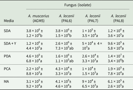
There was a significant effect of media on the conidial concentrations produced after 7 days incubation, while there was no significant effect of fungal isolate nor the interaction between media and isolate (table 1). The conidial concentration of each isolate cultured on different media is shown in table 2. For A. muscarium (AGM5) and A. lecanii (PAL6), the highest condial concentrations were obtained on SDA. No significant difference was observed between the conidial concentration of A. lecanii (PAL7) grown on the various media. However, A. lecanii (PAL8) grown on the PDA medium exhibited the highest conidial concentration compared to other media (table 2).
GLM parameters of main effects and their interaction for five different media and four isolates on colony diameters of the entomopathogenic fungi at 7-days after incubation

Mean of conidial concentrations (conidia ml−1) ± SE (n = 3) of the entomopathogenic fungal isolates grown in five media at 7-days after incubation

SDA, Sabouraud dextrose agar; SDAY, SDA + yeast extract; PDA: potato dextrose agar, PCA, potato carrot agar; MA, malt agar.
The same letters in each column indicate a non-significant difference (LSD test).
The different media significantly affected the fungal colony diameters of each isolate (table 3). For PAL6, PAL8, and AGM5 isolates, the largest value of colony diameter was observed in fungal isolates grown on PDA (table 4).
GLM parameters of main effects and their interaction for five different media and four isolates on the conidial concentration of the entomopathogenic fungi 7-days after incubation

Mean of colony diameter (mm) ± SE (n = 3) of the entomopathogenic fungal isolates cultured in five media at 7-days after incubation

SDA, Sabouraud dextrose agar; SDAY, SDA + yeast extract; PDA, potato dextrose agar; PCA, potato carrot agar; MA, malt agar.
The same letters in each column indicate a non-significant difference (LSD test).
Bioassay trials
The LC50 values of the four isolates at 5–7 days after treatment (DAT) are shown in table 5. There was not a significant difference between the estimated LC50 values of the EPF at each DAT. However, the LC50 value of AGM5 at 7 DAT is significantly lower than other isolates. For A. lecanii isolates (PAL6, PAL7, and PAL8), no significant differences were observed between the LC50 values at 5–7 DAT. Although, the LC50 values of A. muscarius isolate (AGM5) were significantly reduced at days 5–7. Totally, the highest and the lowest LC50 values were 7.3 × 10¹³ and 9.2 × 10⁴ (conidia ml−1) which were observed in A. muscarius at 7 DAT and A. lecanii (isolate PAL6) at 5 DAT, respectively.
The median lethal concentration (LC50) values of five isolates of the entomopathogenic fungi (EPF) to the second instar nymph of B. tabaci

The LT50 values of the EPF in different conidial concentrations are presented in table 6. For all conidial concentrations, the LT50 value of A. muscarius (isolate AGM5) was significantly lower than other isolates. The LT50 values were decreased by increasing conidial concentrations of each isolate. Totally, the highest and the lowest LT50 values were 9.0 and 5.0 days, which was observed in A. lecanii (isolate PAL6) in the concentration 104 conidia ml−1 and A. muscarius (isolate AGM5) in the concentration 108 conidia ml−1, respectively. The bioassay viabilities of A. lecanii and A. muscarius conidia were found as 98 and 99%, respectively.
The median lethal time (LT50) values of various isolates of the entomopathogenic fungi (EPF) to second instar nymph of B. tabaci

Discussion
EPF have been included among microbial biocontrol agents which adversely affect the pest population without any hazardous influence on human and the environment (Butt, Reference Butt and Kempken2002; Thomas and Read, Reference Thomas and Read2007). Due to their pathogenicity process and wide host range, they have been considered as key factors in IPM programs (Khan et al., Reference Khan, Guo, Maimaiti, Mijit and Qiu2012). The two most important and widespread species of EPF, A. lecanii and A. muscarius, have been found as effective biocontrol agents against B. tabaci (Osborne and Landa, Reference Osborne and Landa1992; Faria and Wraight, Reference Faria and Wraight2001; Cuthbertson and Walters, Reference Cuthbertson and Walters2005; Park and Kim, Reference Park and Kim2010; Ren et al., Reference Ren, Ali, Huang and Wu2010). Here we morphologically identified two species of Akanthomyces as the first Iran-originated entomopathogenic species. Phylogenetic analysis based on the ITS region has been extensively applied as a molecular marker to classify fungal species (Hillis and Dixon, Reference Hillis and Dixon1991; Salazar et al., Reference Salazar, Schneider, Julian, Keijer and Rubio1999; Arenal et al., Reference Arenal, Platas, Monte and Peláez2000). Particularly, Akanthomyces spp. have been subjected to a comprehensive phylogenetic analysis according to which A. lecanii and A. muscarius formed separate clusters in the phylogenetic tree (Kouvelis et al., Reference Kouvelis, Sialakouma and Typas2008). Similarly, three isolates of A. lecanii and a single isolate of A. muscarius which had been obtained in this study clustered in two separate clades in the phylogenetic tree. Also, the fungal isolates from Simplicillium spp. were clustered together which was consistent with the results of Kouvelis et al. (Reference Kouvelis, Sialakouma and Typas2008).
It has been shown that culture medium can significantly influence the growth and conidiation of A. lecanii ( Romero and de Romero, Reference Romero and de Romero1986; Sun et al., Reference Sun, Gao, Liu and Wang2009; Prasad and Pal, Reference Prasad and Pal2014; Gao, Reference Gao2018). Based on our results, PDA was found to be the optimal medium for growth and conidiation of A. lecanii. In the case of A. muscarius, however, the highest conidia concentration was observed on the SDA medium. Although there is no specific study on the selection of culture media for vegetative growth and conidiation of A. muscarius, the SDA medium has been used by some researchers (Marshall et al., Reference Marshall, Lester, Glare and Christeller2003; Lazreg et al., Reference Lazreg, Huang, Ali and Ren2009; Luz et al., Reference Luz, Mnyone, Sangusangu, Lyimo, Rocha, Humber and Russell2010; Mohammadipour et al., Reference Mohammadipour, Ghazavi, Bagdadi and Zare2010). In addition to the culture medium, other environmental factors such as pH, water potential, temperature, and light can affect fungal vegetative growth and/or conidiation (Gao et al., Reference Gao, Liu, Sun, Li and Wang2009). Therefore, it is highly recommended that these factors be specifically determined to find an optimum condition for the growth and conidiation of the species of EPF.
The results of the infection bioassays showed that the EPF obtained in this study are pathogenic to second instar nymphs of B. tabaci among which A. muscarius (isolate AGM5) exhibited the highest efficiency as a biocontrol agent of the whitefly.
The LC50 values reported in the present study are relatively lower than the estimated LC50 values of four isolates of A. muscarius (V20, V26, V07, and V17) to B. tabaci nymphs, 1.07 × 10⁶–5.08 × 10⁸ conidia ml−1 (Ren et al., Reference Ren, Ali, Huang and Wu2010). The LC50 values of six Canadian isolates of A. lecanii, (V3450, Vp28, V16063, V0175, V342, and V341) to third instar nymphs of B. tabaci ranged from 2.57 × 10⁵ to 6.03 × 10⁵ conidia ml−1 (Wang et al., Reference Wang, Huang, You and Liu2004) which are relatively lower values than for the isolates of our study. The lower LC50 values may be related to different susceptibility of B. tabaci life stages, second and third instar nymphs. The second instar nymph of B. tabaci has been reported as the most susceptible life stage of the insect (Cuthbertson et al., Reference Cuthbertson, Walters and Northing2005). In our study, progressive mortality of the whitefly nymphs was observed during a time similar to the finding reported by Saito and Sugiyama (Reference Saito and Sugiyama2005).
The LT50 values of A. lecanii and A. muscarius isolates were 6.1–9.5 and 4.9–7.1 days, respectively. The values were relatively higher than those reported for three Chinese isolates of A. lecanii (L22, L14, and L18) on B. tabaci biotype Q, which were 3.4–4.6 days (Zhu and Kim, Reference Zhu and Kim2011). The different responses may be related to many factors including the different fungal isolate, different biotypes (genetic difference of the whiteflies), and different spray methods. Similar to our finding, the LT50 values of an Iranian isolate of A. muscarius, isolated from Zeuzera pyrina L. (Lep., Cossidae), at the concentrations 105, 106, 107, and 108 conidia ml−1 to Trialeurodes vaporariorum Westwood, were 8, 6, 6, and 4 days, respectively (Tabadkani et al., Reference Tabadkani, Askary, Mehrasa and Ashouri2010).
In our study, the mortality of the infected nymphs started on day 3 after treatment. Similarly, the first mortality of third instar nymphs by other isolates of A. lecanii (V3450, Vp28, Vl6063, V0175, V342, and V341) began at day 3 after treatment (Wang et al., Reference Wang, Huang, You and Liu2004). Liu et al. (Reference Liu, Xie, Xue, Gao, Zhang, Zhang and Tan2009) investigated in detail the infection process and histopathological changes of Japanese wax scale, Ceroplastes japonicus Green (Hem., Coccidae) by the A. lecanii. They demonstrated that the death of infected insects occurred 6-days post-inoculation. This variation among infection and death times reported from different studies might be due to several factors, including insect host, environmental conditions, fungal isolate, host plant, etc. (Shah and Pell, Reference Shah and Pell2003).
Totally, the fungal isolates, especially A. muscarius (AGM5), have appropriate potentials as a biological control agent to control B. tabaci in cucumber. Some isolates of Akanthomyces spp. have been applied as commercial microbial biocontrol agents to control various insect pests. Among them, two commercial isolates of A. muscarius, Mycotal®, and Verticillin®, were previously recommended to control whiteflies, especially under greenhouse conditions (Goettel et al., Reference Goettel, Koike, Kim, Aiuchi, Shinya and Brodeur2008). The efficacy of the EPF can be enhanced in greenhouse conditions. However, microbial insecticides may have some limitations including slow effect and high costs (Cuthbertson and Walters, Reference Cuthbertson and Walters2005).
Conclusion
Our finding showed that the fungi A. lecanii (PAL6, PAL7, and PAL8) and A. muscarius (AGM5), isolated from citrus hemipteran pests in the north of Iran, have appropriate potentials for applying as microbial control agents against B. tabaci on cucumber, especially in greenhouse condition. Among the fungi, A. muscarius (AGM5) was the most promising isolate according to its relatively low LC50 and LT50 values. However, further greenhouse and field experiments are required to better understand the EPF–whitefly interactions before the practical use of A. muscarius against B. tabaci in cucumber production can be implemented.
Supplementary material
The supplementary material for this article can be found at https://doi.org/10.1017/S0007485321000298
Acknowledgments
The research was financially supported by the Agricultural Sciences and the Natural Resources University of Khuzestan [grant no. 9628408]. The authors thank Dr S. Aghajanzadeh and the Citrus and Subtropical Fruits Research Center of Iran for their technical assistance.