- AAD
Antibiotic-associated diarrhoea
- CDAD
Clostridium difficile-associated diarrhoea
- SCS
Spent culture supernatant
- MM
microbial mixture
Introduction
The use of probiotics to prevent different gastrointestinal disorders is increasing, since it has been demonstrated that this kind of microorganism is capable of inhibiting the growth or invasion of pathogens, enhance the intestinal barrier, and modulate the mucosal and systemic immune response by regulating secretion of different cytokines (Servin Reference Servin2004; Corthésy et al. Reference Corthésy, Gaskins and Mercenier2007).
Diarrhoea is a common side effect of antibiotic treatment and Clostridium difficile-associated diarrhoea (CDAD) has become a major issue in many countries (Limaye et al. Reference Limaye, Turgeon, Cookson and Fritsche2000). The predominant mechanism of virulence of C. difficile is associated with the production of two large toxins: TcdA (308 kDa) and TcdB (260 kDa). These toxins act as glycosyltransferases, modifying host cell small GTPases involved in actin polymerisation and cytoskeleton assembly (Schirmer & Aktories Reference Schirmer and Aktories2004; Jank et al. Reference Jank, Giesemann and Aktories2007). Castagliuolo et al. (Reference Castagliuolo, Keates, Wang, Pasha, Valenick, Kelly, Nikulasson, LaMont and Pothoulakis1998) reported that TcdA is mainly an enterotoxin whereas TcdB has cytotoxic activity and exerts its biological effect at lower doses than TcdA, even though both toxins have the same enzymatic activity.
The search for the best way to prevent CDAD includes the administration of probiotic microorganisms. Several experimental trials employing either a single strain or a combination of different microorganisms for treatment of patients with CDAD have been performed (Hickson Reference Hickson2011) but currently the results obtained are inconclusive. To date, most evidence in this area involves in vitro and in vivo studies with Saccharomyces boulardii (Pothoulakis et al. Reference Pothoulakis, Kelly, Joshi, Gao, O'Keane, Castagliuolo and Lamont1993; Castagliuolo et al. Reference Castagliuolo, Riegler, Valenick, LaMont and Pothoulakis1999; Qamar et al. Reference Qamar, Aboudola, Warny, Michetti, Pothoulakis, LaMont and Kelly2001) or Lactobacillus rhamnosus GG (Mack et al. Reference Mack, Michail, Wei, McDougall and Hollingsworth1999; Madsen, Reference Madsen2001). In particular, it has been reported that Sac. boulardii interferes with virulence of C. difficile through neutralisation or degradation of toxins (Castagliuolo et al. Reference Castagliuolo, Riegler, Valenick, LaMont and Pothoulakis1999) and inhibition of inflammatory immune response (Chen et al. Reference Chen, Kokkotou, Mustafa, Bhaskar, Sougioultzis, O'Brien, Pothoulakis and Kelly2006). In addition, Trejo et al. (Reference Trejo, Minnaard, Pérez and De Antoni2006) demonstrated that extracellular factors from bifidobacterial species significantly reduce growth of C. difficile and its adhesion to enterocyte-like Caco-2 cells. More recently, the same authors proposed that co-culture with potentially probiotic bacteria affects the synthesis and/or secretion of clostridial toxins reducing the biological activity of C. difficile culture supernatant (Trejo et al. Reference Trejo, Pérez and De Antoni2010).
Kefir is a dairy product obtained by fermentation of milk with kefir grains and its consumption has been associated with several health-promoting properties such as antimicrobial, antitumoral, immunological and hypocholesterolemic effects (Farnworth, Reference Farnworth2005; Vinderola et al. Reference Vinderola, Duarte, Thangavel, Perdigón, Farnworth and Matar2005, Reference Vinderola, Perdigón, Duarte, Thangavel, Farnworth and Matar2006). Kefir grains are clusters of microorganisms that include mainly lactic acid bacteria (lactobacilli, lactococci, leuconostoc), yeasts and acetic acid bacteria which coexist in a symbiotic association in a matrix of polysaccharides and proteins (Garrote et al. Reference Garrote, Abraham and De Antoni2001; Hsi-Chia et al. Reference Hsi-Chia, Sheng-Yao and Ming-Ju2008).
Our workgroup has isolated and characterised more than 100 bacterial and yeast strains from kefir grains which have showed differences in surface and probiotic properties (Garrote et al. Reference Garrote, Delfederico, Bibiloni, Abraham, Pérez, Semorile and De Antoni2004; Golowczyc et al. Reference Golowczyc, Mobili, Garrote, Abraham and De Antoni2007, Reference Golowczyc, Gugliada, Hollmann, Delfederico, Garrote, Abraham, Semorile and De Antoni2008). Among these kefir-isolated microorganisms, it has been demonstrated that yeasts are able to down regulate the pro-inflammatory response induced in intestinal epithelial cells in vitro and in vivo (Romanin et al. Reference Romanin, Serradell, González Maciel, Lausada, Garrote and Rumbo2010), and that different lactobacilli strains show a high inhibitory power against Salmonella spp., Shigella spp. and Escherichia coli O157:H7 in vitro (Golowczyc et al. Reference Golowczyc, Mobili, Garrote, Abraham and De Antoni2007, Reference Golowczyc, Gugliada, Hollmann, Delfederico, Garrote, Abraham, Semorile and De Antoni2008; Hugo et al. Reference Hugo, Kakisu, De Antoni and Pérez2008). Besides, our group demonstrated the ability of surface proteins (S-layer) from Lb. kefir strains to antagonise cytotoxic effects of clostridial toxins on cultured eukaryotic cells (Carasi et al. Reference Carasi, Trejo, Pérez, De Antoni and Serradell2012).
Recently, we have developed a mixture constituted of three bacteria (Lb. plantarum CIDCA 83114, Lb. kefir CIDCA 8348 and Lactococcus lactis CIDCA 8221) and two yeasts (Sac. cerevisieae CIDCA 8112 and Kluyveromyces marxianus CIDCA 8154) isolated from kefir grains which was able to inhibit Sh. sonnei growth in vitro and preserved its viability, antimicrobial capacity and safety after freeze-drying procedure (Bolla et al. Reference Bolla, de Serradell, de Urraza and De Antoni2011). These properties and the potentiality of kefir-isolated microorganisms (or mixtures of them) as starters in probiotic fermented products encouraged us to evaluate the inhibitory effect of these selected strains against other pathogens. Since, as we mentioned above, the use of probiotics to prevent CDAD could be an interesting alternative to the antibiotic treatments, we decided to evaluate the ability of these bacterial and yeast strains isolated from kefir to antagonise the effect of C. difficile toxins on eukaryotic cells in vitro.
Materials and Methods
Bacterial strains and growth conditions
Pure cultures used in this study comprised Lc. lactis subsp. lactis CIDCA 8221, Lb. plantarum CIDCA 83114, Lb. kefir CIDCA 8348, K. marxianus CIDCA 8154 and Sac. cerevisiae CIDCA 8112. These strains were previously isolated from kefir grains and have been identified and characterised by Garrote et al. (Reference Garrote, Abraham and De Antoni2001) and Delfederico et al. (Reference Delfederico, Hollmann, Martínez, Iglesias, De Antoni and Semorile2006). The original reference cultures were maintained in milk at −80 °C. Both lactobacilli and yeasts were propagated in MRS-broth (DIFCO, Detroit, USA) for 48 h at 30 °C. Lc. lactis was grown in 1.1.1 growth media (10 g tryptone/l—Difco, Detroit, USA; 10 g yeast extract/l—Biokard Diagnostic, Beauvais, France and 10 galactose/l—Mann Research Laboratories, NY) Abraham et al. (Reference Abraham, De Antoni and Añón1990) for 24 h at 30 °C. To obtain the microbial mixture (MM), the same volume of each microbial suspension was centrifuged at 10 000 g for 15 min and resuspended together in 1 ml sterile PBS (KH2PO4 0·144 g/l, NaCl 9 g/l, Na2HPO4 0·795 g/l, pH 7·4). The concentrations of bacteria and yeasts in MM were determined by plate counting using MRS agar for lactobacilli, YGC (Yeast extract Glucose Chloramphenicol Agar, Biokard Diagnostic, Beauvais, France) for yeast strains, and 1.1.1. agar for Lc. lactis. Final concentrations of bacteria and yeasts were 109 CFU/ml and 106 CFU/ml respectively.
The clinical isolate of C. difficile strain 117, obtained from the Hospital Dr Muñiz (Buenos Aires, Argentina) and previously characterised as positive for TcdA and TcdB production, was grown for 24 h at 37 °C in Brain Heart Infusion (BHI: Biokar Diagnostic, Beauvais, France) supplemented with 0·5 g cysteine chlorhydrate/l (BHI/cys) in anaerobic conditions (AnaeroPak, Mitshubishi Gas Chemical Co, Inc). Culture was centrifuged at 15 000 g for 15 min and the C. difficile spent culture supernatant (SCS) containing toxins was retained, then passed through a 0·22 μm filter and used in the experiments.
Incubation of SCS with microorganisms or their supernatants
For incubation of C. difficile SCS with each microorganism, microbial cells in stationary phase were harvested by centrifugation and washed three times with PBS. The pellet was resuspended in SCS at a final bacterial concentration of OD550 = 1·0 and incubated for 60 min at 37 °C. Spent culture supernatants pre-incubated with each isolated microorganism were obtained by centrifugation at 10 000 g for 15 min. Similarly, SCS was pre-incubated for 60 min at 37 °C with MM at final concentrations of 109 CFU bacteria/ml and 106 CFU yeasts/ml.
For preparation of microbial supernatants, individual microbial cultures and MM were harvested by centrifugation at 10 000 g for 15 min, washed three times with PBS and resuspended in BHI/cys. After incubation for 1 h at 37 °C in aerobic conditions, microorganisms were centrifuged at 10 000 g for 15 min and the supernatants (SN) were passed through a 0·22 μm filter and conserved at −20 °C until used.
For some experiments, Lc. lactis CIDCA 8221 supernatant was fractionated through an Amicon ultrafiltration StirreCell 8050 Millipore Corporation US, equipped with a regenerated cellulose membrane (cut-off 10 kDa) or was heated at 100 °C for 15 min or was treated with different proteases (trypsin, pepsine, quimiotrypsine or proteinase K) (SIGMA, USA) at 2·5 mg/ml during 1 h at 37 °C based on previous protocols described by Trejo et al. (Reference Trejo, Minnaard, Pérez and De Antoni2006) and Golowczyc et al. (Reference Golowczyc, Mobili, Garrote, Serradell, Abraham and De Antoni2009).
For the incubation of kefir-isolated microorganisms or MM culture supernatants with SCS, supernatants were added to SCS from C. difficile in a 1:1 ratio and biological activity of these samples was compared with SCS obtained from pure C. difficile cultures as described below. In each case, acidity of treated SCS did not show changes compared with control SCS (pH = 6·5).
Cell cultures
Vero cells were grown in Dulbecco's Modified Eagle's Medium (DMEM; Gibco BRL Life Technologies, Rockville, MD, USA) supplemented with 10% (v/v) inactivated (30 min/60 °C) foetal calf serum (BIOSER, Argentina, PAA Laboratories GmbH), 2 g NaHCO3/l, 10 mg streptomycin/l and 10 IU penicillin G/ml. Cells were inoculated (6·25 × 104 cells per well) into 48-well tissue culture plates (Corning, NY) and incubated at 37 °C for 48 h in a 5% (v/v) CO2, 95% (v/v) air atmosphere to allow the formation of a cell monolayer.
Biological effects on cultured cells
The assay was performed as described by Trejo et al. (Reference Trejo, Pérez and De Antoni2010). Briefly, cultured cells were washed twice with 1 ml PBS and 300 μl cell culture medium were added to each well. Control C. difficile SCS, SCS treated incubated with microorganisms (each strain or MM) and SCS treated with microbial supernatants (SCS + SN) were serially two fold diluted in DMEM without foetal calf serum. Two hundred μl of DMEM- diluted samples were added per well and incubated at 37 °C for 16 h in a 5% (v/v) CO2, 95% (v/v) air atmosphere. Biological activity was assessed by evaluation of cell detachment and labelling of F-actin cytoskeleton (Minnaard et al. Reference Minnaard, Humen and Pérez2001, Reference Minnaard, Lievin-Le Moal, Coconnier, Servin and Pérez2004). In some cases, cell detachment assays with SCS + SN Lc. lactis CIDCA 8221 were performed in presence of a cocktail of protease inhibitors (Roche, USA).
For cellular detachment assay, after incubation cells were washed twice with PBS and fixed with 2% (v/v) formaldehyde for 1 min (Minnaard et al. Reference Minnaard, Humen and Pérez2001). The remaining cells were stained with 0·13 g Crystal Violet/l in 5% (v/v) ethanol and 2% (v/v) formaldehyde. Next, an extraction with 50% (v/v) ethanol was performed and OD540 was determined. Biological activity was expressed as the percentage of detached cells (rd), according to the following expression:
 $${\rm rd} = {\rm 100 \times} \left( {{\rm 1} - \left( {{\rm Am} - {\rm Ao}} \right){\rm /}\left( {{\rm Ab} - {\rm Ao}} \right)} \right)$$
where Am, absorbance of sample; Ao, absorbance of well without cells (control of stain adsorption by the well); Ab, absorbance of untreated control cells.
The ratio of detached cells (rd) was modelled as a function of SCS concentration by using a hyperbolic function according to Trejo et al. (Reference Trejo, Pérez and De Antoni2010). This approach allows for the calculation of the dose of SCS that leads to the detachment of 50% of the cells (DD50). This parameter inversely correlates with biological activity of SCS.
Staining of F-actin cytoskeleton was performed using Vero cells grown on sterile glass coverslips (Assistant, Sondheim, Germany) in 24-well culture plates (Greiner Bio One, Germany). After incubation with SCS or SCS + SN Lc. lactis CIDCA 8221, cells were washed twice with PBS and fixed (2 min) with 3% (v/v) paraformaldehyde. Afterwards, cells were treated with NH4Cl (50 mm) and then permeabilised with 0·2% (v/v) Triton X100 solution in PBS before labelling with FITC-phalloidin (SIGMA, Inc., St. Louis, MO, USA) in PBS containing 2 g gelatin/l (SIGMA, Inc. St. Louis, MO, USA) for 45 min in the dark (Minnaard et al. Reference Minnaard, Lievin-Le Moal, Coconnier, Servin and Pérez2004). Cells were observed by fluorescence microscopy.
Dot-blot assay
To test the effect of Lc. lactis CIDCA 8221 SN on TcdA and TcdB present in SCS, a dot-blot assay was performed. After incubation of SN with C. difficile SCS for 60 min at 37 °C, 4 μl of the sample was spotted onto nitrocellulose membranes. After drying for 30 min at room temperature, strips were blocked with 30 g skim milk/l buffer T-TBS: 0·05 m Tris (hydroxymethyl aminomethane Mallinckrodt, Baker Inc.), 0·15 mol NaCl/l, 0·5% (v/v) Tween 20 (Sigma–Aldrich, Inc., St. Louis, MO, USA) pH 7·5, for 90 min at 37 °C. Afterwards, membrane strips were incubated 90 min at 37 °C with mouse monoclonal antibodies anti-TcdA (1/1000) or anti-TcdB (1/500) (Meridian Life Science, Unc.). After washing three times with T-TBS, strips were incubated for 60 min at 37 °C with peroxidase-conjugated anti-mouse IgG (Santa Cruz Biotechnology, Inc., USA) and the reaction was revealed using 4-chloro-1-naphthol (9 g/l) and 18 μl 30% (v/v) H2O2 (E. Merck, Germany) dissolved in 3 ml methanol and 15 ml TBS.
Statistical analysis
Data were analysed by InfoStat Software (Grupo InfoStat, FCA, Universidad Nacional de Córdoba, Argentina). Results were statistically tested using Student t-test to determine any significant difference.
Results and discussion
Effect of microorganisms or microbial supernatants on the biological activity of C. difficile toxins on Vero cells
The ability of kefir-isolated strains to antagonise cytopathic effects of C. difficile SCS was tested performing dose response curves in a cell detachment assay and by determining the dose of SCS that leads to the detachment of 50% of Vero cells (DD50). When Vero cells were incubated with SCS treated with each microorganism or MM (the mixture containing all strains under study), DD50 was not increased compared with control of treatment with SCS (0·063 ± 0·009 vs. 0·065 ± 0·009).
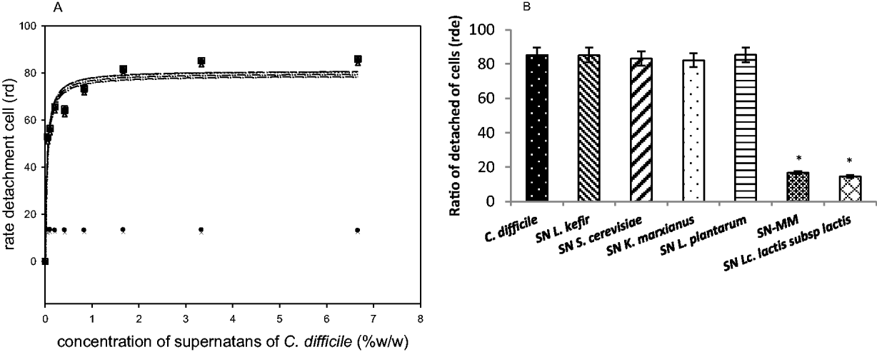
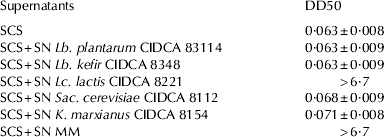
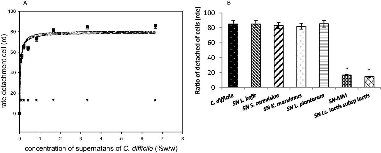
Then, a series of experiments were performed in order to test the effect of microbial supernatants on biological activity of SCS. Figure 1(a) shows the cell detachment curves obtained for SCS and SCS added to supernatants from each strain or MM (SCS + SN). Supernatants obtained from Lc. lactis CIDCA 8221 and MM had the ability to inhibit the cytotoxic effect of C. difficile SCS meanwhile SCS + SN from isolated strains Lb. kefir CIDCA 8348, Lb. plantarum CIDCA 83114, Sac. cerevisiae CIDCA 8112 and K. marxianus CIDCA 8154 did not protect Vero cells against SCS cytopathic effect. Figure 1(b) shows the highest percentage of detached cells determined for all the SCS + SN assessed in the study. There were no significant differences between the results obtained for SCS + SN Lc. lactis CIDCA 8221 and SCS + SN MM, suggesting that the products released by Lc. lactis CIDCA 8221 do not lose its inhibitory activity against clostridial toxins even in the presence of the other microorganisms. The DD50 determined for each SCS + SN is shown in Table 1. Besides, SCS + SN Lc. lactis CIDCA 8221 showed a dose-dependent effect, maintaining its inhibitory ability up to a 10-fold dilution (data not shown).
Biological activity of C. difficile SCS on Vero cells. (a) Dose response curves of cell detachment assay (results expressed as ratio of detached cells) obtained for SCS -○- and SCS treated with microbial supernatants (SN) from Lc. lactis subsp lactis CIDCA 8221 -●-; Lb. kefir CIDCA8348 -△-; Sac. cerevisiae CIDCA 8112 -▲-; K. marxianus CIDCA 8154 -□-; Lb. plantarum CIDCA83114 -■-; MM -X-. (b) Percentage of detached cells at fixed concentration (3·5%) of SCS -fx1- and SCS treated with SN from Lc. lactis subsp lactis CIDCA 8221-fx2-; SN from Lb. kefir CIDCA8348 -fx3-; SN from Sac. cerevisiae CIDCA 8112 -fx4-; SN from K. marxianus CIDCA 8154 -fx5-; SN from Lb. plantarum CIDCA 83114 -fx6-; SN from MM -fx7-. *P < 0·05 compared with SCS
DD50 determined for C. difficile spent culture supernatant (SCS) and SCS incubated with supernatants (SCS + SN) belonging to each strain under study or a mixture of them (MM)
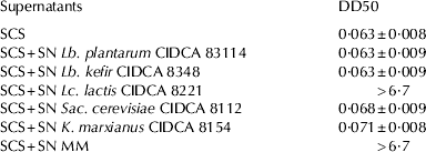
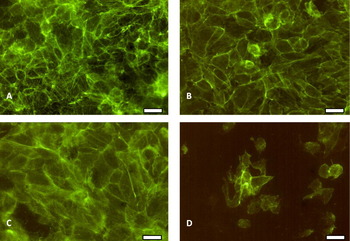
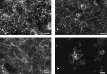
Figure 2 shows phalloidin-FITC labelled F-actin cytoskeleton of Vero cells after treatment with SCS and SCS + SN from Lc. lactis CIDCA 8221. The SCS from C. difficile 117 induced the disruption of the actin network, cell rounding and detachment of Vero cells. However, no damages were observed when cell monolayers were treated with SCS + SN Lc. lactis CIDCA 8221 as compared with untreated controls.
(Colour online) Staining of F-actin cytoskeleton of Vero cells without treatment (panel A), and after treatment with SN from Lc. lactis CIDCA 8221 (panel B), SCS + SN from Lc. lactis CIDCA 8221 (panel C) or SCS (panel D). (Bar = 10 μm)

In this sense, different authors reported that Lactobacillus and Bifidobacterium strains are able to inhibit toxigenic effect of C. difficile on eukaryotic cell cultures (Trejo et al. Reference Trejo, Minnaard, Pérez and De Antoni2006, Reference Trejo, Pérez and De Antoni2010; Banerjee et al. Reference Banerjee, Merkel and Bhunia2009). More recently, Trejo et al. (Reference Trejo, Pérez and De Antoni2010) described that the synthesis and/or secretion of clostridial toxins is reduced by co-culture with potentially probiotic bacteria, including a lactobacilli strain isolated from kefir (Lb. plantarum CIDCA 83 114). Although several authors have been reported the ability of kefir-derived products to antagonise virulence factors of intestinal pathogens in vitro (Garrote, Reference Garrote2000; Londero et al. Reference Londero, Quinta, Abraham, Sereno, De Antoni and Garrote2011) and in vivo (Bekar et al. Reference Bekar, Yilmaz and Gulten2011), to our knowledge this is the first report of protective activity of secreted extracellular factors from kefir-isolated microorganisms against C. difficile toxins.
Effect of Lc. lactis CIDCA 8221 supernatant on the concentration of C. difficile toxins
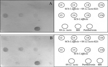
We tested the effect of incubation of SCS with Lc. lactis CIDCA 8221 SN on concentration of clostridial toxins by dot-blot assay. As shown in Fig. 3, the intensity of the spots for both TcdA and TcdB was reduced after incubation of SCS with Lc. lactis CIDCA 8221 SN for 60 min. These findings could suggest a decrease in the concentration of both toxins in SCS treated with Lc. lactis CIDCA 8221 SN.
Dot blot assay with specific monoclonal antibodies against TcdA (panel A) or TcdB (panel B). SCS was incubated with Lc. lactis CIDCA 8221 SN for 60 min at 37 °C, and serially two fold diluted samples were spotted onto nitrocellulose membranes (4 μl/spot). BHI medium, Lc. lactis CIDCA 8221 SN and purified toxins were spotted as controls

Effect of different treatments on the inhibitory ability of Lc. lactis CIDCA 8221 supernatant against C. difficile SCS
To gain further insight into the characteristics of the released products involved in the inhibitory activity of the Lc. lactis CIDCA 8221, the supernatant was fractionated using a 10 kDa cut-off membrane before cell detachment assays. Only the fraction containing components with a molecular mass higher than 10 kDa was able to inhibit the toxigenic effect of SCS on eukaryotic cells (Fig. 4). Moreover, the heat treatment of this fraction at 100 °C for 15 min completely inactivated its inhibitory activity (Fig. 4). These results suggest the involvement of a thermo sensitive component in the inhibitory ability of Lc. lactis CIDCA 8221.
Biological activity of C. difficile SCS on Vero cells. DD50 obtained for SCS -fx1- and SCS treated with supernatant (SCS + SN) from Lc. lactis CIDCA 8221-fx2- ; SCS + SN from Lc. lactis CIDCA 8221 fraction > 10 KDa -fx3-; SCS + SN from Lc. lactis CIDCA 8221 fraction < 10 KDa -fx4-; SCS + SN from Lc. lactis CIDCA 8221 heated at 100 °C for 15 min -fx5-. *P < 0·05 compared with SCS

Based on these findings and taking into account the results obtained by dot-blot assay, we decided to test the presence of proteolytic activity. However, treatment of Lc. lactis CIDCA 8221 supernatant with different proteases (trypsin, chymotrypsin, pepsine and proteinase K) or the presence of protease-inhibitor cocktail did not abrogate the protective effect of Lc. lactis secreted products. There are many reports about the synthesis of different peptidases and serine proteases by Lc. lactis with molecular weights ranging from 40–180 KDa (Sasaki et al. Reference Sasaki, Bosman and Tan1995; Christensen et al. Reference Christensen, Dudley, Pederson and Steele1999; Poquet et al. Reference Poquet, Saint, Seznec, Simoes, Bolotin and Gruss2000; Foucaud-Scheunemann & Poquet Reference Foucaud-Scheunemann and Poquet2003). However, we have found no record of the activity of these enzymes against cytotoxic effects of pathogenic bacteria such as C. difficile. In this sense, Castagliuolo et al. (Reference Castagliuolo, LaMont, Nikulasson and Pothoulakis1996) described a 54-kDa serine protease from Sac. boulardii which digests TcdA and TcdB in vitro. More recently, Banerjee et al. (Reference Banerjee, Merkel and Bhunia2009) reported that Lb. delbrueckii subsp. bulgaricus B-30892 secreted one or more bioactive component which neutralise cytotoxicity of C. difficile probably by inactivating its toxins. In this work, since no abrogation of protective effect was observed after treatment with different proteases or in presence of proteases-inhibitors, we have not been able to demonstrate a proteolytic activity against clostridial toxins in Lc. lactis CIDCA 8221 supernatant, although results of dot-blot assay showed that both the concentration of TcdA or TcdB seemed reduced in treated C. difficile SCS. In order to explain these findings, we hypothesise that there could be a non-covalent interaction between soluble molecules present in Lc. lactis CIDCA8221 supernatant and clostridial toxins which would be responsible for the inhibitory effect on cytotoxicity of C. difficile SCS. This interaction could also interfere with the epitope–paratope reaction, leading to a misreading of the dot-blot assay. Further experiments are needed to confirm the mechanism of action and to determine the nature of Lc. lactis CIDCA 8221 secreted-metabolites.
In conclusion, we demonstrated that kefir-isolated Lc. lactis CIDCA 8221 released heat-sensitive metabolites able to protect eukaryotic cells from cytopathic effect of C. difficile toxins in vitro. These findings provide new insights into the probiotic action of microorganisms isolated from kefir against virulence factors from intestinal pathogens.
This work was supported by Agencia Nacional de Promoción Científica y Tecnológica (PICT 00479/06), CONICET and Universidad Nacional de La Plata (Project 11X/443). P Bolla and P Carasi are fellows of CONICET; M Serradell is a member of the Carrera de Investigador Científico y Tecnológico of CONICET; GL De Antoni is a member of the Carrera de Investigador Científico y Tecnológico of CICPBA.