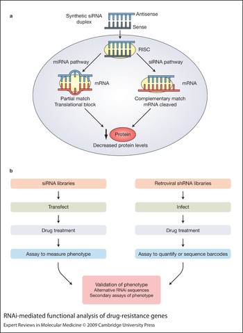
Acquired drug resistance limits the efficacy of cytotoxics used in cancer management (Ref. Reference Longley and Johnston1). The best-studied mediators of drug resistance, the ATP-binding cassette transporter family (ABC transporters), fail to consistently explain in vivo multidrug resistance, and evidence in solid tumours suggests that drug resistance may be mediated by other mechanisms (Ref. Reference Szakacs2). Despite these observations, our understanding of alternative molecular pathways initiating acquired multidrug resistance in vivo remains limited. Following the widespread introduction of RNA interference (RNAi) technology (Fig. 1a), several methods have been developed to enable short- or long-term target silencing in cultured cells in the laboratory (Ref. Reference Downward3), enabling the functional annotation of the human genome. In analysis of drug-resistance pathways, RNAi allows the silencing of genes in a one by one approach to assess the phenotypic effects of each gene on drug sensitivity (Fig. 1b). The complexity of drug resistance and the polygenic nature of this process are emphasised by several high-throughput RNAi screens that have recently implicated hundreds of genes capable of inducing drug resistance in tumour cells. These developments have already highlighted novel pathways of tumour cell survival in response to stress and most importantly provided potential new avenues for therapeutic intervention.
RNAi-mediated functional analysis of drug-resistance genes. (a) The use of RNA interference (RNAi) in gene silencing. Synthetic small interfering RNA (siRNA) duplexes are introduced into the cell. The siRNA associates with the RISC (RNA-induced silencing complex). The antisense portion of the siRNA guides the RISC to its complementary mRNA strand. If it is a partial match, translation is blocked. This normally happens during microRNA (miRNA) processing and could also cause off-target effects of synthetic siRNA. If a complementary match with the antisense strand occurs, the siRNA is cleaved. Potential off-target effects can occur in this pathway if the sense strand of the siRNA duplex is incorporated into the RISC instead of the antisense strand. Both partial and complementary match of the RISC with mRNA result in decreased levels of that mRNA and a corresponding decrease in the levels of protein. (b) RNAi screening approaches for functional assessment of drug-resistance pathways. siRNA and short hairpin (shRNA) libraries can be used to perform high-throughput screens. Synthetic siRNA libraries are normally used in a multiwell format, with each well assigned a different known siRNA. For large-scale retroviral shRNA screens, viral vectors are used to deliver the whole pooled shRNA library (containing unique barcode sequences that can be assayed on microarray platforms) to the cell population of interest. Following delivery of RNAi, cells can be treated with a cytotoxic drug and then assayed for the desired phenotype, such as number of viable cells. If shRNA libraries are used, relative abundance of cells containing the shRNA can be assayed by using microarray technology to measure the unique barcode sequences. Validation steps include using RNAi with alternative sequences against the validation target genes, so as to rule out off-target effects. Secondary assays for the phenotype of interest can also be performed.

This review focuses on developments in our understanding of intrinsic and acquired drug resistance through functional analysis of gene activity, and attempts to define priority areas for future research following the completion of several whole-genome drug-resistance screens. A common theme throughout these studies is the extraordinary complexity of drug resistance and the number of genes capable of influencing this process both within and between cell lines of different tissue origins. We discuss examples of functional RNAi screens in cells lines of different lineages revealing different genes involved in drug resistance that may reflect the lineage dependence of drug-sensitivity pathways. By contrast, studies have also shown common genes influencing drug sensitivity across cancer cell lines of diverse lineage, indicating the existence of common pathways of drug resistance that could be exploited to limit drug resistance in the clinical setting.
Common pathways mediating taxane resistance
Accumulating evidence from several screens from different laboratories implicates the mitotic spindle assembly checkpoint in the response to microtubule-targeted agents. A common effect of all small interfering RNAs (siRNAs) promoting resistance to the taxane paclitaxel in a screen targeting all kinases (kinome library 779 siRNAs) and ceramide pathway regulators (ceramidome library 50 siRNAs) was a reduction in the accumulation of drug-treated cells in mitosis, supporting the concept that spindle checkpoint disruption promotes paclitaxel antagonism (Refs Reference Swanton4, Reference Sudo5). Most siRNAs promoting resistance also induced the appearance of polyploid cells, multinucleation and centrosomal abnormalities without drug treatment. This indicates that morphological differences in cancer cells associated with aberrant mitoses might in the future be exploited to better predict intrinsic taxane sensitivity in vivo. This concept is supported by bioinformatics evidence demonstrating that chromosomal numerical heterogeneity (Refs Reference Swanton4, Reference Roschke6) (which may result from a dysfunctional spindle assembly checkpoint) results in increased resistance to microtubule-targeted agents in breast and colon cancer cell lines.
Microtubule inhibitors are known to induce polyploidy and chromosomal instability (CIN) in checkpoint-deficient or taxane-resistant cells (Refs Reference Cahill7, Reference Shin8, Reference Vogel9, Reference Roberts10), which has prompted the suggestion that polyploidy in response to paclitaxel may be a useful indicator of drug resistance (Ref. Reference Roberts10). Our kinome RNAi screen extends these observations and suggests that genes that are permissive for polyploidy, when targeted by RNAi, influence pathways that taxanes depend upon for cellular cytotoxicity, and that polyploidy or CIN prior to drug treatment may serve as a marker for de novo taxane resistance. Conversely, taxane treatment may select for a higher frequency of CIN in residual tumour. Taxane treatment may result in selection of a population of cells with a partial checkpoint dysfunction that is still sufficient to support long-term growth and survival (as prolonged disruption of the spindle checkpoint is not tolerated by cancer cells because of critical chromosome loss) (Ref. Reference Kops, Foltz and Cleveland11).
This screen also determined that siRNAs inducing polyploidy independently of drug treatment produced sustained activation of the phosphoinositide 3-kinase (PI3K)-regulated kinase AKT following paclitaxel treatment. PI3K pathway deregulation has been shown to correlate with resistance to the taxane docetaxel (Ref. Reference Potti12). A highly significant (P = 1.3 × 10⁻²¹) representation of genes in the PI3K docetaxel- resistance predictor set (cell lines predicted to be resistant to docetaxel are also predicted to show PI3K pathway activation) and genes repressed following targeting of HER2 (human epidermal growth factor receptor 2) are found in the CIN70 signature set (a signature of specific genes whose expression was consistently correlated with total functional aneuploidy in several cancer types) (Ref. Reference Carter13), suggesting a role for HER2/PI3K activation in CIN (Ref. Reference Potti12).
In order to address whether near-diploid tumours are preferentially sensitive to microtubule-stabilising agents, a Phase II clinical trial in colorectal cancer, CINATRA (chromosomal instability and antitubulin response assessment), has been initiated (Refs Reference Swanton15, Reference Swanton, Tomlinson and Downward16). This study exploits the frequent karyotypic separation of mismatch repair (MMR)-deficient tumours, which tend to be diploid, in contrast to the more common CIN+ MMR-proficient colorectal tumours. This Phase II clinical trial aims to compare the sensitivity of MMR-deficient near-diploid tumours to colorectal cancers with CIN following exposure to microtubule-stabilising agents.
In the most extensive analysis of genes enhancing paclitaxel cytotoxicity, a genome-wide siRNA screen was performed in a human non-small-cell lung cancer (NSCLC) cell line, NCI-H1155, in the presence of sublethal doses of paclitaxel (Ref. Reference Whitehurst17). This work identified 87 genes that, when silenced, enhance the cytotoxicity of paclitaxel. The authors identified many proteasome components as synthetic enhancers of paclitaxel cytotoxicity when depleted, in keeping with enhanced efficacy of paclitaxel with proteasome inhibitors (Ref. Reference Hernandez-Vargas18). Four siRNAs identified from their screen preferentially sensitised tumour cell lines in comparison with ‘normal’ lines derived from the same patient.
A common theme emerges from both the paclitaxel kinome screen in three cell lines (Ref. Reference Swanton4) and the whole-genome paclitaxel screen (Ref. Reference Whitehurst17) with respect to the relationship between mitotic arrest and paclitaxel efficacy. Enhancers of paclitaxel cytotoxicity (upon siRNA knockdown) identified in both screens – CERT (ceramide transporter protein; also known as COL4A3BP), ACRBP (acrosin-binding protein) and TUBGCP2 (tubulin, gamma complex associated protein 2) – were capable of augmenting a paclitaxel-induced mitotic arrest, growth inhibition or caspase activity. Intriguingly, the mitotic response to the paclitaxel sensitisers ACRBP and TUBGCP2 was dependent on the cell line, indicating the uncoupling of spindle-checkpoint-induced cell cycle arrest with downstream apoptotic pathways. In the H2126 NSCLC cell lines, gene silencing of ACRBP and TUBGCP2 resulted in the inhibition of proliferation with paclitaxel, whereas the depletion of these genes in H1155 cells enhanced caspase activation induced by paclitaxel. The authors suggest that tumours with a weakened spindle checkpoint may become more dependent on the aberrant expression of genes such as ACRBP that increase the robustness of the spindle assembly checkpoint, thus allowing for efficient mitotic progression.
RNAi has also been used to validate the role of the microtubule-associated protein TAU as a paclitaxel-resistance factor (Ref. Reference Rouzier19). Gene expression data from tumours removed from 82 patients with stage I–III breast cancer prior to paclitaxel treatment were analysed. The gene encoding TAU was one of the most significantly differentially expressed genes in patients with pathological complete response compared with patients with residual disease: lower TAU mRNA expression was found in patients with pathological complete response. When ZR75.1 and MCF-7 cancer cell lines were transfected with siRNA targeting TAU, cell viability was decreased after treatment with paclitaxel compared with control transfected cells. TAU-transfected cells also appeared to take up more paclitaxel. Further data suggested that TAU competes with paclitaxel for binding with microtubules, and therefore results in poorer efficacy of paclitaxel treatment. However, a recent analysis of tumours from the NSABP-28 trial presented at the San Antonio Breast Cancer Symposium by the same group found no significant correlation between TAU expression and benefit from paclitaxel (Ref. Reference Pustzai20). They found that low TAU expression was associated with poor survival, which the authors suggested might result from the association of TAU expression with tumours expressing the oestrogen (estrogen) receptor (ER). These data may reflect the complex and multifactorial nature of drug resistance.
Paclitaxel response and the lysosome
Microtubule-stabilising agents are known to trigger activation of the protease cathepsin B and its release from the lysosome, and cathepsin B inhibition antagonises microtubule-stabilising agents (Ref. Reference Broker21). Furthermore, lysosome-destabilising agents demonstrate synergistic cytotoxicity with either vincristine or paclitaxel (Ref. Reference Groth-Pedersen22). Two screens have identified other lysosomal regulators that influence paclitaxel response. The gene encoding β-glucosidase (GBA) was identified in a screen of ceramide regulators as a candidate paclitaxel-resistance gene following silencing by siRNA (Ref. Reference Swanton4). GBA protein level rises at 12–18 h following paclitaxel treatment, coincident with maximal mitotic arrest of HCT-116 cells. Silencing of GBA prevents a mitotic arrest in response to paclitaxel treatment and triggers endoreduplication both with and without paclitaxel. The vacuolar ATPase subunit ATP6V0D2 was also identified as an enhancer of paclitaxel cytotoxicity following gene silencing (Ref. Reference Whitehurst17). In a pharmacological validation strategy, inhibition of the vacuolar ATPase with a synthetic lysosomal ATPase inhibitor, RTA-203, together with low-dose paclitaxel treatment, significantly reduced the viability of H1155 NSCLC cells. These data indicate the therapeutic potential of lysosome-targeting agents in the augmentation of taxane activity.
Cisplatin, ionising radiation and the BRCA1 pathway
Following a small siRNA screen to identify enhancers of cytotoxicity to paclitaxel and the DNA-targeting drugs cisplatin and gemcitabine in p53-deficient HeLa cells (Ref. Reference Bartz23), a genome-wide screen was carried out to identify enhancers of cisplatin toxicity. This identified genes involved in the DNA-damage repair pathways associated with BRCA (breast cancer susceptibility gene product), and supported previous work demonstrating the association of the signalling pathway involving Fanconi anaemia (FA) genes and BRCA with cisplatin sensitivity (Ref. Reference D'Andrea24). Many of these genes are involved in the regulation of homologous recombination, and they include BARD1 (BRCA1-associated ring domain 1), BRCA1 (breast cancer 1, early onset), BRCA2 (breast cancer 2, early onset) and RAD51 [RAD51 homologue (RecA homologue, E. coli) (S. cerevisiae)]. Corroborating these data, RAD51 was also identified in an RNAi screen for enhancers of cisplatin and radiation sensitivity in Caenorhabditis elegans germ cells (Ref. Reference van Haaften25). Follow-up validation of many of these BRCA pathway hits documented that cisplatin sensitisation is related to the p53 status of the cell. Silencing of BRCA2, RAD51, SHFM1 (split hand/foot malformation (ectrodactyly) type 1) and CHEK1 (CHK1 checkpoint homologue) all resulted in preferential sensitisation to cisplatin in p53-deficient cells compared with p53-isogenic wild-type cell lines, despite equivalent levels of gene silencing.
Functional genomic work combined with microarray analysis of datasets has confirmed the sensitisation of glioblastoma cells to cisplatin following silencing of protein kinase C iota (PKCiota), and further implicated the p38 mitogen-activated protein kinase (MAPK) pathway in cisplatin cytotoxicity (Ref. Reference Baldwin26). Following PKCiota depletion in a human glioblastoma cell line, GMFB (glia maturation factor, beta) expression was selectively increased. GMFB depletion was associated with an increase in cisplatin-mediated p38 MAPK signalling and cisplatin sensitisation that was inhibited by the p38 MAPK inhibitor SKF86002.
Common enhancers of gemcitabine cytotoxicity
Two RNAi screens have probed gene function to identify enhancers of the gemcitabine response – one in HeLa cells (Ref. Reference Bartz23) and the other in the pancreatic cancer cell line MiaPaCa-2 (Ref. Reference Giroux, Iovanna and Dagorn27). In both cases, conventional validation was not performed and only two genes that enhanced gemcitabine cytotoxicity when silenced, CHEK1 and MAP3K1 (MAPK kinase kinase 1), are represented in both datasets (Box 1).
Box 1 Analysis of enhancers of gemcitabine response from two siRNA screens
Gemcitabine response hits from Bartz et al. (Ref.Reference Bartz23)a:
CHEK1, MAP3K1, CARS, TNK1, EPHA1, CFLAR, STAT4, RRMI, FURIN, CD47, PSMB4, RRMI, DVL2, MAPK10, DLGAP4, ASPM, RNASEL, CSEIL, SLC30A2, SMAD4, PKN2, PSMB10, KCND1, RBM12
Gemcitabine response hits from Giroux et al. (Ref.Reference Giroux, Iovanna and Dagorn27)a,b:
CHEK1, MAP3K1, MAK, MAPK12, MAP3K7, PAK4, ADRBK1, GRK4, PIK3CG, PIK3C2A, MAPKAPK1, TGFBR2, DOK1, CSNK1G2, AK1, DCK, TPK1, GUK1, HAK, CSNK2A1, TNNI3K, STK25, HRI, STK22B LOC149420
The 11 pathways identified by GeneGoc in common between the two gemcitabine screens:
G-protein-mediated regulation, p38 and JNK signalling, Ras family GTPase in kinase cascade, regulation of G1–S transition, IL-1 signalling, oncostatin M via MAPK, adenosine A2A receptor signalling, JNK pathway, GDNF family signalling, FGFR signalling pathway, FCɛR1 pathway.
aCommon hits are given in bold.
bHits from Giroux et al. were selected with a threshold of greater than a twofold increase in apoptosis with gemcitabine (and that were not identified as pancreatic survival kinases).
cGeneGo MetaCore is an online pathway analysis tool based on a proprietary manually curated database.
Abbreviations for pathways: FCɛR1, high-affinity IgE receptor; FGFR, fibroblast growth factor receptor; IL-1, interleukin 1; JNK, c-Jun N-terminal kinase; MAPK, mitogen-activated protein kinase; siRNA, small interfering RNA. For full versions of gene names see HUGO Gene Nomenclature Committee (http://www.genenames.org/).
Conceivably, many of the genes identified may simply reflect off-target effects of individual siRNAs. Alternatively, the absence of significant overlap between the two datasets may reflect the dependence of drug-sensitivity pathways on cell line or tissue type (Ref. Reference Swanton28). Questions remain as to how these datasets can be interpreted and whether useful conclusions can be drawn with respect to drug activity and the identification of common pathways implicated in the enhancement of gemcitabine response. In order to identify common features of both datasets, we analysed the genes identified with the GeneGo pathway analysis tool (Box 1). Using this strategy we identified 21 pathways represented in the HeLa gemcitabine siRNA screen and 25 in the pancreatic cell line screen. Ten of these pathways were in common between the two datasets, and some, including MAPK (Ref. Reference Wey29) and Ras (Ref. Reference Gana-Weisz30) pathways, have already been implicated in gemcitabine chemosensitivity. Many of these pathways are amenable to drug targeting with small-molecule antagonists in clinical development.
Identification of drug-resistance pathways influencing response to targeted therapies
RNAi screens have recently illustrated the mechanisms of sensitivity to targeted agents such as trastuzumab, and inhibitors of AKT and poly(ADP-ribose) polymerase (PARP). The serine/threonine kinases AKT1, 2 and 3 act downstream of the PI3K pathway and are frequently activated in cancer due to loss of the phosphatase tumour suppressor PTEN (phosphatase and tensin homologue).
Trastuzumab
An RNAi screen for resistance to the HER2-targeting monoclonal antibody trastuzumab in the HER2-positive BT-474 cell line identified that the predominant regulator of trastuzumab resistance was the activation of the PI3K pathway through the silencing of PTEN (Ref. Reference Berns31). The finding that PTEN silencing decreases sensitivity to trastuzumab was consistent with previous functional work showing that repression of PTEN by antisense oligonucleotides is associated with resistance to trastuzumab (Ref. Reference Nagata32). Studies in metastatic breast cancer demonstrated that PI3KCA (PI3K, catalytic alpha polypeptide) mutations, or PI3KCA mutation together with PTEN loss, predicted poorer patient outcome following trastuzumab monotherapy or in combination with chemotherapy. PI3K pathway activation was an independent negative prognostic factor for progression-free survival with trastuzumab therapy in multivariate analysis. These studies may ultimately influence patient treatment, as HER2-positive breast cancer patients with PI3K pathway activation due to PTEN loss and/or PI3KCA mutations may be better treated with inhibitors of downstream proteins such as AKT or mTOR (mammalian target of rapamycin).
AKT pathway inhibitors
In an effort to improve the efficacy of the AKT ATP-competitive inhibitor A-443654 (AKTi), an RNAi screen was performed to discover kinase-targeting siRNAs that co-operate with this selective AKT inhibitor to induce cell death in the PTEN-deficient renal cell carcinoma cell line 786-O (Ref. Reference Morgan-Lappe33). Genes whose knockdown induced synthetic lethality with AKTi were CSNK1G3 (casein kinase 1, gamma 3), IPMK (inositol polyphosphate multikinase), MAPKAPK2 (MAPK-activated protein kinase 2), RSKB (p90 ribosomal S6 kinase B), MAP4K2 (MAPK kinase kinase kinase 2) and MPP1 (membrane protein, palmitoylated 1, 55 kDa). CSNK1G3 and IPMK siRNAs impaired the activating Ser473 phosphorylation of AKT, which led to the suggestion that these genes may contribute to the activation of AKT by PDK2 (pyruvate dehydrogenase kinase, isozyme 2). siRNAs targeting these kinases also provoked a decrease in S6 ribosomal protein phosphorylation that was further enhanced by using the AKT inhibitor in combination.
PARP inhibitor
CDK5 (cyclin-dependent kinase 5) knockdown was identified as promoting sensitivity to a PARP inhibitor in a kinome siRNA screen (Ref. Reference Turner34). These data emphasised the ability of functional genomics to identify relevant pathways associated with drug sensitivity in unexpected cell types, as CDK5 has been mainly described as a neuronal kinase. CDK5 was determined to be involved in intra-S and G2–M DNA-damage checkpoints in cells of non-neuronal origin. CDK5 genomic loss occurred in 5% of breast cancer patients, thereby identifying patients who may preferentially benefit from PARP inhibitor therapy.
All-trans retinoic acid
Acute promyelocytic leukaemia is associated with translocations involving the retinoic acid receptor α (RARA) gene with the genes PML (promyelocytic leukaemia), NUMA1 (nuclear mitotic apparatus protein 1), ZBTB16 (zinc finger and BTB domain containing 16), NPM1 [nucleophosmin (nucleolar phosphoprotein B23, numatrin)] or STAT5B (signal transducer and activator of transcription 5B), generating fusion products containing the C-terminal region of RARA. All-trans retinoic acid (at-RA), the ligand for RARA, has been used successfully in the treatment of acute promyelocytic leukaemia. An RNAi screen to identify regulators of at-RA-induced differentiation in an at-RA-sensitive acute promyelocytic leukaemia cell line (NB4) identified 26 suppressors of at-RA-mediated cell growth arrest (Ref. Reference Hattori35). The most convincing hit with the most number of colonies matching the same gene encoded an E2 ubiquitin-conjugating enzyme (UBE2D3). UBE2D3 was upregulated at both the mRNA (determined by microarray and qPCR analysis) and protein level following at-RA treatment of NB4 cells, and mediated at-RA-induced cyclin D1 degradation and cell cycle arrest. This study demonstrates the utility of combining microarray expression data with functional genomic data to define the biological pathways associated with drug response. Furthermore, the finding that the proteasome inhibitor MG-132 also suppresses at-RA-induced cell cycle arrest cautions against the use of proteasome inhibitors for treating acute promyelocytic leukaemia in combination with at-RA.
Multidrug-resistance pathways identified by functional genomics
Ceramide pathway mediation of multidrug sensitivity
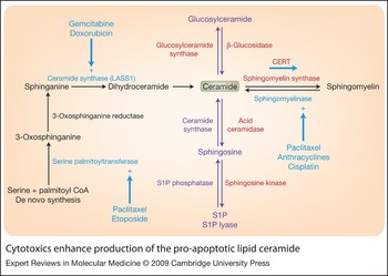
Multidrug-resistance mechanisms have proved difficult to define and to target effectively. Agents targeting the best-described pathway – the multidrug-resistance transporters that promote drug resistance to structurally unrelated cytotoxic agents – have proven disappointing in clinical trials (Ref. Reference Nobili36). In an RNAi screen for regulators of multidrug sensitivity, the ceramide transporter CERT was the strongest hit potentiating multidrug sensitivity to paclitaxel, doxorubicin, cisplatin and 5-FU (fluorouracil) when silenced by RNAi (Ref. Reference Swanton4). CERT transports ceramide, commonly referred to as a ‘pro-apoptotic lipid’, from the endoplasmic reticulum to the Golgi complex, where it is metabolised to sphingomyelin (Ref. Reference Hanada37). Reduction in CERT expression would be expected to result in accumulation of ceramide in the endoplasmic reticulum and increased total cellular ceramide. Ceramide has been associated with apoptosis induction (Ref. Reference Obeid38), although the mechanisms have been difficult to define completely (Ref. Reference Pettus, Chalfant and Hannun39). Several cytotoxic agents have been shown to promote ceramide accumulation, resulting from increased de novo synthesis or sphingomyelinase activity (Fig. 2). This work demonstrates a role for CERT and ceramide metabolism in the endoplasmic reticulum stress pathway. Depletion of CERT by RNAi together with paclitaxel treatment leads to the synergistic induction of endoplasmic stress, as measured by the increase in phosphorylated PERK (protein-kinase-like endoplasmic reticulum kinase) (Ref. Reference Swanton4). Therefore, RNAi technology has the ability to identify novel mechanisms leading to cytotoxic drug sensitisation.
Cytotoxics enhance production of the pro-apoptotic lipid ceramide. Ceramide is a bioactive lipid belonging to the sphingolipid group. Ceramide can be involved in pro-apoptotic signalling and is generated by the breakdown of sphingomyelin by sphingomyelinases or is synthesised de novo by serine palmitoyltransferase and ceramide synthase (black arrows). Activation of the enzymes required for ceramide synthesis occurs following cancer cytotoxic exposure (blue). Ceramide can then be converted to other related bioactive lipids (purple arrows). CERT is a ceramide transporter protein and transports ceramide from the endoplasmic reticulum to the Golgi complex where the ceramide is converted to sphingomyelin. Proteins implicated in mechanisms of drug sensitivity and resistance are highlighted in red. Silencing β-glucosidase (GBA3) results in drug resistance, whereas silencing CERT, acid ceramidase, sphingosine kinase and glucosylceramide synthase results in drug sensitisation. Abbreviations: CERT, ceramide transporter protein; LASS1/CerS1, LAG1 homologue, ceramide synthase 1; S1P, sphingosine 1-phosphate.

Targeting glucosylceramide synthase, an enzyme involved in the conversion of ceramide to glucosylceramide, has been shown to promote sensitivity to doxorubicin, vinblastine and paclitaxel with a concomitant fall in expression of ABCB1 [ATP-binding cassette, subfamily B (MDR/TAP), member 1] (Ref. Reference Gouaze40). When β-glucosidase enzymes (which promote the conversion of glucosylceramide to ceramide) are silenced by GBA1 and GBA3 siRNAs, resistance to paclitaxel and vincristine is promoted (Ref. Reference Swanton4). Consistent with an integral role for the ceramide pathway in influencing multidrug response, several enzymes in this pathway, including glucosylceramide synthase, ceramide synthase, sphingosine kinase and acid ceramidase, have been shown to alter sensitivity to several common cytotoxics (Refs Reference Gouaze40, Reference Itoh41, Reference Liu42, Reference Dawson43, Reference Liu44, Reference Min45).
In keeping with a role for this pathway in influencing drug resistance in vivo, CERT is overexpressed in residual tumour tissue following paclitaxel exposure. The identification of small molecules that may potentiate the generation of ceramide in specific cellular compartments within cancer cells may lead to new therapeutic agents to augment chemosensitivity (Refs Reference Kolesnick46, Reference Reynolds, Maurer and Kolesnick47).
Common pathways of drug response to ‘non-cross-resistant’ cytotoxics
RNAi screening work from our laboratory revealed an unexpectedly high frequency of kinases that alter taxane sensitivity and at least one non-cross-resistant cytotoxic agent (cytotoxic agent with a different mechanism of action) (Ref. Reference Swanton28). By contrast, siRNAs altering drug sensitivity specifically to paclitaxel occurred significantly less frequently than expected. These data provide evidence of common pathways of drug response to non-cross-resistant cytotoxics and a rational explanation for the rapid evolution of drug resistance in solid tumours to non-cross-resistant agents catalysed by genomic instability. It is likely that many of the genes whose loss of function causes multidrug resistance are involved in cell death pathways at nodal points in stress induction pathways. Furthermore, the complexity of multidrug resistance, illustrated by functional genomic screens, provides an explanation for the lack of clinical evidence for the role of multidrug-resistance transporters in acquired multidrug-resistance of cancers (Ref. Reference Borst48) and the failure of Phase III trials targeting P-glycoprotein (Pgp; an ABC transporter) (Ref. Reference Szakacs2).
Conclusions
The polygenic nature of multidrug resistance illustrated by these functional genomic studies, combined with the ability of the tumour population to silence gene expression through epigenetic means (Ref. Reference Glasspool, Teodoridis and Brown49) or activate gene expression through increased gene dosage triggered by chromosome reassortment in aneuploid tumours (Ref. Reference Duesberg, Stindl and Hehlmann50), may explain the relative ease with which tumour populations acquire resistance to non-cross-resistant cytotoxics as treatment lines advance in solid tumours. Although much information remains to be gleaned from these screens, attempts to limit cytotoxic resistance and improve patient survival with ‘non-cross-resistant’ agents are likely to produce only modest benefits unless strategies can be identified to target the underlying genetic propensity of the cancer cell to rapidly disrupt the expression of large numbers of genes potentially involved in multidrug-resistance pathways.
Research in progress and outstanding research questions
This review has highlighted the bewildering complexity of drug resistance and the number of genes that may influence this process. The ‘catalysis’ of multidrug resistance through chromosomal reassortments as proposed by Duesberg and colleagues (Ref. Reference Duesberg, Stindl and Hehlmann51) suggests that identifying cellular survival pathways sustaining distinct patterns of genomic instability will be a more promising strategy to limit drug resistance than targeting single genes associated with drug resistance in the laboratory setting. Continued investigation of nodal pathways impacting on multidrug resistance common to cancer cells of diverse lineage may result in the exploitation of putative targets to resensitise cancers to cytotoxic agents to optimise patient outcome.
Acknowledgements and funding
We thank the peer reviewers for their time and input in reviewing this article, and Cancer Research UK and the Medical Research Council for providing funding for the authors and the Translational Cancer Therapeutics laboratory.