Introduction
Osteomyelitis is an inflammation of bone and bone marrow and has been classically divided into acute and chronic suppurative forms.Reference Bernier, Clermont, Maranda and Turcotte1 Chronic osteomyelitis is deemed to be of more than one month's duration.Reference Marx2, Reference Mercuri3 Osteomyelitis may also result from infection of the bone by tubercular bacilli.Reference Bhatia, Agarwal, Gupta and Khanna4 All these forms of osteomyelitis are much less common in the skull bones than in the long bones and the vertebral column.Reference Bhatia, Agarwal, Gupta and Khanna4, Reference Koorbusch, Fotos and Goll5 The increased availability of effective antibiotics has further reduced the incidence of osteomyelitis.Reference Koorbusch, Fotos and Goll5, Reference Yeoh, MacMohan and Schifter6
The present study retrospectively analysed cases of tubercular and chronic pyogenic osteomyelitis of the cranio-facial bones, which had been managed at a tertiary healthcare centre over the past three and a half years. The study aimed to better understand the presentation, diagnosis and management of this unusual disease entity.
Patients and methods
The study group comprised 14 diagnosed cases of tubercular and chronic pyogenic osteomyelitis which had been managed in the otorhinolaryngology department at Maulana Azad Medical College and the associated Lok Nayak Hospital between May 2002 and December 2005. All the patients were diagnosed on the basis of clinical assessment, radiological investigations and histopathological evaluation.
The case records of the patients were analysed, with special emphasis on the following parameters: age and sex; site of infection; presenting signs and symptoms; causes; personal history; radiological and laboratory investigations (including chest X-rays); treatment (medical as well as surgical); and duration of hospital stay.
Results
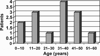
Nine (64 per cent) of the patients were male and five (36 per cent) were female. The youngest patient was four years old and the eldest was 52 years old; the mean age was 30 years (Figure 1).
Age distribution of patients with osteomyelitis.

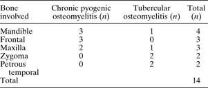
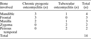
Of the 14 patients, eight had chronic pyogenic osteomyelitis and six had tubercular osteomyelitis. Of the eight patients with chronic pyogenic osteomyelitis, three each had involvement of the frontal and the mandibular bones, whereas two had involvement of the maxilla. Of the six patients with tubercular osteomyelitis, two each had involvement of the zygomatic and the petrous temporal bones, whereas one each had involvement of the maxilla and the mandible (Table I).
Osteomyelitis site distribution
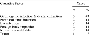
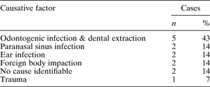
The most common presenting features were pain (71 per cent), swelling (57 per cent), discharging sinus (43 per cent), granulations (21 per cent), neurological symptoms (14 per cent), ear discharge (14 per cent) and ulceration of overlying skin (7 per cent). Odontogenic infections or dental extraction were the most commonly identifiable causative factors (Table II). This association was made purely on clinical grounds, with all these patients giving a clear history of dental extraction or dental infection just preceding the onset of osteomyelitis.
Distribution of causative factors for osteomyelitis
Only one patient with tubercular osteomyelitis had a past history suggestive of pulmonary tuberculosis, having been diagnosed and adequately treated four years prior to his presentation to us. None of the patients gave a history suggestive of active tubercular infection elsewhere in the body. None of the patients had a chest X-ray suggestive of pulmonary tuberculosis. Six patients had a history of tobacco chewing, whereas a history of alcohol abuse and smoking was present in three patients each. Two patients had diabetes mellitus and two were found to be hypertensive. No other significant comorbidity was identified.
Amongst the eight patients with chronic pyogenic osteomyelitis, four had an elevated erythrocyte sedimentation rate (ESR), whereas all six patients with tubercular osteomyelitis had an elevated ESR and four had a positive Mantoux test. Serum enzyme-linked immunosorbent assay for Mycobacterium tuberculosis was performed in three patients with suspected tubercular osteomyelitis and was found to be reactive in all of them. None of the patients were found to have seropositivity to human immunodeficiency virus (HIV). None of the patients' chest X-rays revealed any abnormality.
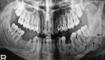
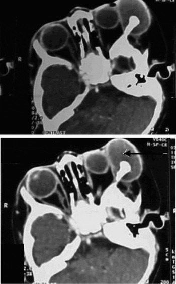
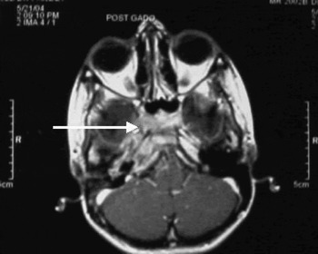
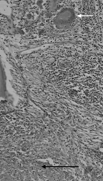
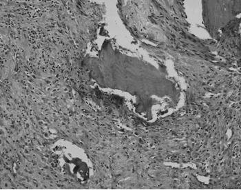
Ten patients were subjected to plain radiography. Computed tomography (CT) scans were ordered for eight patients and magnetic resonance imaging (MRI) scans for two (cases with petrous apex osteomyelitis). Radiological features were consistent with osteomyelitis in 12 (86 per cent) of the cases (Figures 2, 3 and 4). A histopathological diagnosis was established in all cases of tubercular osteomyelitis (Figure 5), and staining for acid-fast bacilli was positive in four cases (67 per cent). Histopathological confirmation of chronic inflammation was established in all cases of chronic pyogenic osteomyelitis (Figure 6); however, microbial cultures revealed no pathological organism in any case.
Radiograph showing lytic area involving the mandible (multiple arrows), suggestive of osteomyelitis.

Axial, contrast-enhanced computed tomography scans showing an abscess associated with sclerosis of the zygoma (black arrow), suggestive of osteomyelitis.

Axial, post-gadolinium magnetic resonance imaging scan showing bony destruction with sclerosis involving the petrous apex (white arrow).

Photomicrograph showing epithelioid granulomas with Langhan's type giant cells (white arrow) and an area of caseation (black arrow) (H&E; ×200).

Photomicrograph showing fragments of dead bone lying within chronic inflammatory granulation tissue, suggestive of chronic pyogenic osteomyelitis (H&E; ×200).

All the patients were initially commenced on a combination of oral antimicrobial chemotherapy, consisting of metronidazole combined with amoxycillin (seven cases), with cephalosporins (four cases) or with clindamycin (three cases), on an out-patient basis. The treatment was continued for two weeks in all cases, except in those diagnosed with tubercular osteomyelitis. All the patients with tubercular osteomyelitis were successfully treated with anti-tubercular chemotherapy. All the patients with chronic osteomyelitis required surgical intervention in the form of debridement (six cases) or resection with plating (two cases). One of the cases required repeat surgery for residual disease.
The patients' average hospital stay was eight days, and their average follow up was 18 months for cases with tubercular osteomyelitis and 10 months for cases with chronic pyogenic osteomyelitis.
Discussion
The incidence of osteomyelitis of the cranio-facial bones has declined in the past owing to the availability of effective antibiotics. This entity is uncommon in developed countries, but cases have been repeatedly reported from developing countries.Reference Bhatia, Agarwal, Gupta and Khanna4, Reference Mahindra, Bais, Sohail and Maheshwari7–Reference Bhandari, Mandowara and Joshi10 This relatively higher incidence may be attributable to the lack of primary healthcare infrastructure and low literacy rates in developing countries. The present study was undertaken to better understand the presentation, diagnosis and management of this rare entity in our population. Very few previous studies have taken into account this rare condition. In our literature review, we could not identify any such study from this part of the world.
The mean age group of patients in our study was 30 years. This is similar to the age group reported in a previous study.Reference Joseph, Mishra and Singh9 However, two previous studies reported a higher mean age group.Reference Bernier, Clermont, Maranda and Turcotte1, Reference Koorbusch, Fotos and Goll5 This discrepancy may have arisen because of the fact that all the previously reported studies included only cases of chronic pyogenic osteomyelitis, and not tubercular osteomyelitis. Since our study also included cases with tubercular osteomyelitis, which typically affects young individuals,Reference Meher, Singh and Raj8 the average age group in our study was relatively lower. The sex ratio of the patients in our study was approximately two to one, with a male predominance. One previous study reported an equal sex distribution,Reference Adekeye and Cornah11 whereas another found a sex ratio of three to one, with a male predominance.Reference Koorbusch, Fotos and Goll5
The most commonly involved bone in our study was the mandible, followed by the frontal bone. The mandible has been reported as the most commonly involved bone in most previous studies.Reference Bhatia, Agarwal, Gupta and Khanna4, Reference Koorbusch, Fotos and Goll5, Reference Adekeye and Cornah11, Reference Daramola and Ajagbe12
The most common attributable cause in our study was odontogenic infection. Similar results have been reported by most previous studies.Reference Koorbusch, Fotos and Goll5, Reference Adekeye and Cornah11, Reference Daramola and Ajagbe12
In our patients, the most common presenting features were pain, swelling and a discharging sinus, consistent with previous studies.Reference Koorbusch, Fotos and Goll5 Two of our patients (cases with petrous temporal osteomyelitis) had neurological features, in the form of lateral rectus palsy and retro-orbital pain. Chronic bone infection may be associated with chronic systemic diseases, immunocompromised states and conditions associated with bone ischaemia.Reference Koorbusch, Fotos and Goll5 Systemic conditions such as diabetes mellitus, malignancy, malnutrition, anaemia, acquired immunodeficiency syndrome, osteopetrosis, autoimmune disorders, and tobacco and alcohol abuse have been implicated to be associated with an increased risk of chronic osteomyelitis.Reference Mercuri3, Reference Saunders13–Reference Wald17 In our study, four patients had a history of tobacco chewing, whereas three each had a history of alcohol abuse and smoking. Two patients had diabetes mellitus and two were hypertensive. None of our patients was found to be seropositive for HIV. Ten patients were found to have an elevated ESR, whereas four, out of the six with tubercular osteomyelitis, had a positive Mantoux test.
The chest X-rays of all the patients were found to be normal. Of the six patients with tubercular osteomyelitis, only one had a history of tuberculosis for which adequate treatment had been sought. None of the cases was found to have an active focus of tuberculosis anywhere else, as assessed by clinical examination and chest X-rays. The diagnosis of primary tuberculosis of the facial bones can be arrived at only by excluding a preceding or concomitant tubercular infection elsewhere in the body. The most reliable method for such exclusion is a proper history, clinical evaluation and a chest skiagram. In our study, the diagnosis of primary tuberculosis was established after excluding tubercular infection elsewhere, using the above mentioned modalities. Thus, all the patients in our study had primary tubercular osteomyelitis. This is unusual as, in the majority of cases, tubercular osteomyelitis is caused by haematogenous dissemination of bacilli from an active focus, usually in the lungs.Reference Bhatia, Agarwal, Gupta and Khanna4, Reference Joseph, Mishra and Singh9 However, other cases with primary tubercular involvement of the cranio-facial bones have been previously reported.Reference Joseph, Mishra and Singh9
Plain radiographs were obtained for all the cases, whereas CT scans were obtained for eight patients. Magnetic resonance imaging scans were ordered for two patients with petrous temporal osteomyelitis. Radiologically, 86 per cent of our patients showed lytic lesions with or without areas of sclerosis. This feature, although suggestive of osteomyelitis, is not diagnostic.Reference Bhatia, Agarwal, Gupta and Khanna4, Reference Bhandari, Mandowara and Joshi10 A similar radiological picture may be seen in congenital syphilis, eosinophilic granuloma or secondary malignant deposits.Reference Bhandari, Mandowara and Joshi10 Some authors suggest that a bony reaction in a lytic lesion is usually suggestive of tubercular infection, whereas chronic osteomyelitis is characterised by an absence of bony reaction.Reference Bhatia, Agarwal, Gupta and Khanna4 However, in our study, such demarcation was not observed, and cases of tubercular osteomyelitis were diagnosed only on the basis of histopathological confirmation. The MRI scans performed in two patients also showed bone destruction associated with an abscess, suggestive of osteomyelitis. Another imaging modality which can be used to diagnose osteomyelitis is bone scintigraphy using technetium 99 and gallium citrate scans.Reference Koorbusch, Fotos and Goll5, Reference Sato, Indo, Kawabata, Agarie, Ishigami and Noikura15 Positron emission tomography scanning has also been used for diagnosis, with great accuracy.Reference Zhiang, Duarte, Pourdehand, Shnier and Alavi18 These modalities can also be used to evaluate patients' therapeutic response.Reference Koorbusch, Fotos and Goll5 However, due to the unavailability of these imaging modalities for routine diagnostic use in our institution, none of the patients in our study were subjected to them.
The cases of tubercular osteomyelitis were diagnosed on the basis of pathological evaluation of fine needle aspirates and tissue biopsies (granulation and sinus excisions) from the infected bones. In all cases, histopathological evaluation showed epithelioid, caseating granulomas. The presence of caseating granulomas is strongly suggestive of tuberculosis.Reference Bhatia, Agarwal, Gupta and Khanna4, Reference Meher, Singh and Raj8, Reference Joseph, Mishra and Singh9, Reference Daramola and Ajagbe12 The diagnosis was further supported by positive acid-fast bacilli staining in four cases, enzyme-linked immunosorbent assay seropositivity for Mycobacterium tuberculosis in three cases and an elevated ESR with positive Mantoux test in four case. Another test which can be used in the diagnosis of tuberculous infection is polymerase chain reaction, which is considered to have a high sensitivity and specificity.Reference Daniel, Isselbacher, Braunwald, Wilson, Martin, Fausi and Kasper19
All our patients were initially commenced on oral combination chemotherapy, in the form of metronidazole combined with amoxicillin, with cephalosporin or with clindamycin, without obtaining a culture and sensitivity report. Penicillins, cephalosporins or clindamycin in combination with metronidazole (for effective anaerobic coverage) have been traditionally considered as drugs of choice in treating chronic pyogenic osteomyelitis.Reference Yeoh, MacMohan and Schifter6, Reference Mader, Shirtliff, Bergquist and Calhoun20 Some authors recommend a minimum of two weeks' treatment, whereas others suggest a minimum of four weeks.Reference Marx2, Reference Bamberger21 The use of hyperbaric oxygen has also been reported to reduce the treatment time and morbidity associated with osteomyelitis.Reference Mader, Shirtliff, Bergquist and Calhoun20, Reference Madder, Adams, Wallace and Calhoun22
• Osteomyelitis of the cranio-facial bones is a rare entity which may be pyogenic or tubercular in origin
• Pyogenic and tubercular osteomyelitis have a similar clinical presentation and may thus be difficult to differentiate
• Radiology plays an important role in the diagnosis, but cannot distinguish between pyogenic and tubercular aetiologies
• Histological examination plays a very important role in the diagnosis and differentiation of the two types of osteomyelitis
All our patients with chronic pyogenic osteomyelitis underwent surgical treatment, in the form of debridement or resection with reconstruction, depending upon the extent and site of involvement. Combined surgical and medical treatment is the current protocol in the management of chronic pyogenic osteomyelitis. Thus, the management of patients in our study was similar to the current protocol.Reference Koorbusch, Fotos and Goll5, Reference Yeoh, MacMohan and Schifter6 The treatment of tubercular osteomyelitis is primarily medical, in the form of anti-tubercular chemotherapy. Surgery is reserved for cases with extensive destruction, secondary infection or intracranial involvement.Reference Meher, Singh and Raj8 All our patients with tubercular osteomyelitis were managed medically with anti-tubercular chemotherapy, consisting of isoniazid, rifampicin, pyrazinamide and ethambutol, with complete cure.
Conclusion
We would like to emphasise that osteomyelitis of the cranio-facial bones is an uncommon entity that requires a high index of clinical suspicion for diagnosis. Radiological evaluation plays an important diagnostic role. The differentiation between chronic pyogenic and tubercular osteomyelitis cannot be made radiologically and requires histopathological correlation. The management of chronic pyogenic osteomyelitis involves both medical and surgical treatment, whereas tubercular osteomyelitis is primarily managed medically.