Introduction
Dementia is a syndrome that is characterized by impairment of memory and at least one other cognitive domain (aphasia, apraxia, agnosia, executive function). The disorder mainly affects the elder and the published prevalence of dementia doubles with every five-year increment in age (World Alzheimer Report, 2014). Worldwide prevalence is estimated at 47.5 million, is expected to double up by 2030 and triple by 2050. Alzheimer's Disease (AD) is the most common form of dementia in the elderly, accounting for 60% to 70% of cases (World Health Organization, 2015). A definitive diagnosis of AD requires post-mortem histopathologic examination, which is rarely done in life. Clinical criteria for the diagnosis of probable AD have been established by the National Institute of Neurological and Communicative Disorders and Stroke and the AD and Related Disorders Association (NINCDS-ADRDA) (McKhann et al., Reference Mckhann, Drachman, Folstein, Katzman, Price and Stadlan1984) and American Psychiatric Association (DSM-IV-TR) (American Psychiatric Association, 2000).
The second most common form of late onset dementia and the most common form of secondary dementia is Vascular Dementia (VaD). VaD is heterogeneous, due to different, small, and large vessel related causes (Bowler, Reference Bowler2007). The National Institute of Neurological Disorders and Stroke and Association Internationale pour la Recherché et l'Enseignement en Neurosciences criteria (NINDS-AIREN) are most detailed and most widely used (Roman et al., Reference Roman1993). One of the earliest clinical criteria for the differential diagnosis of VaD is the Hachinski Ischemic Scale (HIS), which was established specifically to distinguish VaD from AD. Number of studies have shown that current clinical criteria do not separate AD from VaD patients with high specificity (Wetterling et al., Reference Wetterling, Kanitz and Borgis1996; Holmes et al., Reference Holmes, Cairns, Lantos and Mann1999; Wallin et al., Reference Wallin, Milos, Sjogren, Pantoni and Erkinjuntti2003).
The current neurochemical background of AD is characterized by the reduction of essential neurotransmitters, such as acetylcholine, serotonin, noradrenaline, and glutamate, within specific regions of the cortex and the hippocampus, with the concurrent neuropathological presence of amyloid plaques and neurofibrillary tangles. On the other hand, VaD is conceptualized by the aggregated impact of small and large strokes leading to brain cell death and overall neuron and axon loss (Jacoby et al., Reference Jacoby, Oppenheimer, Dening and Thomas2008).
Given that different pathological entities contribute to AD and VaD, different pharmacological treatments have been proposed (e.g. Acetylcholinesterase inhibitors are only effective in AD). Early intervention is highly desirable, since it is most likely to be effective, but requires differentiating patients with different risk profiles. Moreover, the identification and differentiation of prodromal stages of dementia into homogeneous groups of patients will facilitate new drug studies (Jack et al., Reference Jack2011; European Medicines Agency, 2012; Dubois et al., 2014). Importantly, it has been proposed that delaying the onset of dementia by five years without affecting mortality would reduce the population prevalence by 50%, greatly reducing its impact on the general population (World Alzheimer Report, 2014). Several cognitive instruments have been evaluated for their ability to detect and differentiate early AD (Lonie et al., Reference Lonie2009; Thompson et al., Reference Thompson, Brodaty, Trollor and Sachdev2010; Tan et al., Reference Tan, Herrmann, Mainland and Shulman2015).
There are two different visual streams (parvocellular and magnocellular) that are involved in color perception (Livingstone and Hubel, Reference Livingstone and Hubel1987). Although aging has been related to the miosis and the yellowing of the lens, that may induce color perception disturbances (Verriest, Reference Verriest1963), differences in performance are not consistently linked to age. Recent findings in AD patients suggest that there is a close link between macular degeneration and β-amyloid deposits in the retina (Ratnayaka et al., Reference Ratnayaka, Serpell and Lotery2015), while others observed a thinning of the RNFL and dysregulation of the magnocellular pathway (Parisi et al., Reference Parisi, Restuccia, Fattapposta, Mina, Bucci and Pierelli2001; Sartucci et al., Reference Sartucci2010). The loss of visuospatial skills is an early feature of AD that is sometimes prominent at presentation (Mendez et al., Reference Mendez, Mendez, Martin, Smyth and Whitehouse1990; Guerin et al., Reference Guerin, Belleville and Ska2002) and the performance of AD patients in color discrimination tests is reduced both due to vision structures like the primary cortex and the retinal gaglionar cells (Salamone et al., Reference Salamone2009).
We investigated whether the performance of the visual perception between AD and VaD patients could improve the differential diagnosis for these two diseases. Interestingly, although there have been studies investigating the correlation between dementia and color blindness (Kaeser et al., Reference Kaeser, Ghika and Borruat2015), the complete 38 plate version, one of the most popular color blindness test had never been evaluated. Thus, we decided to estimate the usefulness of the extended Ishihara Color Blindness test, as an easy and quick test for the differential diagnosis between AD and VaD in their early stages.
Methods
Measures and procedure
Participants in the study were patients of the Memory Clinic during the time period from 2007 to 2011, First Neurological Department, AHEPA University Hospital, Thessaloniki, Greece. The study adhered to the principles of the Declaration of Helsinki and the protocols employed had been approved by the Hospital's Research and Ethics Committee. All participants provided informed consent before the evaluation. Demographic data (age, gender, level of education) were recorded and each participant underwent dementia evaluation, including a detailed clinical history, neurological examination, MRI, complete dementia blood screening test, and EEG. Cognitive impairment was screened with the MMSE (Folstein et al., Reference Folstein, Folstein and Mchugh1975) by the associate neuropsychologist of the memory clinic and patients were diagnosed by a board certified Neurologist (V.C.) and classified according to the Reisberg scale (Reisberg et al., Reference Reisberg, Ferris, De Leon and Crook1982). Afterwards, all potential participants had a detailed ophthalmological exam to exclude patients with ocular pathology. Participants with MRI findings of large occipital ischemic lesions or ocular pathology and history of ocular disease, trauma, surgery, or laser treatment were excluded from the study. All participants used refractory corrections, appropriate for the viewing distances employed in the various tests. None suffered from any systemic or ocular conditions known to affect color vision. The inclusion criteria were met by 68 individuals: AD (n = 32, 11 males, mean age 73.3 years) according to DSM-IV-TR, NINCDS-ADRDA criteria and VaD (n = 36, 12 males, mean age 75.6 years) based on ΝΙΝDS-AIREN, as well as MRI results. In addition, a healthy control group (n = 35, 16 males, mean age 74.2 years) was recruited.
Visual perception test
The most common test for screening for color blindness is the Ishihara Color deficiency test. It consists of 38 pseudoisochromaric plates (PIP). The PIP could represent a digit or a pathway. In addition, within the test, there are plates that reveal figures or trail-tracing plates that are only identified by color deficient individuals. People with normal color vision can make up to two errors. Previous studies have proved that the cut-off value (CI 99%) of errors in the general population is >36 while six errors suggest definitely that the participant suffers from color blindness (Verriest, Reference Verriest1982). Visual perception of all participants was evaluated with the use of Ishihara plates: “Ishihara Color Vision Test - 38 Plate.” The test was performed in a pleasant, quiet environment, and the plane of the plate was adequately illuminated. Detailed instructions were given and the patients were asked to confirm their ability to recognize and detect basic colors, through a basic primary and secondary color wheel. A paper version of the 38 plate version of the Ishihara Test was performed.
Statistics
Statistical analysis was performed with SPSS (Scientific Package for Statistic Science Version 23 of SPSS Inc., USA). Analysis of variance (ANOVA) was used to estimate any significant variation between the three groups within their age, MMSE, Reisberg Scale, and total Ishihara score. In addition, stepwise multiple analysis, using the Scheffé’s Alpha procedure, estimated the mean differences. Possible differences in the distribution of gender and education were calculated between all groups with Pearson's χ 2 test of significance (χ 2). Univariate ANOVA test was used, to compare the VaD with AD patient's performance in the Ishihara test, when controlling for Reisberg category, MMSE, age, and education. A Receiver Operating Curve (ROC) analysis was used to estimate the discriminative validity of the Ishihara test as a diagnostic test between AD and VaD patients.
Results
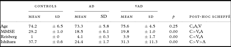
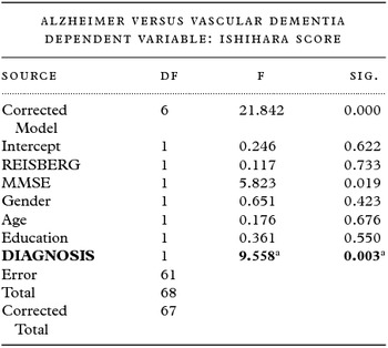
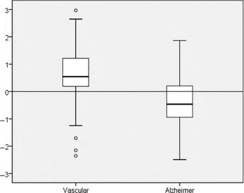
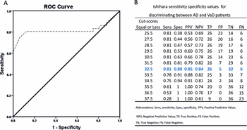
At total of 171 individuals agreed to participate in our research. Sixty-eight (M:43, F:25) were excluded based on the selection criteria. Data on 103 participants diagnosed with AD (n = 32), VaD (n = 36), and healthy controls (n = 35) were analyzed in the context of the present study. There was no difference in categorical variables (gender, level of education) between the three groups (Table 1). We were interested to assess total Ishihara score as a test for the differential diagnosis between the three groups. ANOVA was used to estimate any significant variation between the three groups for age, MMSE, Reisberg Scale, and total Ishihara score (Table 2). This showed no significant differences in the age, but indicated differences in MMSE and Reisberg Scale (as expected, since healthy controls were included) and importantly, total Ishihara scores. To further investigate this finding, we applied a stepwise multiple analysis, using the Scheffé’s Alpha procedure. The post hoc analysis with the Scheffé’s method, showed differences in the MMSE and Reisberg Scale between Controls and AD, as well as VaD patients, but not statistically significant differences between AD and VaD with these tests. Interestingly, Ishihara score was significantly different between AD and VaD participants. Based on this finding, we performed a post-hoc ANOVA comparing VaD with AD participants, controlling for Reisberg, MMSE, gender, age, and education, which gave us a significant difference in Ishihara scores between types of dementia (F(1,61) = 9.558, p = 0.003; Table 3; Figure 1). Finally, an ROC curve was used to estimate the discriminative power of the Ishihara test as diagnostic test (Figure 2). The optimal cut-off value for discriminating AD from VaD participants was 32.50 with a sensitivity of 80.6% and specificity 87.5%, AUC = 0.819, SD = 0.59 which results in a CI 95% = (0.703, 0.934) (Figure 2).
Pearson's chi-square test (χ 2) for the distribution of gender and education between the three groups

ANOVA and post-hoc Scheffé method of the three study groups

Univariate ANOVA of the Ishihara test performance between AD and VaD patients, controlling for diagnosis, reisberg, MMSE, gender, age, and education

R-Squared = 0.682 (Adjusted R-Squared = 0.651).
aThe significance value of the table is p < 0.05.
Box plots of standardized residuals of Ishihara score, controlling for age, education, MMSE, and the Reisberg Scale for both patients groups.

ROC showing sensitivity and 1-specificity of unadjusted Ishihara scores in discriminating AD from VaD(A); data points (B).

Discussion
The World Health Organization and the G8 Dementia Summit have identified dementia and AD prevention as a major public health priority (World Health Organisation, 2012, G8 Dementia Summit Declaration, 2013). Early intervention to modifiable risk factors for AD and VaD is highly desirable, since it is most likely to be effective, but requires differentiating patients with different risk profiles. The US Food and Drug Administration (FDA) and the European Medicines Agency (EMA), is moving in the direction of developing new diagnostic criteria, that will facilitate the identification and differentiation of early stages of dementia into homogeneous groups of patients and will optimize new drug studies (Jack et al., Reference Jack2011; European Medicines Agency, 2012; Dubois et al., 2014).
We wanted to develop a method to differentiate AD and VaD patients on clinical grounds, based on previous studies that were promising on that aspect (De Jager et al., Reference De Jager, Hogervorst, Combrinck and Budge2003). Cronin-Colomb et al. suggested that there is a deficit in color discrimination in AD that is specific to blue hues, and in the absence of such damage to anterior visual structures, peristriate, and inferotemporal visual cortical neuropathological lesions are most likely the substrate for the deficit (Cronin-Golomb et al., Reference Cronin-Golomb, Sugiura, Corkin and Growdon1993). A disruption of the visual signals mediated by area 17 and more specifically in the magnocellular or the M pathway of visual processing was supported by other studies (Hof and Morrison, Reference Hof and Morrison1990; Gilmore et al., Reference Gilmore, Wenk, Naylor and Koss1994). Optic nerve and retinal ganglion cell degeneration is not a specific characteristic of AD according to Curcio et al. (Curcio and Drucker, Reference Curcio and Drucker1993), while Davies et al. also, support that optic nerve degeneration is not a feature of AD and the deficit result from cortical dysfunction (Davies et al., Reference Davies, Mccoubrie, Mcdonald and Jobst1995). Independent of the pathophysiological basis, visual dysfunctions in AD has been recently studied as a predictor of early dementia, as well as the progression of the disease (Crutcher et al., Reference Crutcher, Calhoun-Haney, Manzanares, Lah, Levey and Zola2009; Zola et al., Reference Zola, Manzanares, Clopton, Lah and Levey2013). One of the most difficult tasks in the assessment of vision in AD is the selection of the appropriate tool that will facilitate communication and perception of the test (Cronin-Golomb and Hof, Reference Cronin-Golomb and Hof2004). We decided to use Ishihara plates, because the process of the test is easy to understand and indeed perception was confirmed by our patients. Additionally, it uses arithmetic (Arabic) digits, a common international code and can be applied worldwide. The test is short and patients can cooperate and remain focused. In contrast to sophisticated exams, our method can be easily applied in an outpatient visit from specialized medical doctors and educated general practitioners that might have a key role in the accurate assessment and early diagnosis of dementia (Tang et al., Reference Tang, Birdi and Robinson2016). Interestingly, our study revealed statistically significant differences between AD and VaD patients’ performance in the Ishihara test, even in the earliest stages of dementia, which are the most important for pharmaceutical intervention (European Medicines Agency, 2012; Dubois et al., 2014; Suri et al., Reference Suri, Topiwala, Mackay, Ebmeier and Filippini2014). Thus, one of the most important practical application of our findings is the use of Ishihara test in future cognitive battery tests, in order to facilitate earlier intervention in the prodromal stages of the diseases. To this aspect, Ishihara test will also facilitate differentiation of early stages of dementia into homogeneous groups of patients and will optimize new drug studies.
Methodological considerations
The usefulness of Ishihara plates in the differential diagnosis of AD and VaD does have limitations. First, earlier studies have shown that the prevalence of congenital color blindness in Caucasians of European ancestry is estimated around 8% in men and 0.4% in women (Birch, Reference Birch2012). The aforementioned prevalence vary depending on the origin of the participant, but still implies an important limitation to approximately 10% of the population. Second, chronic illnesses, trauma, infarcts, lesions, medication, or chemicals that affect the retina or the cerebrum, may be some of the causes of acquired color blindness. Furthermore, we had to exclude all participants suffering from any systemic or ocular conditions known to affect color vision, as well as patients with MRI findings of large occipital ischemic lesions. Another important limitation that should be taken under consideration is the small sample size of our research and the exclusion of the Hachinski scales from our analysis. Last, despite the ease of the process, cooperating with dementia patients is always a challenge and a pleasant, quiet, and trusted environment must be secured.
Conclusion
Ishihara Color Vision Test - 38 Plate is a promising method as an easy and quick test for the differential diagnosis between AD and VaD. Further studies are required to confirm and extend our findings and determine the reliability and clinical usefulness of our method in other samples.
Conflict of interest
None.
Description of author's roles
Dr Arnaoutoglou, N.A. participated in setting the research questions, designing the study, carrying it out, analyzing the data, and writing of the paper. Assist Prof Arnaoutoglou M. participated in setting the research questions, designing the study and writing of the paper. Dr Nemtsas P. participated in analyzing the data and writing of the paper. Prof Costa V. participated in carrying out the study, supervised the data collection. Prof Em. Baloyannis S.J. participated in formulating the research questions, designing the study. Prof Ebmeier K.P. participated in formulating the research questions, supervised the analysis of the data, and the writing of the paper.
Acknowledgments
Dr Arnaoutoglou N.A. was supported by the Greek Scholarship Foundation (4893), as part of a PhD research project. The funders had no role in the design of the study, the collection, the analysis, and interpretation of data.