Introduction
Phthalate acid esters, commonly called phthalates, used for a variety of purposes including the widely used plasticizer di-(2ethylexyl) phthalate (DEHP), are components of many plastic products, such as food wraps, toys and some medical devices.Reference Schettler1 DEHP is loosely held with the plastic polymer and can be easily released into the environment. While exposure via house dust is extensive, at least for some phthalates (e.g. DEHP), foodstuff and, to a lesser extent, the use of oral drugs present the major uptake pathways.Reference Wormut2
Because phthalates are found ubiquitously in the environment, humans have chronic and repetitive exposure to phthalates. Phthalates have a short life in the body; however, exposure assessment can be based on a single urine sample in epidemiological studies since exposure is relatively constant over time.Reference Hoppin, Brock, Davis and Baird3–Reference Teitelbaum, Britton and Calafat5
Experiments on animal models have demonstrated that diverse phthalate isomers cause reproductive toxicity in both prenatal and postnatal stages of development, particularly for dibutyl phthalate, di-2-ethylhexyl phthalate, benzylbutyl phthalate and di-isononyl phthalate.Reference Masutomi, Shibutani and Takagi6 Through their metabolites, phthalates act as endocrine disruptors during the developmental stage in male animals through an anti-androgen pathway, causing damage to the reproductive system.Reference Parks, Ostby and Lambright7 Some effects on development include reduction in androgen-dependent tissues, such as seminal vesicles, epididymis and prostate, and reduction in the anogenital distance (AGD).Reference Ema and Miyawaki8–Reference Nakahara, Shono and Suita10
Studies of phthalate exposure in humans are recent and scarce; however, results suggest that the stages of development affected by phthalates may be consistent throughout the species. AGD is an indicator sensitive to the hormonal effects of chemical tests. A study conducted by Swan et al. in the United States reported a reduction in AGD with higher phthalate exposure in children younger than 2 years of age.Reference Swan, Main and Liu11, Reference Swan12 More recent research found that urinary mono-2-ethylhexyl phthalate (MEHP) was inversely correlated with AGD in a Japanese population.Reference Suzuki, Yoshinaga, Mizumoto, Serizawa and Shiraishi13
Given that phthalates have acted as endocrine disruptors during the developing stage in male animals,Reference Parks, Ostby and Lambright7, Reference Ema and Miyawaki8, Reference Nakahara, Shono and Suita10 and in young children in the United States and Japan,Reference Swan, Main and Liu11, Reference Suzuki, Yoshinaga, Mizumoto, Serizawa and Shiraishi13, Reference Huang, Kuo, Chou, Lin and Lee14 we sought to evaluate the relationship between prenatal phthalate exposure and AGD measurements in a group of Mexican male newborns to evaluate the consistency of this association and added penile and anthropometric measurements in this evaluation.
Methods
Study design and population
We conducted a hospital-based cohort study among Mexican pregnant women during their last prenatal visit at the Hospital Materno Infantil of the Instituto de Seguridad Social del Estado de México y Municipios. As we aimed to assess antiandrogenic effects, only male newborns were included in this analysis.
The women in this study were all 18 years of age or older (mean age of 29.5 years), nonsmokers, in their last trimester of pregnancy, middle class, employees of the state of Mexico with health insurance coverage and residents of the city of Toluca and surrounding areas. Only women with singleton pregnancies were included in the study, and most of the women had had three or more previous full-term pregnancies. Women with serious health problems (e.g., diabetes, hypertension, pre-eclampsia and renal or hepatic pathologies) were excluded from participation in the study.
The study was approved by the Investigation and Ethics Committee of the Hospital Materno Infanti. After the study was explained in detail and women signed the informed consent form, women were asked to donate a urine sample and participate in a personal interview using a baseline standardized questionnaire.
The questionnaire elicited information on the participating women's sociodemographic, reproductive and medical histories, phthalate exposure (e.g., utilization of plastics in daily life, use of cosmetics and perfumes, etc.), smoking and alcohol use and phytoestrogen consumption.
All the urine samples were collected at a doctor's office during the third trimester of pregnancy. All samples were frozen at −20°C until processing. Because the urine samples were collected at different times of the day, all the samples were adjusted for creatinine during the statistical analyses. We measured four primary phthalate metabolites – mono-2-ethylhexyl phthalate (MEHP), mono-benzyl phthalate (MBzP), mono-ethyl phthalate (MEP) and mono-butyl phthalate (MBP). In addition, we summed up the four phthalates measured in an index called total prenatal phthalate levels.
Anthropometric and genital measurements
Newborns were weighed and measured at birth (supine length and head, thoracic and abdominal circumference), and three AGD (anoscrotal distance, anus to posterior and anterior base of penis) and two penile measurements (width and stretched length) were obtained between 24 and 48 h after birth. AGD and penile measurements were taken with a plastic Vernier caliper (Swiss Precision Calimax) from the Bel Art (Pequannock, NJ, USA) product line. To prevent skin lesions on the newborn, the edges of the calipers were removed.
All AGD measurements were taken by two trained nurses following the methodology used by Salazar-Martinez et al. Reference Salazar-Martínez, Romano-Riquer, Yáñez-Márquez, Longnecker and Hernandez-Avila15 in a study conducted among newborns in Mexico. Briefly, newborns were placed in a dorsal decubitus position; one nurse flexed both of the infant's hips and exerted light pressure on the infant's thighs, and the other nurse measured the distance from the center of the anus to the junction of the smooth perineal skin and the rugated skin of the scrotum, and from the center of the anus to the posterior base of the penis and to the anterior base of the penis.
Penile width, penile circumference and stretched penile length were each measured in duplicate. The first measurement was the most reliable, as penile erection and scrotal sac retraction usually occurred during consecutive measurements in response to temperature changes when infants remained uncovered for prolonged periods; thus, penile width was used in this analysis. The method proposed by Capurro et al. Reference Capurro, Konichezky, Fonseca and Caldeyro-Barcia16 was used to estimate gestational age.
Analytical method for phthalate analysis
Phthalates were identified and quantified at the Laboratorio de Toxicología, Facultad de Química of the Universidad Autónoma del Estado de México in Toluca by gas chromatography–mass spectroscopy (GC–MS), using a modification of the high-performance liquid chromatography(HPLC)–tandem MS (MS/MS) method reported by Albro et al. Reference Albro, Jordan, Schroeder and Corbett17 The rationale for modifying the methodology proposed by Albro et al. Reference Albro, Jordan, Schroeder and Corbett17 was twofold: first, our laboratory did not possess an HPLC–MS/MS, and second, we wanted to develop a more accessible and inexpensive method for phthalate analysis.
For the purpose of avoiding contamination during the extraction process, the modifications proposed by Blount et al. Reference Blount, Milgram and Silva18 were incorporated, and a derivatization step was included to make the metabolites more volatile and conducive to GC–MS. A VARIAN gas chromatograph was used with the following conditions: temperature program: 60°C for 3 min, increased to 300°C at 10°C/min and held for 10 min; injection volume 2 μl using a split injection (split ratio 20:1); injection port temperature 280°C; and carrier gas (He) flow 1 ml/min. The mass spectrometer was operated in the negative ion mode with electron impact ionization, and full scan spectra were obtained (from 50 to 400 amu).
To determine the validity of the method, the following method characteristics were determined: linearity, limits of detection (LOD), limits of quantification (LOQ), precision and relative recovery (spiked recovery). For this purpose, standard curves were made for a mixture of MEP, MBP, MEHP and MBzP phthalate metabolites, preparing solutions with final concentrations of 3, 5, 10, 20, 30 and 50 μg/l each.
To determine the analytical precision, an analysis of the concentration of the metabolite mixture (20 μg/l) was performed in triplicate by two analysts on two different days. The standard deviation of the measurements divided by the mean provided a relative standard deviation (RSD) that we could use to evaluate the precision of the method. The relative recovery was made adding the same concentration of a solution of deionized water, phosphate buffer with a pH of 6.8 and 0.1% of sodium chloride, extracted and quantified using GC–MS. The RSD for extracting the tested metabolites and LODs and LOQs were determined using standard formulas.Reference García, Soberón, Cortez, Rodríguez and Herrera19
Metabolite extraction from urine samples
One hundred milliliters of urine collected from each study participant was used to carry out the metabolite extraction. As phthalate metabolites are excreted as conjugates of glucuronides or sulfates, it was necessary to liberate the monoesters by adding 50 μl of a solution of β-glucuronidase (12 μg/ml; Escherichia coli) to the urine samples that had been previously adjusted to a pH of 6.8 with a 0.2 M phosphate buffer. The samples were then incubated in an oven at 37°C for 12 h. After incubation, the hydrolysates were adjusted to pH 2 using concentrated HCl then 0.1 g of NaCl and 1 ml of HPLC grade MeOH were added to facilitate the extraction of the monoesters. The phthalate metabolites were extracted via liquid–liquid extraction using 300 ml of dichloromethane (equivalent to three times the urine volume). The extract was then passed in a bed of anhydrous sodium sulfate (0.5 g) to eliminate excess water and particulates, and then it was evaporated at ambient temperature to a volume of 5 ml.
Metabolite derivatization
One milliliter of recently prepared diazomethane (from diazonium salts) and 1 ml of HPLC grade methanol were added to convert the nonvolatile phthalate monoester metabolites into volatile methylated metabolites. The extract was then evaporated to dryness at ambient temperature and resuspended with 100 μl of methanol, and then transferred to an amber vial and stored at −20°C until analyzed.
Creatinine determination
Creatinine in the urine samples was determined using the RANDOX brand colorimetric immunoassay kit, as recommended by Adibi et al. Reference Adibi, Whyatt and Paige20
Method characteristics
Table 1 includes the LODs, LOQs for each analyte and demonstrates the linearity of the analytical method throughout the calibration range. All r 2 values were >0.98 and the confidence intervals for the gradient and for the ordinate at the source complied with the recommended acceptance criteria by García et al. Reference García, Soberón, Cortez, Rodríguez and Herrera19 After combining the data from the two analysts, the RSD of the method was <2% and a standard deviation of <1%.
LOD and LOQ for each metabolite under study and linearity of the analytical method

LOD, limits of detection; LOQ, limits of quantification; MBzP, mono-benzyl phthalate; MEP, mono-ethyl phthalate; MBP, mono-butyl phthalate; MEHP, mono-2-ethylhexyl phthalate; MOP, mono-n-octyl phthalate.
Initial metabolite concentration = 20 μg/l; y = area under the curve obtained from the chromatogram; x = metabolite concentration in μg/l and r 2 = determination coefficient.
Statistical analyses
All analyses were performed with the statistical software Stata (version 8.0, 2003, Stata Corp., College Station, Texas). Intra- and inter-observer anthropometric and genital measurement reliability tests were conducted on all measurements by three standardized observers, and results were not statistically different.
Because phthalate concentration is a left-censored independent variable owing to the limit of detection (LOD), to assess linear regression models we imputed values for women below the LOD with LOD/square root of 2.
Linear regression models were first fit to evaluate the association between all the measurements under study and mean phthalate exposure levels, adjusted for creatinine. In addition, linear regression models were also fit to evaluate the association between prenatal phthalate exposure levels and the measurements adjusted for creatinine level and supine length at birth, as the length of the newborn may influence the AGD and penile measurements.Reference Suzuki, Yoshinaga, Mizumoto, Serizawa and Shiraishi13
Results
The majority of the 73 women included in the analysis resided in the city of Toluca (75.4%). The mean age was 30.0 ± 5.4 years. The mean number of years of education was 12.2, which correspond to a middle-upper educational status or a technical career in México. More than half of the women were homemakers (54.4%); the next most common occupations were university professor (21.7%) and civil service occupations (19.9%; not shown).
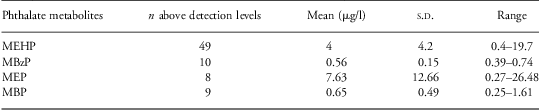
The primary phthalate metabolite and total phthalate levels obtained during the last trimester of pregnancy among the 73 women who delivered male infants are summarized in Table 2. The exposure level was highest for MEP and lowest for MBP. However, only a few women had exposure to MBzP, MEP and MBP above the detection levels. Therefore, the statistical models only assessed the association with AGD and penile measurements with MEHP and total phthalate levels, in order to maintain sufficient statistical power to masses the associations.
Mean prenatal phthalate metabolite concentrations among 73 Mexican women with male newborns
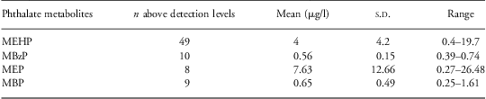
MEHP, mono-2-ethylhexyl phthalate; MBzP, mono-benzyl phthalate; MEP, mono-ethyl phthalate; MBP, mono-butyl phthalate.
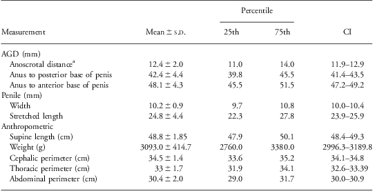
The AGD, penile and anthropometric measurements of the 73 male newborns are summarized in Table 3. All of the measurements were normally distributed.
AGD, penile and anthropometric measurements among 73 Mexican male newborns
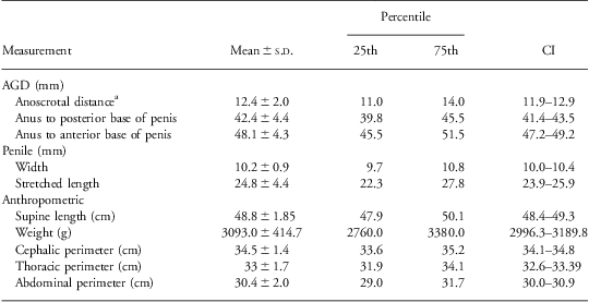
AGD, anogenital distance.
aDistance from center of anus to junction of smooth perineal skin and rugated skin of scrotum.
Crude associations between prenatal MEHP and total phthalate exposure and male newborns’ AGD, penile and anthropometric measurements, adjusted for creatinine showed significant inverse associations only between prenatal MEHP levels and the width of the penis (β = −0.0372 mm/1 μg/l, P = 0.044). In addition, a significant inverse association was observed between total prenatal phthalate levels and penile width (β = −0.0388 mm/μg/l, P = 0.010).
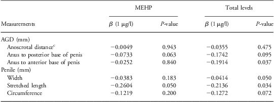
Table 4 shows the results of the linear regression models of the association between prenatal phthalate exposure and male newborns’ AGD and penile measurements, adjusted for creatinine levels and supine length of the newborns at birth. We also assessed models with other variables not shown (i.e. consumption of foods rich in phytoestrogens, exposure to pesticides, tobacco and alcohol use) and found no statistically significant effects. Exposure to MEHP was associated with a reduction in the stretched length of the penis (β = −0.2604 mm/1 μg/l, P = 0.050). Total phthalate prenatal exposure level was inversely associated with anus to anterior base of penis, penile width and stretched length.
Linear regression models of the association between prenatal MEHP and total phthalate exposure levels (μg/l), and AGD, and penile measurements, adjusted for creatinineFootnote a and supine length at birthFootnote b, among 73 Mexican male newborns
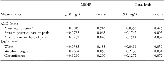
AGD, anogenital distance; MEHP, mono-2-ethylhexyl phthalate.
a To compensate for urine collection at different times of the day.
b The size (length) of the newborn could be related to the AGD and penile measurements of the newborns.
c Distance from center of anus to junction of smooth perineal skin and rugated skin of scrotum.
Discussion
In this study, we assessed the relationship between prenatal exposure to phthalates and AGD and genital and anthropometric measurements in Mexican male newborns. Exposure levels are consistent with reports from Mexican populations.Reference Svensson, Hernández-Ramírez and Burguete-García21, Reference Romero-Franco, Hernández-Ramírez and Calafat22 Our findings are consistent with two previous reports by SwanReference Swan, Main and Liu11, Reference Swan12 who found inverse association between prenatal maternal urinary DEHP metabolites, AGD and penile measurements, and a more recent report by Suzuki et al.Reference Suzuki, Yoshinaga, Mizumoto, Serizawa and Shiraishi13 However, Huang et al.Reference Huang, Kuo, Chou, Lin and Lee14 did not find this association for male newborns. Our results also provide new insights into the relationship between prenatal phthalate exposure and the male reproductive system, as we also observed inverse association between prenatal phthalate exposure and penile width and length among the newborns.
In a previous study conducted on newborns in Chiapas, Mexico,Reference Romano-Riquer, Hernández-Ávila, Gladen, Cupul-Uicab and Longnecker23 the mean penile length (27.1 mm) was similar to the one observed in our study (24.8 mm). The newborns in our study had significantly shorter penises than non-Mexican newborns. Mexican in our study 24.8 mm v. Chinese 30 mm, Caucasians 34 mm, Indians 36 mm,Reference Fok, Hon and So24 Argentinians 33 mm,Reference Anigstein25 French 36.5 mm,Reference Damon, Berlier, Durozier and Francois26 Turks 36.5 mm,Reference Carmudan, Oz and Ilhan27 Saudis 35.5 mmReference Al-Herbish28 and Japanese 29 mm.Reference Fujieda and Matsuura29
Given that the newborns in our study had smaller penises than non-Mexican newborns, we further investigated whether any of the penises observed in our newborn population could be classified as micropenises. A micropenis is a very small penis with a normal configuration and is considered a sign and not a diagnosis. The etiologies of micropenis are classified as hypogonadotropic hypogonadism, primary hypogonadism, androgen insensitivity or idiopathic. According to the formula set by Lee et al. Reference Lee, Mazur and Danish30 a micropenis is classified as a penis smaller than the population mean minus 2.5 standard deviations (s.d.). For a Mexican newborn to be classified as having a micropenis, the penile length would have to be 13.9 mm (24.8 mm − 2.5 s.d.). The smallest penile length in our newborn population was 14.3 mm, and it was observed among only 1% of the newborns (not shown). In addition, in our study we used the same technique to measure penile length as the one used previously by Romano-Riquer et al. Reference Romano-Riquer, Hernández-Ávila, Gladen, Cupul-Uicab and Longnecker23 (measured at the anterior base of the penis, where the penis meets the pubic area) among Mexican male newborns in Chiapas, Mexico, and the findings in their study were similar to ours (ours: 10.2 ± 0.9 mm; Chiapas (Romano-Riquer et al. Reference Romano-Riquer, Hernández-Ávila, Gladen, Cupul-Uicab and Longnecker23: 10.5 ± 0.9 mm). These findings indicate that the small penile length observed in the two Mexican newborn populations may not be indicative of a micropenis, as micropenis is usually associated with hypothalamic disorders.Reference Anhalt, Neely and Hintz31
It is also possible that the observed differences in penile measurements between newborns from different countries may be because of differences in measurement techniques, measurement instruments or the environments where the newborns were measured. Nevertheless, the findings from the study conducted by Swan et al. Reference Swan, Main and Liu11 among American newborns and toddlers and the findings from our study demonstrate that phthalates can affect AGD and penile measurements.
Available scientific evidence on phthalate exposure in humans, although limited, has generated great concern because of the possible adverse effects on masculine gonads from development until adulthood. This concern continues to increase as results from recent studies have associated phthalate exposure with adverse health effects in children.Reference Swan, Main and Liu11
As male rodent modelsReference Ema and Miyawaki8, Reference Bowman, Barlow, Turner, Wallace and Foster33–Reference Welsh, Saunders and Fisken36 can be applied to humans, it is plausible that the same biological mechanisms observed in rodents can act as endocrine disruptors in humans. In rodents, perineal development is determined by the androgens (dihydrotestosterone) during sexual differentiation.Reference Bowman, Barlow, Turner, Wallace and Foster33 Ema and MiyawakiReference Ema and Miyawaki8 demonstrated that testosterone metabolites are reduced by prenatal exposure to MBP, suggesting an antiandrogenic effect. In addition, other in vitro studies have shown that phthalates exert a negative influence on Leydig cells,Reference Ge, Chen, Tanrikut and Hardy34 disrupting testosterone production and increasing cell proliferation. These effects potentially contribute to testicular dysgenesis syndrome, which includes a variety of conditions that involve the male reproductive system, including undescended testes, hypospadias (an abnormality of the penis in which the urethra opens on the underside), changes in the timing of puberty, testicular cancer and reduced fertility.Reference Chen, Dong, Ge and Hardy35
As previously stated, the study conducted by Swan et al. Reference Swan, Main and Liu11 demonstrated that phthalate exposure was associated with reduced AGD but not reduced penile size, as we demonstrated in our study. In contrast to the methodology used by Swan, the newborns in our study were measured within 24–48 h of birth. We also did not utilize an AGD index but rather performed a reliability analysis on each of the AGD measurements under study. In addition, we adjusted for the supine length of the newborns at birth, as AGD was associated with the length of the male infants at birth in a previous study conducted in Mexico.Reference Salazar-Martínez, Romano-Riquer, Yáñez-Márquez, Longnecker and Hernandez-Avila15 Nonetheless, residual confounding may still exist in our study as we only obtained a single urine sample, during the third trimester of pregnancy; moreover, urine was not collected during the first trimester, the window of greatest risk for phthalate effects.
Conclusions
Human exposure to phthalates is a public health concern, and the system most vulnerable to its potential effects seems to be the immature male reproductive tract. As studies on phthalate exposure in humans are few and recent, especially among vulnerable populations, additional epidemiologic, basic and clinical research is needed to further elucidate the toxic effects of these chemicals and to improve environmental management and policy decision making. In addition, one of the goals of environmental health programs worldwide is to identify and evaluate environmental health risks among vulnerable populations, especially children, as owing to their size and developmental stage children are more susceptible than adults to the effects of environmental toxins.37
This is the first study conducted in Mexico to explore the toxic effects of phthalates in male newborns using AGD and penile measurements as markers of reproductive toxicity. Even though our findings are consistent with those observed by Swan et al. Reference Swan, Main and Liu11, Reference Swan12 in United States and Suzuki et al.Reference Suzuki, Yoshinaga, Mizumoto, Serizawa and Shiraishi13 in Japanese infants, it is important to further investigate whether the observed effects will be maintained over time or whether mechanisms of compensation exist among populations with higher phthalate levels. To further confirm our study findings, we suggest conducting a larger cohort study using the same parameters of reproductive toxicity among male newborns whose mothers are occupationally exposed to phthalates.
Acknowledgments
The authors thank the administration of the Hospital Materno Infantil of the Instituto de Seguridad Social del Estado de México y Municipios (ISSEMYM) in Toluca, Mexico, where the study was conducted, particularly Fausto Manuel Pinal González, MD, and Yecenia Velázquez de la Sancha, Licensed Nurse, for their collaboration in the recruitment of study participants; Guadalupe Jiménez Martínez, Chemist, for her collaboration in the phthalate analyses; and Ms Stephanie Deming from The University of Texas M. D. Anderson Cancer Center for her editorial comments.
Financial Support
The study was funded by the Universidad Autónoma del Estado de México (UAEM) with Grant 2245/Contract 2006, and with ‘Fondos de Consolidación 2009’ project number S81109. This study was also supported by the Fulbright-García Robles Scholars Program through the scholarship provided to Dr María A. Hernández-Valero to cover her travel expenses and research in Mexico, and through the research support provided to Dr Hernández-Valero by the National Institutes of Health, National Center for Minority Health and Health Disparities (NCMHD P60-MD000503).
Conflicts of Interest
None.
Ethical Standards
The study was approved by the Investigation and Ethics Committee of the Hospital Materno Infantil.