Introduction
Truncus arteriosus is a conotruncal anomaly in which a single great artery arises from the heart to supply both systemic and pulmonary circulation, usually associated with increased pulmonary blood flow. Its incidence is approximately 0.07 per 1000 live births. Reference Liu, Chen and Zühlke1 It is classified according to the origin of the pulmonary arteries from the truncal vessel. Approximately one-third of patients with truncus arteriosus carry a 22q11.2 deletion. Tricuspid atresia is a complex CHD characterized by the absence of a connection between the right atrium and right ventricle, resulting in single ventricle physiology. Its prevalence is approximately 0.11 per 1000 live births. Reference Liu, Chen and Zühlke1 Survival requires the presence of both an atrial septal defect and a ventricular septal defect. It is classified based on the ventricular septal defect and the relationship between the great arteries. The coexistence of these two complex cardiac anomalies is extremely rare and has only been reported in isolated case reports. Here, we present a preterm infant with the rare combination of truncus arteriosus and tricuspid atresia.
Case report
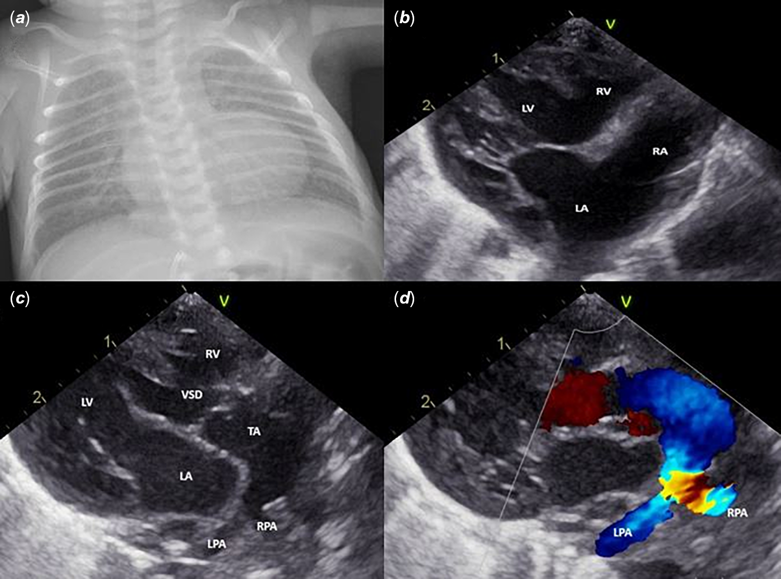
A male infant was born at 30 weeks of gestation with a birth weight of 1090 g. Paediatric cardiology consultation was requested shortly after birth because of persistent hypoxaemia. On physical examination, no cardiac murmur was noted. Additionally, right thumb agenesis and increased corneal opacity were observed. Ophthalmologic evaluation was consistent with congenital glaucoma. Oxygen saturation was measured at 90%. Chest X-ray revealed a narrowed pulmonary conus and a widened mediastinum (Figure 1). Electrocardiography showed left axis deviation without other notable findings. Echocardiographic evaluation revealed normal atrial situs and ventricular looping. The tricuspid valve was atretic, and the right ventricle appeared hypoplastic. The main pulmonary artery could not be visualized. A truncal artery originating from the heart was identified, along with a large subtruncal ventricular septal defect and a non-restrictive atrial septal defect with left-to-right shunting. Confluent and hypoplastic pulmonary arteries were seen arising from the proximal portion of the truncal artery (Collett-Edwards Type I), with mild stenosis of the right pulmonary artery. The truncal valve was tricuspid in morphology with minimal regurgitation. No interruption or coarctation of the aortic arch was observed. The ductus arteriosus was not visualized. A persistent left superior vena cava was present. Chromosomal analysis revealed a normal 46,XY karyotype without a 22q11.2 deletion. During follow-up, the patient developed tachypnoea, respiratory distress, and elevated lactate levels. Furosemide treatment was initiated. The patient was evaluated for pulmonary artery banding, but due to concurrent respiratory distress syndrome and neonatal sepsis, he died on the 8th postnatal day.
(a): Chest X-ray demonstrating a narrowed pulmonary conus and widened mediastinum. (b):Apical four-chamber echocardiographic view demonstrating the characteristic findings of tricuspid atresia with a hypoplastic right ventricle (RV). A non-restrictive atrial septal defect (ASD) and a large ventricular septal defect (VSD) are also present. RA, right atrium; LA, left atrium; LV, left ventricle. (c): Parasternal long-axis view demonstrating a single truncal artery (TA) overriding a large subtruncal ventricular septal defect (VSD). The right (RPA) and left (LPA) pulmonary arteries arise as confluent branches from the proximal truncal artery. (d): Colour Doppler imaging demonstrating mild stenosis of the right pulmonary artery (RPA).

Figure 1. Long description
Panel A: A chest X-ray image showing a narrowed pulmonary conus and a widened mediastinum. Panel B: An apical four-chamber echocardiographic view displaying a hypoplastic right ventricle (RV) characteristic of tricuspid atresia. The image also shows a non-restrictive atrial septal defect (ASD) and a large ventricular septal defect (VSD). The right atrium (RA), left atrium (LA), and left ventricle (LV) are labeled. Panel C: A parasternal long-axis view showing a single truncal artery (TA) overriding a large subtruncal ventricular septal defect (VSD). The right pulmonary artery (RPA) and left pulmonary artery (LPA) arise as confluent branches from the proximal truncal artery. Panel D: A color Doppler imaging view demonstrating mild stenosis of the right pulmonary artery (RPA).
Discussion
The coexistence of truncus arteriosus and tricuspid atresia is a rare and complex anomaly, first reported in 1974. Reference Tandon, Moller and Edwards2 Since then, only isolated case reports and a limited number of small case series have been published. In addition to tricuspid atresia, other forms of functionally univentricular hearts reported in association with truncus arteriosus include unbalanced atrioventricular septal defect, double-inlet left ventricle, and hypoplastic left heart syndrome. Reference Chatila, Houyel, Hily and Bonnet3,Reference Elhedai, Yong and Chaudhari4 Recent reports have demonstrated that although staged single-ventricle palliation may be feasible in selected patients, overall outcomes remain poor because of the complexity of the cardiac anatomy and the frequent presence of associated cardiac and extracardiac anomalies. Reference Elhedai, Yong and Chaudhari4,Reference Weld, Lee and Loomba5
In truncus arteriosus, increased pulmonary blood flow frequently leads to early pulmonary hypertension and heart failure in the neonatal period, necessitating timely surgical intervention. A 22q11.2 deletion is present in approximately 35.5% of patients. Reference Peyvandi, Lupo and Garbarini6 In our patient, no 22q11.2 deletion was identified, and chromosomal analysis demonstrated a normal karyotype, despite the presence of multiple extracardiac anomalies.
The current surgical strategy for patients with truncus arteriosus and single-ventricle physiology is staged palliation aimed at achieving Fontan circulation. Initial palliation may include pulmonary artery banding or separation of the pulmonary arteries from the truncal vessel followed by establishment of controlled pulmonary blood flow using a systemic-to-pulmonary artery shunt. Reference Kobayashi, Sano and Narumiya7 Subsequent bidirectional cavopulmonary anastomosis and Fontan completion are performed when anatomical and haemodynamic conditions permit. In cases with associated interrupted aortic arch or coarctation, simultaneous repair or hybrid procedures may be necessary. Weld et al. successfully performed a hybrid procedure involving ductal stenting and pulmonary banding in a premature infant with truncus arteriosus, tricuspid atresia, and interrupted aortic arch. Reference Weld, Lee and Loomba5
To our knowledge, this is the lowest birth weight infant reported with this rare combination. Despite planned staged palliation, the outcome was unfavourable because of extreme prematurity, neonatal sepsis, and associated comorbidities. Our case further highlights the considerable challenges in managing this exceptionally rare anomaly in very low birth weight infants.
Conclusion
This case describes the rare coexistence of truncus arteriosus and tricuspid atresia in an extremely low-birth-weight preterm infant. Despite planned staged palliation, the outcome was unfavourable because of severe prematurity and associated comorbidities.
Financial support
This research received no specific grant from any funding agency, commercial, or not-for-profit sectors.
Competing interests
None.
Ethical standard
Written informed consent for publication was obtained from the patient’s parents. All procedures were performed in accordance with the ethical standards of the institutional and national research committee and with the Declaration of Helsinki and its later amendments.