Introduction
Telomeres are nucleoprotein complexes that are present at both ends of chromosomes and contain telomeric DNA, which consists of long tandem repeats of the sequence TTAGGGn. Telomeres protect the ends of the chromosomes from sequence erosion and fusion with neighbouring chromosomes. A progressive shortening of telomeres occurs with each cellular division and, when a critical length is reached, cessation of cell division and cellular senescence ensue.
Telomeres, and particularly the 5′-GGG triplet, are very sensitive to oxidative stress-induced DNA damage (Kawanishi and Oikawa, Reference Kawanishi and Oikawa2004). Mitochondrial dysfunction may cause progressive shortening of telomeres by promoting the generation of reactive oxygen species (ROS). Telomere shortening, in turn, is a key mechanism leading to cell senescence, which has also been associated with decreased oocyte quality through disruption of chromosome alignment and spindle structure during meiosis (Keefe and Liu, Reference Keefe and Liu2009). In addition, telomere shortening in mouse and human oocytes triggers apoptosis in embryos.
Mice with oocyte-specific deletion of mitofusin-2 (Mfn2, a GTPase localized in the outer mitochondrial membrane and essential for mitochondrial fusion) have subfertility and accelerated follicular depletion (Zhang et al., Reference Zhang, Bener, Jiang, Wang, Esencan, Scott, Horvath and Seli2019b). Mfn2-deficient oocytes have mitochondrial dysfunction, increased ROS, and shorter telomeres are suggestive of an ageing phenotype. Mitofusin-1 is another protein that is required for mitochondrial fusion. Mice with Mfn1 deletion in the oocyte are characterized by accelerated follicular depletion and infertility associated with defective oocyte maturation and follicular development (Zhang et al., Reference Zhang, Bener, Jiang, Wang, Esencan, Scott Iii, Horvath and Seli2019a).
In the current study, we aimed to determine whether mitochondrial dysfunction in oocytes with targeted deletion of Mfn1 caused telomere shortening.
Materials and methods
Germinal vesicle (GV) stage oocytes (collected from six mice for each genotype at each timepoint, and pooled as two mice per sample analyzed) were collected from 3-, 6- and 9-month-old Mfn1 −/− mice and compared with wild-type (WT). Granulosa cumulus cells and white blood cells (WBC) were also evaluated as somatic controls.
GV-stage oocytes, oocytes at metaphase of second meiotic division (MII), and cumulus cells were obtained by superovulation of mature 2-, 6-, and 9-month-old Mfn1 −/− and WT mice. To obtain oocytes at the GV stage, mice were euthanized by CO2 inhalation 44 h after intraperitoneal injection of 5 IU PMSG. Ovaries were removed and punctured in M2 medium (Sigma, St. Louis, MO) with 10 µM milrinone (Sigma, St. Louis, MO) under a dissecting microscope (Olympus SZH-ILLK) with a 26½-gauge needle to isolate cumulus–oophorus complexes containing GV-stage oocytes and cumulus cells. Oocytes were stripped from cumulus cells using a mouth pipette and collected in individual tubes. DNA was extracted from oocytes and cumulus cells using the QIAmp DNA Micro Kit (Qiagen, Valencia, CA) and quantified. Approximately 0.5 ml of blood sample was collected from each mouse and blood DNA was extracted using the DNA isolation kit for mammalian blood (Roche, Basel, Switzerland). A standard curve for polymerase chain reaction (PCR) was generated by serial dilutions of known amounts of DNA from somatic (WBCs and granulosa) cells (Fig. 1A). The telomere/single-copy gene ratio (T/S) was used as an indicator of telomere length (Liu et al., Reference Liu, Bailey, Okuka, Muñoz, Li, Zhou, Wu, Czerwiec, Sandler, Seyfang, Blasco and Keefe2007). Immunofluorescence for TRF1 and H2A.X was quantified using a rat anti-TRF1 monoclonal antibody (Abcam, Cambridge, UK, cat. no. ab192629) and a mouse anti-H2A.X monoclonal antibody (Sigma-Aldrich, St. Louis, MO, USA, cat. no. 05-636-25UG) as the primary Alexa Fluor 568-conjugated goat anti-rat antibody and Alexa Fluor 488-conjugated goat anti-mouse antibody and secondary antibodies, respectively.
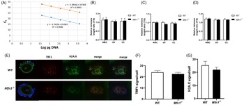
Telomere length in Mfn1 −/− female mice. The standard curve was generated by serial dilution of known amounts of DNA to calculate relative DNA concentrations (log DNA) from Ct values of the qRT-PCR products. Blue dots, telomere gene; orange dots, 36B4 single-copy gene control. The correlation regression equation and coefficients (R2) of Ct versus log DNA are shown (A). The relative telomere length of white blood cells (WBC), GV oocytes and cumulus cells (CC) are represented as ratio of T/S, in 3-, 6- and 9-month-old wild-type (WT) and Mfn1 −/− female mice respectively (B–D). Immunofluorescence double staining of TRF1 (red) and H2A.X (green) in cumulus–oophorus complexes of Mfn1 −/− and WT mice. 4′,6-Diamidino-2-phenylindole (DAPI) was used to stain nuclei (blue) (E). Quantitative analysis of TRF1 and H2A.X immunofluorescence in Mfn1 −/− and WT GV oocytes (F, G). Data are presented as mean ± SEM using a t-test.

Results and Discussion
A standard curve for PCR was generated by serial dilution of known amounts of DNA from somatic (WBCs and granulosa) cells (Fig. 1A). Telomere length in oocytes from 2-month-old Mfn1 −/− mice was not different compared with WT. The number of PCR cycles (21.8 ± 1.79 vs 21.4 ± 1.68, P = 0.84) and telomere/single-copy gene ratio (T/S), used as an indicator of relative telomere length (1019 ± 0.02 vs 1027 ± 0.06, P = 0.93), were also similar (Fig. 1B). Telomere length of granulosa cells and WBCs in 2-month-old Mfn1 −/− mice were also unchanged compared with WT (Fig. 1B). To determine if there was an age-related effect, 6- and 9-month-old Mfn1 −/− and WT mice were similarly tested. There was no statistically significant difference in telomere length in oocytes, granulosa cells and WBCs compared with WT at 6 and 9 months (Fig. 1C, D). Also, Mfn1 −/− oocytes had a similar expression of telomere protective protein TRF1 (Fig. 1E, F). DNA repair through histone H2A.X phosphorylation also did not seem to be activated in Mfn1 −/− (Fig. 1E, G) as there was no significant increase in the co-localization of TRF1 and H2A.X (Fig. 1E).
In conclusion, mitochondrial dysfunction due to the deletion of Mfn1 does not seem to affect telomere length in mouse oocytes.
Author contributions
MC and ES conceived the idea, wrote the manuscript and provided approval for the version to be published. All authors agreed to be accountable for all aspects of the article.
Funding information
This work was supported by the GR109707.CC1122.PG00032.PJ000001.EUS2 grant to ES from the Foundation for Embryonic Competence.
Conflicts of interest
MC declares no conflict of interest. ES is a consultant for and receives research funding from the Foundation for Embryonic Competence; he is also co-founder and a shareholder of ACIS LLC and co-holds the patent US2019/055906 issued for using electrical resistance measurement for assessing cell viability and cell membrane piercing.
Ethics approval for animal study
Mice care, breeding, and experimental procedures were conducted according to Yale University animal research requirements, using protocols approved by Institutional Animal Care and Use Committee (protocol #2020-11207).