Introduction
Double-outlet right ventricle is a complex CHD in which both great arteries—the aorta and the pulmonary artery—originate totally or predominantly (more than 50%) from the right ventricle.Reference Walters, Mavroudis, Tchervenkov, Jacobs, Lacour-Gayet and Jacobs1 With an incidence of 1 in 6,000 to 10,000 live births, double-outlet right ventricle accounts for 1–3% of all CHDs.Reference Hoffman and Kaplan2 Double-outlet right ventricle presents with considerable anatomical variability, primarily determined by the location of the ventricular septal defect, the spatial relationship of the great arteries, and the presence of additional structural abnormalities. These differences arise from variations in embryological development, particularly involving the conotruncal region and the migration of the great arteries. As a result, double-outlet right ventricle can be classified into subtypes such as subaortic, subpulmonary, doubly committed, or remote ventricular septal defects, each with distinct surgical and haemodynamic implications. Given this complexity, precise diagnosis and thorough morphological assessment are crucial to guide appropriate surgical planning and improve patient outcomes.Reference Pang, Meng and Hu3,Reference Obler, Juraszek, Smoot and Natowicz4
The double-outlet right ventricle type of ventricular septal defect is characterised by a subaortic ventricular septal defect without pulmonary stenosis that is functionally similar to an isolated large ventricular septal defect. This variant results in pulmonary overflow from the systemic circulation, which can cause symptoms of heart failure. Transposition of the great arteries type of double-outlet right ventricle, also known as Taussig–Bing anomaly, is defined by a subpulmonary ventricular septal defect without pulmonary stenosis. This form has anatomic similarities to transposition of the great arteries, and severe cyanosis may develop depending on the direction of the shunt. Tetralogy of fallot type of double-outlet right ventricle is characterised by a subaortic ventricular septal defect and pulmonary stenosis with haemodynamic features similar to tetralogy of fallot.Reference Öcal, Demirci and Kahramanoğlu5 The univentricular type of double-outlet right ventricle is associated with mitral atresia, unbalanced atrioventricular septal defects, or severe ventricular hypoplasia and typically requires univentricular palliation. Accurate classification of these anatomic subtypes is essential for assessing surgical eligibility, selecting the most appropriate surgical technique, and predicting long-term prognosis.
The first-line imaging modality to evaluate for double-outlet right ventricle is transthoracic echocardiography, which is preferred because of its widespread availability, low cost, and lack of radiation exposure. Echocardiography is an effective imaging modality for assessing cardiac anatomy and function. However, the diagnostic accuracy of transthoracic echocardiography can be limited in some patients due to operator dependence, limited acoustic window, and challenges in the evaluation of small anatomical features. In adult patients with a history of multiple surgical procedures, the limited acoustic window may additionally complicate detailed visualisation of extracardiac structures.
In recent years, technological developments have made CT a widely used non-invasive imaging modality for the diagnosis and evaluation of double-outlet right ventricle. Data acquisition now requires only a portion of the cardiac cycle or at most several cardiac cycles. The highest pitch scan modes and volumetric scanners provide full anatomic coverage of a paediatric thorax in less than a second or a single heartbeat, freezing respiratory motion. This rapid image acquisition eliminates or reduces the need for sedation and anaesthesia in those unable to cooperate with a short breath-hold. The prospective delivery of radiation to a limited portion of the cardiac cycle and post-processing approaches such as iterative reconstruction allow for significantly reduced radiation doses while maintaining or improving image quality.Reference Han, Rigsby and Hlavacek6 Advanced technologies, such as multidetector CT, allow the acquisition of high-resolution anatomical images, which significantly contribute to surgical planning. Electrocardiography-gated CT scans reduce motion artefacts caused by cardiac motion, allowing for clearer and more accurate anatomical visualisation. In addition, CT imaging is more reliable, especially for children, because automated voltage adjustment, current modulation, and wide detector arrays minimise radiation exposure.Reference Kumar and Bhatia7
CT angiography provides high resolution in identifying the location of the ventricular septal defect, the relationship of the great arteries and associated anatomical abnormalities, making it a critical pre-operative planning modality.Reference Goo8 The fast scan time minimises the need for anaesthesia, especially in children, thereby improving procedural comfort. In addition, CT three-dimensional modelling and virtual surgical planning capabilities provide a comprehensive assessment of anatomical structures, facilitating precise surgical correction.Reference Brüning, Kramer and Goubergrits9 Despite the inherent limitations associated with ionising radiation exposure and contrast administration, recent advances in low-dose imaging protocols, improved image processing algorithms, and next-generation technologies designed to mitigate the long-term effects of radiation have significantly improved the safety profile of CT imaging.
In conclusion, the combined use of CT and transthoracic echocardiography plays a critical role in the diagnosis, surgical planning, and post-operative follow-up of double-outlet right ventricle. CT angiography, as a highly accurate and non-invasive imaging modality, has significantly improved the evaluation of this complex congenital defect. Advances in low-dose imaging, AI-driven processing, and three-dimensional modelling have further increased its role. Given the complex anatomy of double-outlet right ventricle, accurate pre-operative assessment is essential. While transthoracic echocardiography remains the first-line modality, its limitations highlight the value of complementary techniques. In this regard, CT angiography provides high-resolution anatomical detail that improves both pre-operative planning and post-operative monitoring. The aim of this study was to compare pre-operative anatomic assessments using transthoracic echocardiography and CT angiography with definitive post-operative findings, evaluating their accuracy and performance in pre-operative anatomical evaluation.
Materials and methods
Our study was a single-centre, retrospective study. This study included infants younger than 1 year with double-outlet right ventricle who were hospitalised in the paediatric cardiac ICU of our hospital for pre-operative management between January 1, 2022, and September 1, 2024. The study was designed in accordance with the tenets of the Declaration of Helsinki and obtained the necessary approval from the local ethics committee. Patients over 1 year of age, double-outlet right ventricle patients with hypoplastic ventricles, patients who had previously undergone palliative cardiac surgery, patients with impaired renal function tests, patients who were unsuitable for surgical repair, and patients with critical comorbidities were excluded from the study.
First, all patients were evaluated with transthoracic echocardiography during the diagnostic and follow-up process. In addition to echocardiography, other imaging modalities such as CT angiography was used to provide a detailed anatomical assessment before surgery. Demographic variables including age, sex, height, weight, and body surface area (0.007184 × height0.725 × weight0.425; calculated using the Du Bois method) were documented.
Echocardiographic evaluation was performed using the Philips Affiniti 50 cardiac ultrasound system (Philips Affiniti 50 Cardiac Ultrasound, Bothell, WA, USA) with a 9 and 12 MHz probe. All patients underwent echocardiography using standardised protocols according to the guidelines of the American Society of Echocardiography. Standard views of the paediatric echocardiogram were recorded, including parasternal (long and short axis), apical (four and five chambers), subcostal, and suprasternal views. Cardiac morphology was evaluated in the direction of blood flow using a segmental approach. Atrial situs, venoatrial connection (systemic and pulmonary venous return), atrium-ventricular connections, ventricles, ventricular-great artery connection, the spatial position of great arteries, intracardiac defects, and extracardiac vascular anomalies were examined as the main components of this approach. The pattern of coronary artery anatomy in patients with double-outlet right ventricle was carefully identified in the parasternal short-axis views as described in the American Society of Echocardiography report. In the ideal imaging position, both semilunar valves, as seen in cross-sectional views, and both coronary artery origins were visualised. Two-dimensional transthoracic echocardiography (Philips Epix 3D device, Amsterdam, Netherlands) was performed by two paediatric cardiologists with more than 10 years of experience in the evaluation of CHD. During the two-dimensional transthoracic echocardiographic evaluation of the infants, no additional sedative medication was administered.
Cardiac CT was performed on a single-source 640-slice CT (Aquilion ONE, GENESIS Edition Canon Medical Systems, Tochigi, Otawara, Japan) using a wide detector aperture (16 cm), an iterative reconstruction algorithm, adaptive iterative dose reduction three-dimensional enhanced. A prospective electrocardiography-gated technique was performed during one cardiac cycle in all patients. All images were acquired in volume axial mode (rotation time: 0.35 s, scan range: 80–120 mm). The tube current was set by automatic exposure control. A low kV value (80 kV) was selected to maximise the iodine contrast-to-noise ratio for all children. All patients were injected intravenously with an iodinated contrast agent (Kopaq 300 mgI/mL, Onko&Kocsel Pharmaceuticals, Kocaeli, Turkey) at a rate of 1.5 mL per kg, followed by 10–20 mL of saline solution with a dual-head power injector (Bayer HealthCare, MEDRAD Europe, Beek, Netherlands). The injection rate was 0.7–0.9 mL/kg, depending on the catheter and patient size. The contrast agent was used undiluted for the procedure. All cardiac CT images were obtained without breath-hold and without sedation for the study. All examinations were performed under the direction of an attending cardiac radiologist at our hospital. All images were acquired during the first pass of contrast through the anatomical structures of interest. Targets for the centre of the acquisition window were set at 45% of the R–R interval in patients (all patients with heart rates higher than 90 beats/min). During other phases of low interest, the operation is stopped. The best motion-free cardiac phase adjacent to the predefined cardiac phase was individually selected from the raw data for each patient by the attending radiologist. Axial images for all cardiac CT images were transferred to a post-processing workstation. Image reconstruction was performed with a slice thickness of 0.5 mm and the standard reconstruction kernel. Iterative reconstruction was performed with adaptive iterative dose reduction three-dimensional enhanced. Reconstructed images were acquired using multiplanar reconstruction, maximum intensity projection, and three-dimensional volume rendering techniques. Radiation exposure parameters, including imaging area, dose-length product, and volumetric CT dose index, were recorded for each patient that underwent cardiac CT. The CT dose index, dose-length product, and conversion factor were expressed for a 32 cm body phantom reference. The effective dose was calculated from the dose-length product multiplied by 2 to apply to the 16 cm phantom. The cardiac CT effective dose was calculated from the dose-length product multiplied by the conversion factor. The specific dose-length product conversion coefficients for neonates and infants were 0.039 mSv/(mGy.cm) DI for each age group. CT angiography was performed by a paediatric cardiovascular radiologist with at least 15 years of experience in congenital heart imaging.
Ventriculoarterial connection was defined as double-outlet right ventricle if more than 50% of the aorta originated from the right ventricle, and only these patients were included in the study. Great artery relationship was assessed as normal, D malposition, L malposition, side-by-side, or anterior-posterior. The ventricular septal defect was evaluated as subaortic, subpulmonic, doubly committed, or remote. Outflow tract stenosis, atrioventricular valve abnormalities, and coronary artery abnormalities were considered present or absent. The classification of coronary arteries was conducted according to the Leiden convention.
The two imaging modalities were used to evaluate the parameters of ventricular septal defect localisation and great vessel relationship, coronary artery anomalies, aortic arch hypoplasia, ınterrupted aortic arch, aortopulmonary collateral vessels, anomalous pulmonary venous connection, addition ventriculer septal defect (minimal, < 1.5 mm).
In the pre-operative period, both transthoracic echocardiography and cardiac CT angiography were performed. Imaging findings were compared with intraoperative surgical findings, which served as the gold standard. Definitive diagnoses were established through direct surgical visualisation and systematically documented using a standardised checklist, ensuring consistent and reliable assessment of key anatomical features such as ventricular septal defect localisation, great artery relationships, coronary and aortic arch anomalies, collateral vessels, and venous connections.
This study aimed to compare the diagnostic accuracy and efficacy of transthoracic echocardiography and cardiac CT angiography in the pre-operative evaluation and surgical planning of double-outlet right ventricle patients.
Descriptive and inferential statistical methods were used for data analysis. The normality of continuous variables was tested using the Kolmogorov–Smirnov and Shapiro–Wilk tests. Normally distributed variables were analysed using parametric tests, whereas non-parametric tests were applied for non-normally distributed variables. Categorical variables were presented as frequency and percentage. Anthropometric measurements, including age, weight, and height, were analysed using percentile and standard deviation score values. These calculations were performed based on the assumption of normal distribution, using mean and standard deviation to obtain z-scores. Sensitivity, specificity, positive predictive value, negative predictive value, accuracy, area under the curve, and Cohen’s Kappa coefficient were calculated to assess the performance of diagnostic tests. Cross-tabulations were created to compare the agreement between echocardiography and CT angiography methods with surgical diagnosis. The statistical significance of associations was evaluated using the chi-square test to assess the independence between the two methods. Fisher’s exact test was applied for contingency tables with cell frequencies less than 5. The classification performance of diagnostic tests was evaluated using receiver operating characteristic analysis, and the area under the curve was calculated. The consistency of diagnostic tests was measured using Cohen’s Kappa coefficient. The balance of classifications was assessed using a Shannon entropy-based uniformity analysis. All statistical analyses were performed using SPSS version 25.0 (SPSS Inc., Chicago, IL, USA), and a p-value of < 0.05 was considered statistically significant.
Result
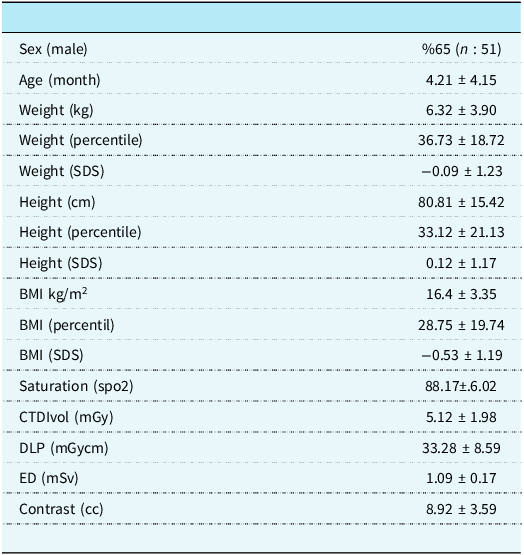
A total of 78 patients were included in the study, of which 65% (n = 51) were male. The mean age of the patients was 4.21 ± 4.15 months. The mean body weight was 6.32 ± 3.90 kg, with a weight percentile of 36.73 ± 18.72 and a weight standard deviation score of −0.09 ± 1.23. The mean height was 80.81 ± 15.42 cm, with a height percentile of 33.12 ± 21.13 and a height standard deviation score of 0.12 ± 1.17. The mean body mass index was 16.4 ± 3.35 kg/m2, with a body mass index percentile of 28.75 ± 19.74 and a body mass index standard deviation score of –0.53 ± 1.19. The mean body surface area was 0.28 ± 0.13 kg/m2, and the mean oxygen saturation (SpO2) was 88.17 ± 6.02. Radiation dose parameters were measured during CT angiography with a mean volume CT dose index (CTDI.vol) of 5.12 ± 1.98 mGy and a dose-length product of 33.28 ± 8.59 mGy-cm. The estimated effective dose was 1.09 ± 0.17 mSv. In addition, the mean contrast volume used during the procedure was 8.92 ± 3.59 cc. The baseline clinical characteristics of the 78 double-outlet right ventricle patients who underwent surgery were summarised in Table 1.
The baseline clinical characteristics

SDS = standard deviation score, BMI = body mass index, ED = effective dose, DLP = dose-length product, CTDIvol = volume computed tomography dose index.
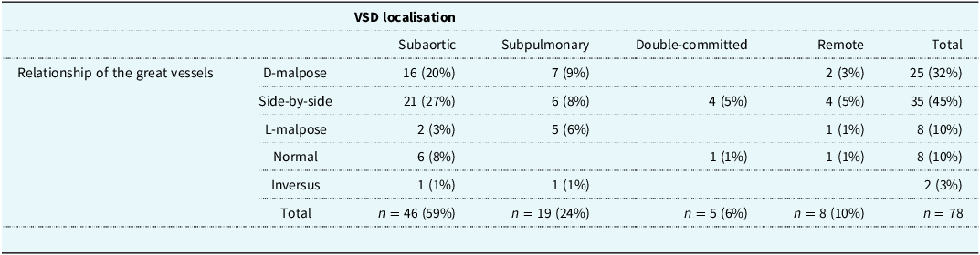
Among the 78 patients with double-outlet right ventricle, the great vessel relationship was side-by-side in 45% (n = 35), D-malposition in 32% (n = 25), L-malposition and normal alignment in 10% (n = 8), and inversus in 3% (n = 2). With regard to ventricular septal defect location, subaortic ventricular septal defect was found in 59% (n = 46), subpulmonary ventricular septal defect in 24% (n = 19), double-committed ventricular septal defect in 6% (n = 5), and remote ventricular septal defect in 10% (n = 8). The most common great vessel relationship in patients with subaortic ventricular septal defect was side-by-side, while in patients with subpulmonary ventricular septal defect, D malposition was the most common. In patients with double-committed and remote ventricular septal defect, the most common great vessel relationship was side-by-side position. The location of the ventricular septal defect and its relationship to the great vessels was shown in Table 2.
VSD localisation and great vessel relationship

VSD = ventricular septal defect.
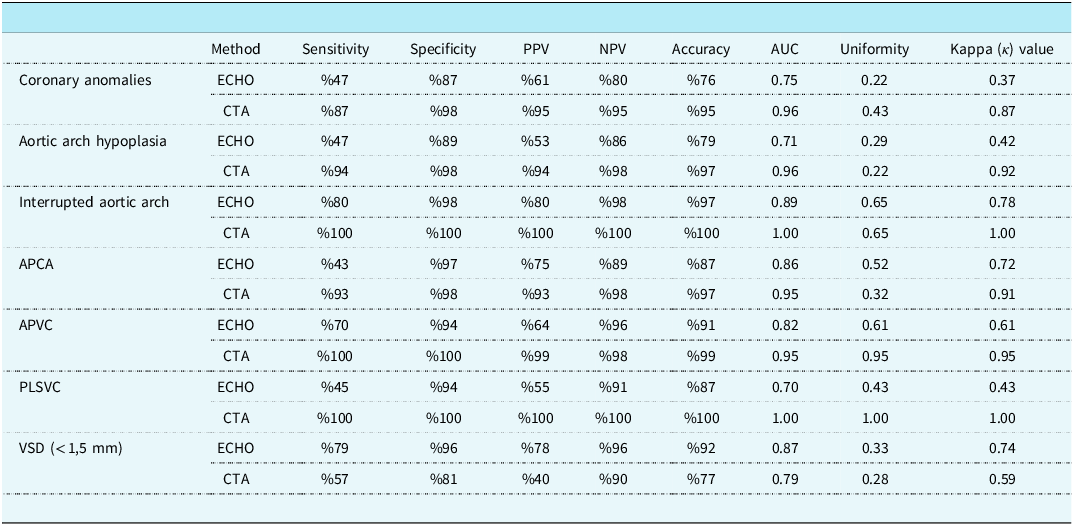
Comparison of imaging modalities after anatomic definition based on post-operative surgical outcomes in double-outlet right ventricle patients evaluated pre-operatively with transthoracic echocardiography and CT angiography. The identification of coronary artery anomalies showed a sensitivity of 47%, specificity of 87%, positive predictive value of 61%, negative predictive value of 80%, and an overall accuracy of 76% with echocardiography. CT angiography showed a sensitivity of 87%, specificity of 98%, positive predictive value of 95%, negative predictive value of 95%, and accuracy of 95%. Chi-square test showed a statistically significant difference (χ2 = 6.07, p = 0.014). In conclusion, cardiac CT was statistically significantly more effective than transthoracic echocardiography in identifying coronary abnormalities (p < 0.05). A three-dimensional model demonstrating the inverted origin of coronary arteries in a patient with a coronary artery anomaly is shown in Figure 1. The diagnosis of aortic arch hypoplasia with echocardiography had a sensitivity of 47%, specificity of 89%, positive predictive value of 53%, negative predictive value of 86%, and overall accuracy of 79%. CT angiography had sensitivity of 94%, specificity of 98%, positive predictive value of 94%, negative predictive value of 98%, and accuracy of 97%. The chi-square test showed a statistically significant difference (χ2 = 5.06, p = 0.024). Cardiac CT angiography was statistically significantly more effective than transthoracic echocardiography in diagnosis of aortic arch hypoplasia. In addition, the sensitivity, specificity, positive predictive value, negative predictive value, and overall accuracy for the detection of interrupted aortic arch with echocardiography were 80%, 98%, 80%, positive predictive value, negative predictive value, and 98%, respectively. CT angiography had a sensitivity of 100%, specificity of 100%, positive predictive value of 100%, negative predictive value of 100%, and accuracy of 100%. Chi-square test showed no statistically significant difference (χ2 = 0.00, p = 1.00). A patient with a aortic arch hypoplasia was shown in Figure 2.
A patient with a coronary artery anomaly (inverted origin of coronary arteries).

A patient with a aortic arch hypoplasia.

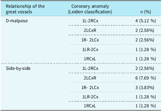
The sensitivity of echocardiography for detecting aortopulmonary collateral vessels was 43%, the specificity was 97%, the predictive value was 75%, the negative predictive value was 89%, and the accuracy was 87%. CT angiography had a sensitivity of 93%, specificity of 98%, predictive value of 93%, negative predictive value of 98%, and accuracy of 97%. Chi-square test showed a statistically significant difference (χ2 = 1.617, p = 0.039). CT angiography was statistically significantly more effective than transthoracic echocardiography in detecting aortopulmonary collateral vessels. A patient with a aortopulmonary collateral artery was shown in Figure 3. The identification of anomalous pulmonary venous connection by echocardiography showed a sensitivity of 70, specificity of 94%, positive predictive value of 64%, negative predictive value of 96%, and accuracy of 91%. CT angiography showed a sensitivity of 100%, specificity of 100%, positive predictive value of 100%, negative predictive value of 100%, and accuracy of %. Chi-square test showed a statistically significant difference CT angiography was statistically significantly more effective than transthoracic echocardiography in detecting anomalous pulmonary venous connection (χ2 = 24.53, p = 0.001). A patient with a total anomalous pulmonary venous connection was shown in Figure 4. Echocardiography showed a sensitivity of 45%, specificity of 94%, positive predictive value of 55%, negative predictive value of 91%, and accuracy of 87% for the detection of persistent left superior vena cava. CT angiography showed a sensitivity of 100%, specificity of 100%, positive predictive value of 100%, negative predictive value of 100%, and accuracy of 100%. Chi-square test showed a statistically significant difference. CT was statistically significantly more effective than transthoracic echocardiography in detecting persistent left superior vena cava (χ2 = 10.82, p = 0.001). A patient with a persistent left superior vena cava was shown in Figure 5. Addition ventricular septal defect less than 1.5 mm detection with echocardiography showed a sensitivity of 79%, specificity of 96%, positive predictive value of 78%, negative predictive value of 96%, and accuracy of 92%. CT angiography showed a sensitivity of 57%, specificity of 81%, positive predictive value of 40%, negative predictive value of 90%, and accuracy of 77%. Chi-square test showed a statistically significant difference (χ2 = 3.52, p = 0.035). For the detection of ventricular septal defects less than 1 mm, transthoracic echocardiography was statistically significantly more effective than CT angiography. The diagnostic accuracy of echocardiography and CT angiography was shown in Table 3, while the comparative diagnostic performance of imaging modalities was shown in Table 4. The distribution of coronary artery anomalies in double-outlet right ventricle according to the Leiden classification is presented in Table 5.
A patient with a aortopulmonary collateral artery.

A patient with a total anomalous pulmonary venous connection.

A patient with a persistent left superior vena cava.

Sensitivity and specificity of echocardiography and CT angiography

AUC = area under receiver operating characteristic curve; CTA = CT angiography; Echo = echocardiography; PPV = positive predicative value, NPV = negative predicative values, APCA = aberrant pulmonary collateral artery, APVC = anomalous pulmonary venous connection, PLSVC = persistent left superior vena cava, VSD = ventricular septal defect.
Compare diagnostic performance of imaging modalities

(+) = positive finding, (-) = negative findings.
Leiden classification of coronary artery anomalies in double-outlet right ventricle

Ao = aorta; PA = pulmonary artery; Cx = circumflex coronary artery; L = left anterior descending coronary artery; R = right coronary artery.
Discussion
The effectiveness of imaging modalities is critical in the diagnosis and pre-operative surgical planning of double-outlet right ventricle. Transthoracic echocardiography is the primary imaging modality for the pre-operative evaluation of patients with double-outlet right ventricle. Due to its low cost, rapid accessibility, and lack of radiation exposure, transthoracic echocardiography is the primary imaging modality of choice. Although transthoracic echocardiography provides excellent visualisation of intracardiac morphology, its ability to evaluate extracardiac structures is limited. In contrast, cardiac CT angiography demonstrates better sensitivity and specificity in identifying extracardiac structures.Reference Kumar and Bhatia7 With recent technological advances, cardiac CT angiography has emerged as the imaging modality of choice due to its high-quality imaging, reduced radiation exposure, and reduced contrast requirements.Reference Priya, Nagpal and Sharma10
One of the most critical steps in the perioperative evaluation of patients with double-outlet right ventricle is the accurate identification of its subtype. The classification of the subtypes is highly dependent on the location of the ventricular septal defect and its relationship to the great arteries. The position of the ventricular septal defect and the relationship to the great arteries are essential points of intracardiac assessment, for which transthoracic echocardiography has demonstrated high sensitivity and specificity. In our study, the most commonly found double-outlet right ventricle subtype was subaortic double-outlet right ventricle, identified in 36% of cases, which is consistent with previously reported rates in the literatüre. Similarly, subpulmonary double-outlet right ventricle, also known as Taussig–Bing variant, was the second most common subtype. The double-committed and remote ventricular septal defect subtypes were identified at lower and similar frequencies. In a study of 47 patients by Poe et al, subaortic ventricular septal defect type double-outlet right ventricle was identified as the most common subtype.Reference Shi, Yang and Chen11
The anatomical relationship between the great arteries plays a critical role in the surgical planning of patients with double-outlet right ventricle. One of the most essential steps in biventricular repair is to ensure that the ventricular septal defect patch effectively incorporates the great artery to establish a proper outflow tract, whether the ventricular septal defect is closed directly or redirected through tunnelling.Reference Meng, Pang and Li12 In our study, the most commonly identified great artery relationship was the side-by-side pattern. D malposition and L malposition were identified at lower frequencies. Similarly, the study by James p Spaeth reported a similar great artery relationship.Reference Spaeth13 Among the great artery relationships, the D malposition was the most common, followed by the side-by-side configuration. In our study, the findings were similar to the literature, with side-by-side and D malposition being the most commonly identified aortic relationships.
Transthoracic echocardiography is the first-line imaging modality for the evaluation of complex CHD. However, this imaging modality may not be sufficient to identify accurately the origin and course of the coronary arteries, thereby limiting its utility in surgical risk stratification. In patients with double-outlet right ventricle undergoing arterial switch operation, coronary artery anatomy is of critical importance for surgical outcomes. One of the most important factors in the success of arterial switch operation is the accurate and reliable transfer of the coronary arteries. Therefore, pre-operative identification of coronary artery anomalies is critical. In a study by J. Wetter et al and Shyh-Jye Chen et al, coronary artery anomalies were identified in approximately 30% of patients diagnosed with double-outlet right ventricle who underwent arterial switch surgery. In our study, 32% of patients had coronary artery anomalies, a finding consistent with previous literatüre.Reference Wetter, Sinzobahamvya and Blaschczok14,Reference Chen, Lin and Lee15 A study by Woo et al. showed that CT angiography was statistically better than transthoracic echocardiography in evaluating coronary anatomy in the pre-operative evaluation of double-outlet right ventricle patients.Reference Goo16 Similarly, our study showed that CT angiography was significantly more effective than transthoracic echocardiography in the pre-operative identification of coronary arteries. However, in three cases in our study, CT angiography was not able to accurately identify the anatomy of the coronary arteries. Post-operative evaluation revealed that these patients had single and inverted coronary origins.
The presence of a persistent left superior vena cava is of significant importance for surgical strategy and post-operative outcomes in patients with double-outlet right ventricle undergoing cavopulmonary anastomosis after palliative treatment. In pre-operative evaluation, CT angiography was found to be statistically better than transthoracic echocardiography in detecting the persistent left superior vena cava. Similarly, the study by Shei et al. reported that CT angiography was more effective than transthoracic echocardiography in identifying persistent left superior vena cava in the pre-operative evaluation of double-outlet right ventricle patients, which is similar to our findings. Aortic arch hypoplasia and interrupted aortic arch are common cardiovascular anomalies in double-outlet right ventricle patients, and accurate pre-operative identification is crucial because of its effect on surgical strategy.Reference Gressani, Aynetdinova, Kostolny, Schievano, Cook and Belitsis17 In our study, CT angiography was found to be statistically significantly better than transthoracic echocardiography in the pre-operative assessment of interrupted aortic arch and aortic arch hypoplasia. Similarly, the study by Shei et al. reported that CT angiography was more effective than transthoracic echocardiography in detecting interrupted aortic arch in the pre-operative evaluation of double-outlet right ventricle patients.
In patients undergoing congenital heart surgery, aortopulmonary collaterals increase the length of stay in the ICU, the need for mechanical ventilation, and mortality and morbidity.Reference Kamalı and Tanıdır18,Reference Doulamis, Marathe and Oh19 In patients with double-outlet right ventricle, transthoracic echocardiography may provide diagnostic difficulties in identifying collateral vessels originating from the aortic arch. In our study, CT angiography was found to be statistically better than transthoracic echocardiography in detecting aortopulmonary collateral artery in the pre-operative evaluation of double-outlet right ventricle patients. Similarly, a study by Shei et al. reported that CT angiography was more effective than transthoracic echocardiography in identifying aortopulmonary collateral artery in the pre-operative evaluation of double-outlet right ventricle patients, which is consistent with our findings. Similarly, the presence of an additional ventricular septal defect is a critical factor in post-operative patient management. Additional ventricular septal defects have the potential to cause haemodynamic instability. Our study showed that transthoracic echocardiography was statistically better than CT angiography in detecting small ventricular septal defects (< 1–1.5 mm). In four patients in our study, additional ventricular septal defects were not detected by CT angiography. CT angiography may have encountered difficulties in detecting small muscular ventricular septal defects in patients due to several factors. Compared with echocardiography, its limited spatial resolution may make it more difficult to identify very small defects within the trabeculated muscular septum. Motion artefacts resulting from rapid cardiac motion, especially in children, can reduce image clarity and obscure fine structural details. Inadequate contrast opacification during certain cardiac phases can also contribute to reduced visibility of small defects. In addition, partial volume averaging in thin-slice CT imaging may obscure or underestimate the size of small ventricular septal defects. In addition, the lower volume of contrast used in CT angiography, along with images acquired predominantly in the systolic phase due to electrocardiography gating, may further limit the detection of small intracardiac shunts. While CT is generally an excellent modality for anatomical assessment, its performance in the evaluation of functional parameters and intracardiac flow is less than that of echocardiography.
In addition, CT angiography allows evaluation of the lung parenchyma and additional pulmonary pathologies. In our study, 16% of patients had abnormal lung parenchyma, with subsegmental atelectasis being the most common finding.
In recent years, advances in medical imaging technologies have revolutionised the diagnosis and treatment of CHD. Three-dimensional CT angiography has emerged as a critical diagnostic method, particularly in patients with double-outlet right ventricle, by optimising surgical planning.Reference Milano, Kostolny and Pajaziti20 Surgical treatment of double-outlet right ventricle requires a detailed anatomical assessment, and three-dimensional CT angiography enables precise localisation of the ventricular septal defect and great arteries, allowing surgeons to select the most appropriate surgical technique.Reference Yoo and van Arsdell21 In addition, three-dimensional CT angiography improves surgical success rates by providing detailed visualisation of the outflow tracts and associated anomalies. In the pre-operative period, three-dimensional printed models and holographic imaging provide surgeons with the ability to examine the patient’s cardiac anatomy in detail, minimising potential intraoperative challenges.Reference Yıldız, Köse and Tanıdır22 With the advancement of minimally invasive surgical techniques, three-dimensional CT angiography serves as a valuable guide, allowing for safer and more effective procedures, particularly in small incision surgery. With the continuous advancements in medical imaging technology, three-dimensional CT angiography is expected to become more widely adopted as a standard tool in surgical planning. Its ability to provide detailed anatomical assessment and pre-operative visualisation will facilitate safer, more personalised, and minimally invasive approaches in congenital heart surgery.Reference Hoffman and Kaplan2Reference Pang, Meng and Hu3,Reference Goo, Park and Yoo24
Conclusions
Transthoracic echocardiography remains the primary imaging modality for the initial evaluation of patients with double-outlet right ventricle. While transthoracic echocardiography is optimal for the assessment of intracardiac morphology and functional parameters, its limitations in the evaluation of extracardiac structures and vascular anomalies necessitate the complementary use of CT angiography. Our study demonstrated that CT angiography has superior sensitivity and specificity compared to transthoracic echocardiography in the pre-operative evaluation of great artery relationships, aortic arch anomalies, aortopulmonary collaterals, and coronary artery abnormalities. In particular, electrocardiography-gated imaging techniques improve synchronisation with cardiac motion, reducing motion artefacts and providing more accurate anatomical visualisation. Advantages of CT angiography include low-dose radiation (80 kV), shorter scan times, reduced radiation exposure, and fewer access-related complications. In addition, volume CT imaging allows comprehensive assessment of large anatomical regions in a single scan without the need for anaesthesia, thereby improving patient safety and comfort. In conclusion, the combined use of transthoracic echocardiography and CT angiography in the pre-operative evaluation of double-outlet right ventricle patients increases the accuracy of surgical planning and contributes to improved post-operative outcomes. With continued advances in imaging technology, CT angiography is expected to become a widely accepted standard tool in the management of CHD, optimising both diagnostic accuracy and surgical decision-making.
Acknowledgements
We would like to thank Dr Berivan Guzelbag for her valuable contribution in preparing the coronary artery illustrations used in this manuscript.
Authors contribution
ANG: Conception or design of the work, Drafting the work, Final approval of the version to be published, any part of the work are appropriately investigated and resolved; SB: Conception or design of the work, Drafting the work, Final approval of the version to be published, any part of the work are appropriately investigated and resolved; EO: Acquisition and analysis, Final approval of the version to be published, any part of the work are appropriately investigated and resolved; ACH: Acquisition and analysis, Final approval of the version to be published, any part of the work are appropriately investigated and resolved; HDÖ: Acquisition and analysis, Final approval of the version to be published, any part of the work are appropriately investigated and resolved; EDY: Acquisition and analysis, Final approval of the version to be published, any part of the work are appropriately investigated and resolved; HZG: any part of the work are appropriately investigated and resolved; ER: any part of the work are appropriately investigated and resolved; ŞÖ: Acquisition and analysis, Final approval of the version to be published, any part of the work are appropriately investigated and resolved; MHHT: Acquisition and analysis, Final approval of the version to be published, any part of the work are appropriately investigated and resolved; DK: Acquisition and analysis, Final approval of the version to be published, any part of the work are appropriately investigated and resolved; BÇ: Acquisition and analysis, Final approval of the version to be published, any part of the work are appropriately investigated and resolved.
Financial support
This research received no external funding.
Competing interest
The authors declare no conflicts of interest.
Institutional review board statement
This study was conducted in accordance with the Declaration of Helsinki and approved by the Ethics Committee of Yeni Yüzyıl University Health Sciences Research Ethics Committee (Protocol Code: 220/07-483 and Date: 09 July 2020).
Informed consent statement
Patient consent was waived due to the retrospective nature of this study.
Data availability statement
The data that support the findings of this study are available from the corresponding author upon reasonable request.