INTRODUCTION
The demosponge family Cladorhizidae Dendy, 1922 has gained special interest due to the discovery of carnivorous feeding on minute animals (Vacelet & Boury-Esnault, Reference Vacelet and Boury-Esnault1995, Reference Vacelet and Boury-Esnault1996). The aquiferous system, which in a typical sponge is used to filter seawater for nutrients, is reduced or absent (Vacelet & Boury-Esnault, Reference Vacelet and Boury-Esnault1995; Kübler & Barthel, Reference Kübler and Barthel1999). The carnivorous feeding habit is now considered general for all cladorhizid species (Vacelet, Reference Vacelet, Custódio, Lôbo-Hajdu, Hajdu and Muricy2007).
In recent years, the family Cladorhizidae has been given increased attention, and the more recent studies have revealed a high degree of diversity within the family (e.g. Vacelet, Reference Vacelet2006a; Rios et al., Reference Rios, Kelly and Vacelet2011). Genera have been re-established or erected, so that in the World Porifera Database seven genera with over 120 species are recorded, and new species are continually being described (van Soest et al., Reference van Soest, Boury-Esnault, Hooper, Rützler, de Voogd, Alvarez de Glasby, Hajdu, Pisera, Manconi, Schoenberg, Janussen, Tabachnick, Klautau, Picton, Kelly, Vacelet, Dohrmann and Díaz2013). The new genera, as well as the large number of new species, have re-actualized the question of whether the family Cladorhizidae is monophyletic. Carnivory has been described, or is suspected also in deep water species of Esperiopsidae Hentschel, 1923 and Guitarridae Dendy, 1924, and it is possible that this adaptation has evolved in several distinct lineages as a response to poor nutrient access in parts of the deep sea (Lopes et al., Reference Lopes, Bravo and Hajdu2011).
Cladorhizids have been collected during a number of expeditions in the Atlantic and Antarctic seas, ranging back to the late 1800s. Mostly, sampling has been concentrated in the northern Atlantic (e.g. Hansen, Reference Hansen1885; Fristedt, Reference Fristedt and Nordenskiöld1887; Lambe, Reference Lambe1900; Lundbeck, Reference Lundbeck1905; Topsent, Reference Topsent and Richard1913; Arnesen, Reference Arnesen1932), with some sampling in the Arctic (e.g. Levinsen, Reference Levinsen and Lütken1887; Koltun, Reference Koltun1959), the Mid-Atlantic (e.g. Topsent, Reference Topsent1909; Tendal, Reference Tendal1973), and a few expeditions in the Southern Ocean (e.g. Topsent, Reference Topsent1901b; Hentschel, Reference Hentschel and von Drygalski1914; Göcke & Janussen, Reference Göcke and Janussen2013). More recently, some species have also been described from the south-west Atlantic and outside Africa (e.g. Lévi, Reference Lévi, Spärck, Greve and Madsen1964; Vacelet & Boury-Esnault, Reference Vacelet and Boury-Esnault2002; Cristobo et al., Reference Cristobo, Urgorri and Rios2005; Lopes & Hajdu, Reference Lopes and Hajdu2013). New species are regularly encountered when collecting samples in most parts of the Atlantic Ocean and large areas are still poorly known (Lopes & Hajdu, Reference Lopes and Hajdu2013).
In this paper we present descriptions of five new species as well as redescriptions of 10 already known Cladorhizidae from bathyal and abyssal areas of the Atlantic Ocean, from material collected during a number of Ifremer cruises during 1981–2004. While some well-known species were collected at a depth of 670–1010 m, the majority of the specimens examined are rare or previously undescribed species from middle to lower bathyal and abyssal depths (~2000–5000 m), including two species of the genus Abyssocladia, only recently reported from the Atlantic Ocean (Lopes & Hajdu, Reference Lopes and Hajdu2013).
MATERIALS AND METHODS
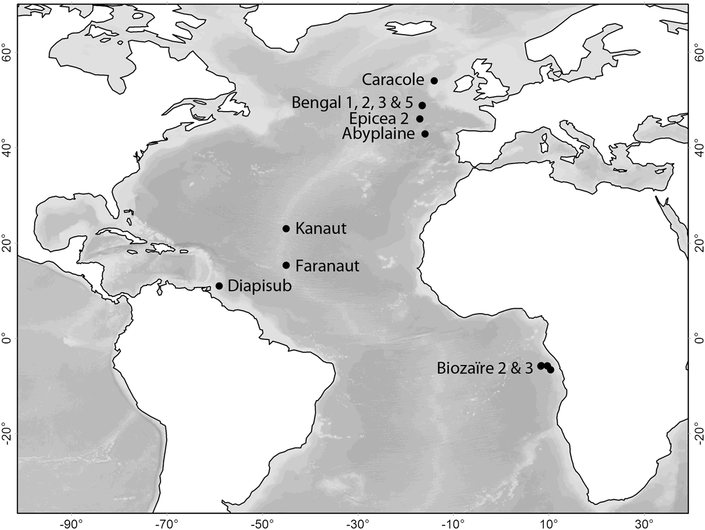
The material was collected during the Ifremer cruises Abyplaine (Cryos, 1981), Epicea 2 (‘Le Suroît’ and ‘Jean Charcot’, 1988), Faranaut (‘L'Atalante’, 1992), Kanaut (‘L'Atalante’, 1992), Diapisub (‘Le Nadir’, 1992–1993), Caracole (‘L'Atalante’, 2001), Biozaïre 2 and 3 (L'Atalante', 2001 and 2003–2004) and during the EC MAST III programme cruises Bengal 1, 2, 3 and 5 (RSS ‘Discovery’, 1996–1998) (Fourt et al., Reference Fourt, Vacelet and Boury-Esnault2006) (Figure 1) (see also http://www.ifremer.fr/sismer/UK/catal/campagne/campagne_UK.htm). In all, 36 specimens representing 15 species were examined from the genera Abyssocladia, Asbestopluma, Cladorhiza and Chondrocladia. Specimens were collected using different means, including beam trawl, box corer, remotely operated vehicle (ROV) and the manned submersible ‘Nautile’. Specimens were fixated in formalin and preserved in 70% ethanol. Types are deposited at the Muséum National d'Histoire Naturelle (MNHN) in Paris.
Map of cruise locations for the Ifremer samples. Collection depths: Caracole (670–1010 m); Bengal 1, 2, 3, and 5 (4842–4846 m); Epicea 2 (~4700 m); Abyplaine (4330 m); Kanaut (~4000 m); Faranaut (2600–3578 m); Diapisub (1947 m); Biozaïre 2 and 3 (2627–3964 m).

Many of the samples are fragmentary or tiny single specimens, conditions which in some cases have limited the degree of information available for species descriptions. Some whole specimens had to be used for making spicule preparations, and sometimes the number of spicules measured is less than ideal as they represent all the unbroken spicules of a certain type in that specimen.
Spicule preparations were made by dissolving sponge tissue in nitric acid using the standard method (Boury-Esnault & Rützler, Reference Boury-Esnault and Rützler1997). In many cases, small specimens or specimen parts were mounted directly on slides. Histological sections were made by embedding the specimen in resin before sectioning. Pictures were taken of the complete specimen, details of interest, skeletal structure and individual spicules using standard light microscopy and/or SEM. In some cases given the minute size of the specimens or fragments, SEM pictures of certain spicule types could not be obtained and are represented by optical slide pictures. Minimum, maximum and average measurements, standard deviations and number measured are given for each spicule type.
SPICULE NOMENCLATURE
We have followed Lopes et al. (Reference Lopes, Bravo and Hajdu2011) in dividing isochelae with alae and central teeth touching or nearly touching into cleistochelae and abyssochelae: Cleistochelae are defined as isochelae with central teeth nearly in contact or touching (Boury-Esnault & Rützler, Reference Boury-Esnault and Rützler1997), whereas abyssochelae in addition are defined as ‘stout isochelae’ with a height × width ratio which ought to be small, in the vicinity of 1.0, and in which both alae and teeth are nearly in contact or touching (van Soest & Hajdu, Reference van Soest, Hajdu, Hooper and van Soest2002).
SYSTEMATICS
SYSTEMATIC INDEX
Class DEMOSPONGIAE Sollas, 1885
Order POECILOSCLERIDA Topsent, 1928
Suborder MYCALINA Hajdu, van Soest & Hooper, 1994
Family CLADORHIZIDAE Dendy, 1922
Genus Abyssocladia Lévi, Reference Lévi, Spärck, Greve and Madsen1964
Abyssocladia faranauti sp. nov.
Abyssocladia tecta sp. nov.
Genus Asbestopluma Topsent, Reference Topsent1901b
Subgenus Asbestopluma Topsent, Reference Topsent1901b
Asbestopluma (A.) belgicae (Topsent, Reference Topsent1901a)
Asbestopluma (A.) infundibulum (Levinsen, Reference Levinsen and Lütken1887)
Asbestopluma (A.) lycopodium (Levinsen, Reference Levinsen and Lütken1887)
Asbestopluma (A.) parvula sp. nov.
Asbestopluma (A.) pennatula (Schmidt, Reference Schmidt1875)
Asbestopluma (A.) rastrichela sp. nov.
Asbestopluma (A.) sp.
Genus Chondrocladia Thomson, Reference Thomson1873
Subgenus Chondrocladia Thomson, Reference Thomson1873
Chondrocladia (C.) vaceleti Cristobo, Urgorri & Rios, Reference Cristobo, Urgorri and Rios2005
Genus Cladorhiza Sars, Reference Sars1872
Cladorhiza abyssicola Sars, Reference Sars1872
Cladorhiza acanthoxea sp. nov.
Cladorhiza aff. gelida Lundbeck, Reference Lundbeck1905
Cladorhiza methanophila Vacelet & Boury-Esnault, Reference Vacelet and Boury-Esnault2002
Cladorhiza thomsoni Topsent, Reference Topsent1909
LIST OF SPECIES
Family CLADORHIZIDAE Dendy, 1922
DIAGNOSIS
Sponges, usually small, symmetrical, mostly in deep water, with diagonal, radiating supporting processes and basal root adaptations for living in soft sediments. Axial skeleton composed of monactinal or diactinal megascleres, from which radiating extra-axial tracts diverge to the ectosome. Microscleres include (an)isochelae, sigmas, forceps or micro(subtylo)styles (microspined, spear-shaped in a few cases). Considerable reduction to complete loss of the choanocyte layer is known for some species, being associated with an adaptation to carnivory, preying on relatively large food items (Hajdu & Vacelet, Reference Hajdu, Vacelet, Hooper and van Soest2002).
Genus Abyssocladia Lévi, Reference Lévi, Spärck, Greve and Madsen1964
TYPE SPECIES
Abyssocladia bruuni Lévi, Reference Lévi, Spärck, Greve and Madsen1964 (by monotypy).
DIAGNOSIS
Cladorhizidae most often pedunculate, carrying a disciform or flabelliform body with a radial architecture. Microscleres are a combination of abyssochelae, cleistochelae, arcuate chelae, and/or sigmancistras (Lopes et al., Reference Lopes, Bravo and Hajdu2011).
Abyssocladia faranauti sp. nov.
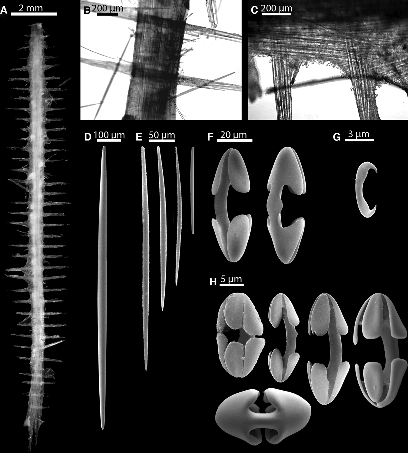
(Figure 2A–H)
TYPE MATERIAL
Holotype: MNHN DJV157—Faranaut FR 13 no.1 (1992–03, Dive 13, 15°32′N 046°33′W, 3578 m).
The holotype was recovered during the Faranaut cruise at the intersection of the Mid-Atlantic Ridge with the fracture zone at 15°20′N using the ‘Nautile’ submersible.
ETYMOLOGY
From Faranaut, the cruise where the holotype was collected.
DIAGNOSIS
Elongate, pinnate sponge, bearing thin, regularly spaced lateral filaments. Megascleres are four size-classes of mycalostyles. Microscleres are arcuate isochelae, smaller arcuate isochelae/cleistochelae and sigmancistras.
DESCRIPTION
The holotype consists of four incomplete pieces of a straight, flattened spicule axis in all about 50 mm long and about 1 mm in diameter. Presumably attached to the substrate with a basal plate, but this part is lacking. The axis bears numerous thin, short filaments, about 1 mm long and 100 µm in diameter. The filaments are regularly situated perpendicular to the axis in two symmetrically arranged lateral rows, about 0.4 mm apart. No aquiferous system was observed. The colour is yellowish-white in alcohol (Figure 2A).
Abyssocladia faranauti sp. nov.: (A) largest fragment of the holotype; (B, C) detail from axis and lateral filaments; (D, E) mycalostyles; (F) larger type of arcuate isochela; (G) sigmancistra; (H) smaller type of arcuate isochela/cleistochela.
SKELETON
The central axis consists of a dense core of large, longitudinally arranged mycalostyles, most of them straight, but some of the shorter ones slightly curved. The filaments are composed of smaller mycalostyles anchored symmetrically in and crossing the main axis. Arcuate isochelae and cleistochelae are arranged teeth outwards covering the surface of the axis, densest at the junction where the filaments protrude from the axis. While sigmancistras are present, the lateral filaments lack the covering of isochelae and cleistochelae, though this might in part be because of damage to the specimen (Figure 2B, C).
SPICULE TYPES
1. Mycalostyles. In the main axis. Straight and fusiform. 1020–1640 (1364 ± 166) µm long, 31.4–55.0 (43.8 ± 7.4) µm wide (N = 30) (Figure 2D).
2. Mycalostyles. In the main axis and lateral filaments. Straight and fusiform. 530–940 (689 ± 102) µm long, 14.1–29.8 (20.7 ± 3.4) µm wide (N = 30) (Figure 2E).
3. Mycalostyles. Straight or slightly curved. 280–560 (395 ± 85) µm long and 7.9–17.3 (13.4 ± 2.4) µm wide (N = 30) (Figure 2E).
4. Mycalostyles. Straight or slightly curved. 130–270 (193 ± 37) µm long and 3.1–7.9 (5.7 ± 1.4) µm wide (N = 30) (Figure 2E).
5. Arcuate isochelae. Covering main axis and base of filaments. The middle part of the shaft may be laterally expanded to various degrees. 48.7–67.5 (61.2 ± 4.6) µm long (N = 30) (Figure 2F).
6. Arcuate isochelae to cleistochelae. Covering main axis and base of filaments. 22.0–33.0 (25.6 ± 2.4) µm long (N = 38). In some cases, the smaller sizes (22.0–23.6) may partly or wholly approach cleistochelae. The arcuate chelae approaching cleistochelae are not very numerous, and given the presence of intermediate forms, are most likely variations of the smaller type of arcuate isochelae (Figure 2H).
7. Sigmancistras. Covering surface of filaments. In a single plane, shaft enlarged on both sides. 6.3–7.9 (6.4 ± 0.4) µm long (N = 30) (Figure 2G).
REMARKS
The chelae of this species include arcuate isochelae with some forms approaching cleistochelae (Figure 2F, H) but no abyssochelae. As shown by Lopes et al. (Reference Lopes, Bravo and Hajdu2011) however, several of the already described species placed into the genus do not actually have isochelae with touching or nearly touching frontal teeth, and on this basis the diagnosis of the genus was amended. Abyssocladia faranauti sp. nov. clearly fulfils the criteria for inclusion as an Abyssocladia according to the new genus diagnosis. Basal substrongyles are common in the genus, but the base was not recovered for A. faranauti sp. nov. and no substrongyles were found. This species has a pinnate morphology which is known in several species of Abyssocladia: A. naudur Vacelet, 2006 and A. myojinensis Ise & Vacelet, Reference Ise and Vacelet2010; partly in A. diegoramirezensis Lopes et al., Reference Lopes, Bravo and Hajdu2011 and A. umbellata Lopes et al., Reference Lopes, Bravo and Hajdu2011. In addition to A. faranauti sp. nov., three known Abyssocladia-species, A. myojinensis, A. umbellata and A. tecta sp. nov., have two types of chelae and one type of sigma/sigmancistra. Abyssocladia myojinensis spicules sizes are the most similar to A. faranauti sp. nov., but are larger cleistochelae and smaller abyssochelae rather than larger arcuate isochelae and smaller arcuate isochelae/cleistochelae as in A. faranauti sp. nov.
Abyssocladia tecta sp. nov.
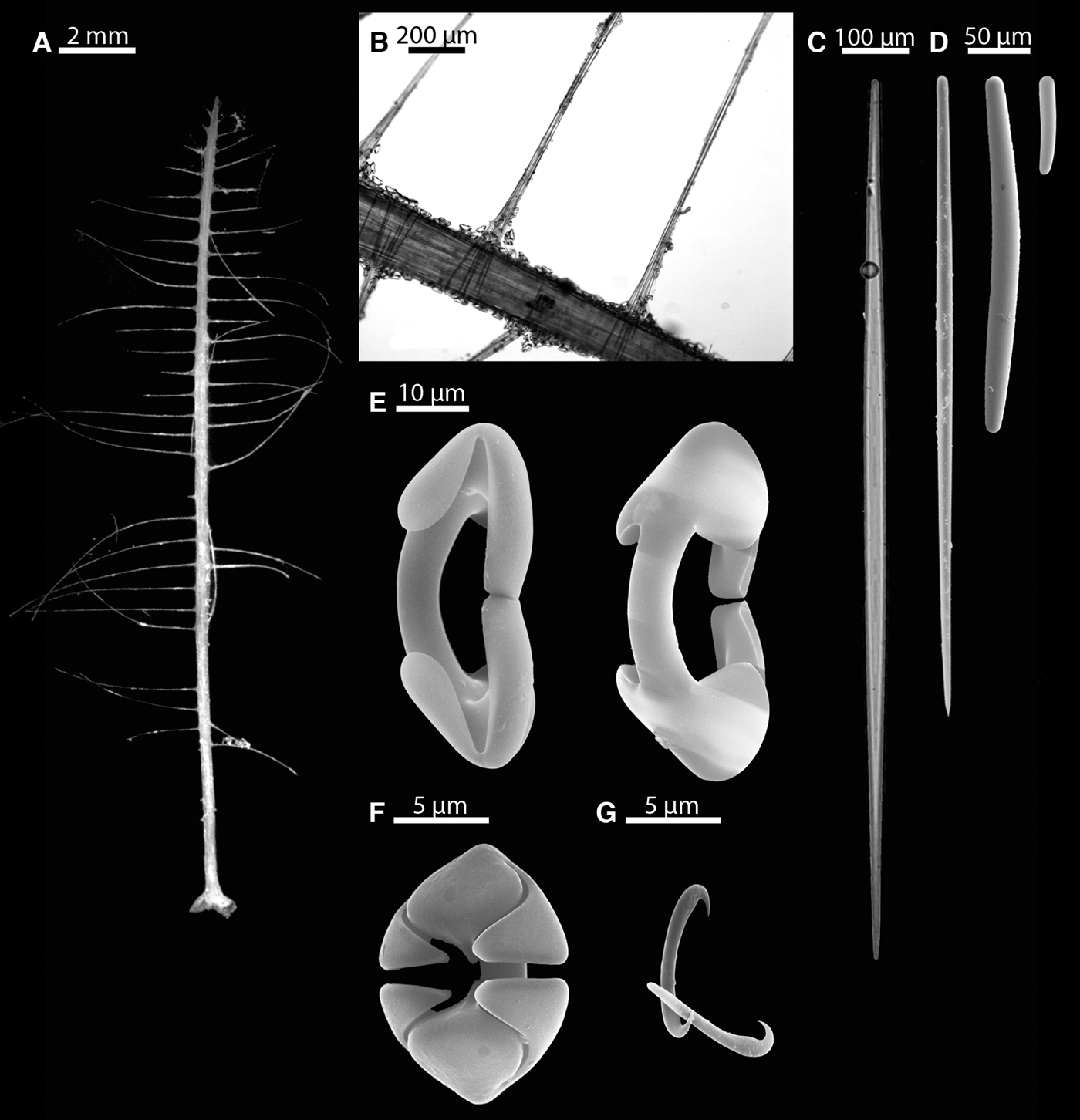
(Figure 3A–G)
TYPE MATERIAL
Holotype: MNHN DJV158—Faranaut FR 13 no.3bisa (1992–03, Dive 13, 15°32′N046°33′W, 3578 m).
The holotype was recovered during the Faranaut cruise, at the intersection of the Mid-Atlantic Ridge with the fracture zone at 15°20′N using the ‘Nautile’ submersible.
ETYMOLOGY
From tecta Latin, adj. fem. form, meaning covered, hidden or guarded, referring to the shape of the abyssochelae with the alae partially covering the frontal teeth.
DIAGNOSIS
Elongate, pinnate sponge attached by basal plate, bearing thin lateral filaments regularly spaced. Megascleres are two size classes of mycalostyles and two size-classes of substrongyles. Microscleres are cleistochelae, abyssochelae and sigmancistras.
DESCRIPTION
The holotype is a small, erect, pinnate sponge, consisting of a thin, straight, flattened axis, 30 mm long and 0.2 mm in diameter, attached by an enlarged base 1 mm in diameter. The axis bears numerous thin filaments, up to 5 mm long and 30 µm in diameter, shorter at the top of the sponge, and sometimes broken. The filaments are regularly arranged perpendicularly to the axis in two lateral rows alternating on each side, with a spacing of approximately 0.6 mm. The basal part of the axis is devoid of filaments on the first 4 mm. The colour is white in ethanol (Figure 3A).
Abyssocladia tecta sp. nov.: (A) the holotype; (B) detail from axis and lateral filaments; (C) large mycalostyle; (D) small mycalostyle and substrongyles; (E) cleistochelae; (F) abyssochela; (G) sigmancistras.
SKELETON
The skeleton of the attachment base consists of two different types of substrongyle (45–180 and 220–430 µm long). The main axis is composed of a dense core of large, longitudinally arranged mycalostyles. The axis of the filaments consists of large and small mycalostyles, point outwardly directed, fanning out in their anchoring point which entirely crosses the main axis. The chelae form a dense lining along the main axis. The microsclere lining of the filaments is less dense, possibly due to preservation problem, with chelae mostly found near their base, and sigmancistras more numerous along the filaments (Figure 3B).
SPICULE TYPES
1. Mycalostyles. In the main axis. Straight and fusiform. 1040–1490 (1241 ± 114) µm long and 20.4–34.5 (27.8 ± 3.7) µm wide (N = 30) (Figure 3C).
2. Mycalostyles. In the main axis and lateral filaments. Straight and fusiform. 370–860 (617 ± 178) µm long and 6.3–17.3 (11.5 ± 3.2) µm wide (N = 30) (Figure 3D).
3. Substrongyles. In the attachment base. Straight or feebly curved, with the two ends slightly different. 220–430 (294 ± 63) µm long and 15.7–31.4 (25.2 ± 4.9) µm wide (N = 15) (Figure 3D).
4. Substrongyles. In the attachment base. Straight or feebly curved, with the two ends slightly different. 45–180 (84 ± 34) µm long and 7.9–20.4 (12.6 ± 3.3) µm wide (N = 30) (Figure 3D).
5. Cleistochelae. Lining main axis and base of filaments. Shaft curved, frontal teeth touching or nearly in contact. 42.4–55.0 (47.4 ± 3.8) µm long (N = 30) (Figure 3E).
6. Abyssochelae. Lining main axis and base of filaments. Nearly spherical, frontal teeth strongly recurved toward the shaft and tapering toward the point, alae also strongly recurved and partially covering the frontal teeth. 11.0–14.1 (12.1 ± 1.0) µm long (N = 30) (Figure 3F).
7. Sigmancistras. Lining surface of filaments. Contort, with an acerate point strongly curved. 6.3–7.9 (6.6 ± 0.7) µm long (N = 30) (Figure 3G).
REMARKS
Like Abyssocladia faranauti sp. nov., this species has a pinnate morphology. Based on morphology and spicule complement, the likely closest relatives are the species A. naudur, A. myojinensis, A. diegoramirezensis and A. umbellata as well as A. faranauti sp. nov. Like A. myojinensis, A. umbellata and A. faranauti sp. nov., A. tecta sp. nov. has two types of chelae and one type of sigma/sigmancistra, and while there are differences in chela shape, the microsclere sizes, while smaller, most closely resemble those of A. myojinensis and A. faranauti sp. nov. The species is well characterized by its spicule complement, especially by the shape of the abyssochelae with recurved palms.
Genus Asbestopluma Topsent, Reference Topsent1901b
TYPE SPECIES
Cladorhiza pennatula Schmidt, Reference Schmidt1875 (by subsequent designation).
DIAGNOSIS
Cladorhizidae with palmate and/or arcuate (an)isochelae, and, in some cases, the latter in combination with tridentate anchorate anisochelae (Lopes & Hajdu, in press).
Subgenus Asbestopluma Topsent, Reference Topsent1901b
TYPE SPECIES
Cladorhiza pennatula Schmidt, Reference Schmidt1875 (by subsequent designation).
DIAGNOSIS
Asbestopluma without spear-shaped microtylostyles (Lopes et al., Reference Lopes, Bravo and Hajdu2011).
Asbestopluma (Asbestopluma) belgicae (Topsent, 1901)
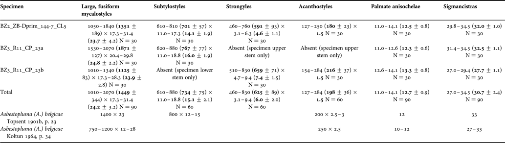
(Table 1; Figure 4A–J)
ORIGINAL DESCRIPTION
Cladorhiza (Asbestopluma) belgicae Topsent, Reference Topsent1901a: VIII.
EXAMINED MATERIAL
BZ2_ZB-Dprim_144-7_CL5 (2001-11-25, Dive 144-7, 05°43.83′S 008°27.19′E, 3898 m; Victor 6000 ROV), BZ3_R11_CP_23a and BZ3_R11_CP_23b (2004-01-03, Station CP23, loc. R11, 05°45.92′S009°43.99′E, 3147 m; beam trawl).
The specimens were recovered from outside the western coast of Africa at Gabon–Congo during the Biozaïre 2 and Biozaïre 3 cruises.
DESCRIPTION
One complete and two incomplete specimens of a pinnate sponge made of a single main axis carrying filaments or spicule tufts. The axis is thickest by the base, where the sponge is secured to the substrate with rhizoids. The lower part of the axis is light brown, solid and smooth. The upper part of the axis is white or yellowish and appears grooved. Lateral spicule tufts or filaments (up to 10 mm long) protrude from the main axis in series along lines that follow the grooves. The filaments are thin and very numerous, but may be reduced or otherwise appear as small tufts. The complete specimen recovered is 35 cm tall (Figure 4A, B).
Asbestopluma (A.) belgicae (Topsent, 1901): (A) complete specimen (BZ2 144-7 CL5); (B) detail of axis showing grooves and filament tufts from specimen BZ3_R11_CP_23a; (C) cross-section of axis showing spicule bundles and filaments; (D) mycalostyle; (E) subtylostyle, strongyle and acanthostyle; (F) detail from mycalostyle; (G) detail from subtylostyle; (H) detail from acanthostyle; (I) palmate anisochelae; (J) sigmancistra.
SKELETON
The central axis consists of bundles of large mycalostyles arranged in a ring around the middle, giving the axis a grooved structure (Figure 4C). Filaments or tufts project from the grooves at regular intervals perpendicular to the axis, and consist of bundles of subtylostyles tip outwards, and covered by sigmancistras and palmate anisochelae. The rhizoids consist of mycalostyles, flexuous strongyles and smaller acanthostyles (Figure 4D–G).
SPICULE TYPES
1. Mycalostyles. In the main axis. Blunt, straight and fusiform. 1010–2070 (1449 ± 344) µm long and 17.3–31.4 (24.2 ± 3.2) µm wide (N = 90). Generally shorter in the lower part of the axis and base (1010–1340 µm long) (Table 1; Figure 4D, F).
2. Subtylostyles. In the upper main axis and lateral filaments. Straight and fusiform. 610–880 (734 ± 75) µm long and 11.0–18.8 (15.1 ± 2.1) µm wide (N = 60) (Table 1; Figure 4E, G).
3. Strongyles. In the rhizoids only. Thin and non-fusiform, straight or curved. 460–830 (659 ± 71) µm long and 3.1–9.4 (6.0 ± 2.0) µm wide (N = 60) (Table 1; Figure 4E).
4. Acanthostyles. In the basal part of the axis and the rhizoids, with numerous small recurved spines. 127–250 (198 ± 36) µm long and ~1.5 µm wide (N = 60) (Table 1; Figure 4E, H).
5. Palmate anisochelae. Covering the filaments. Shaft curved. With upper lateral alae large (5/6 of total length), curving and completely fused to the shaft. Small lower alae and a central lower tooth ending in a point. 11.0–14.1 (12.7 ± 0.9) µm long (N = 90) (Table 1; Figure 4I).
6. Sigmancistras. Covering surface of axis and filaments. Mostly contort. 27.0–34.5 (30.7 ± 2.4) µm long (N = 90) (Table 1; Figure 4J).
Asbestopluma (A.) belgicae Topsent, 1901. Spicule dimensions (µm). Minimum, maximum and average dimensions are given, as well as the number of spicules measured (N).
PREVIOUSLY REPORTED DISTRIBUTION
The Antarctic. Previously from the Davis Sea, Antarctica (71°19′S 087°37′E, 450 m; 70°15′S 084°06′E, 569 m) (Topsent, Reference Topsent1901b; Koltun, Reference Koltun, Pavlovskii, Andriyashev and Ushakov1964b) and the South Shetland Islands (Koltun, Reference Koltun, Pavlovskii, Andriyashev and Ushakov1964b). Depth range 370–569 m (Koltun, Reference Koltun, Pavlovskii, Andriyashev and Ushakov1964b).
REMARKS
Asbestopluma (A.) belgicae has previously been reported from shallower (370–569 m) areas in the Antarctic, while the specimens described here were collected from the deep sea (3147 and 3898 m) close to the equator. While admittedly the geographical and depth range is quite different, the morphology, spicule complement and spicule sizes are all virtually identical to previous descriptions, with the exception that strongyles have not previously been reported for the species. Strongyles are found only in the rhizoids however, and Topsent states that the basal parts are missing from the specimens he examined, while Koltun does not mention any basal parts at all (Topsent, Reference Topsent1901b; Koltun, Reference Koltun, Pavlovskii, Andriyashev and Ushakov1964b).
The morphology and spicule complement is similar to A. (A.) pennatula and related species, as opposed to the forceps-bearing Asbestoplumas. It differs from most related species by lacking a second type of larger palmate anisochela. The size of the sigmas is slightly larger than other described species. The greatest similarity is to the species A. (A.) quadriserialis Tendal, Reference Tendal1973, A. (A.) biserialis (Ridley & Dendy, Reference Ridley and Dendy1886) and A. (A.) pennatula var. bihamatifera (Carter, Reference Carter1876).
Asbestopluma (Asbestopluma) infundibulum (Levinsen, Reference Levinsen and Lütken1887)
(Figure 5A–D)
ORIGINAL DESCRIPTION
Esperella infundibulum Levinsen, Reference Levinsen and Lütken1887: 366, pls XXIX, XXXI.
EXAMINED MATERIAL
DIA_DS 10a and DIA_DS 10b (1993-01-03, Station 10, 11°N059°W, 1947 m).
The specimens were recovered from the Barbados Accretionary Prism using a Sanders dredge during the Diapisub cruise.
COMPARATIVE MATERIAL EXAMINED
Asbestopluma (A.) infundibulum: Boury-Esnault, 1994: Balgim CP10-180 (SEM); unpublished specimens from the Norwegian shelf (SEM).
DESCRIPTION
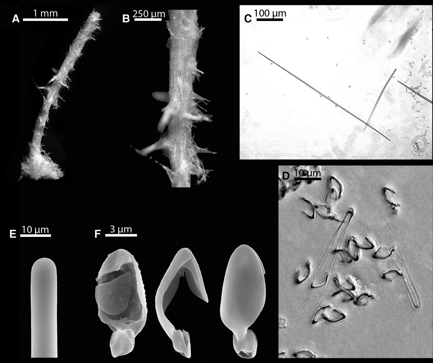
The specimens are two small sponge fragments from the same station. Asbestopluma (A.) infundibulum is described as a stipitate sponge with a cup-shaped body in earlier literature (e.g. Lundbeck, Reference Lundbeck1905). The first fragment (a) is a fluffy, white and amorphous piece of the sponge body. The second fragment (b) is pale, long, thread-like, 3 mm long and 0.5 mm wide, probably containing parts of both the peduncle and body (Figure 5A).
Asbestopluma (A.) infundibulum (Levinsen, Reference Levinsen and Lütken1887): (A) specimen fragments; (B) palmate/arcuate anisochelae; (C) large subtylostyle, curving, slender fine-pointed style, small subtylostyles; (D) large subtylostyle detail.
SKELETON
Given the fragmentary nature of the specimens, their skeletal structure is not possible to describe accurately.
SPICULE TYPES
1. Subtylostyles. Slightly fusiform, straight or slightly curved. 280–570 (397 ± 76) µm long and 4.7–7.9 (6.2 ± 1.1) µm wide (N = 30) (Figure 5C, D).
2. Subtylostyles. Slightly fusiform, straight or slightly curved. 132–274 (180 ± 36) µm long and 3.1–6.3 (5.1 ± 1.1) µm wide (N = 30) (Figure 5C).
3. Styles. Curving and slender with a long, very fine point. 450–910 (695 ± 124) µm long and 3.1–6.3 (4.8 ± 1.0) µm wide (N = 30) (Figure 5C).
4. Palmate/arcuate anisochelae. Shaft curved. Upper lateral alae and frontal tooth about half the length of the spicule, lateral alae fused to the shaft about 2/3 of total ala length. Foot large, about a third of total spicule length, with frontal tooth tapering into a point, and plate-like, denticulate lateral alae. 15.7–20.4 (18.6 ± 1.2) µm long (N = 30) (Figure 5B). A single anisochela was 25.1 µm long.
There is some overlap between the two different straight forms of subtylostyle, but in general the majority of the measured spicules fall into one or the other category. The tyles at the head of the subtylostyles are in some cases easy to recognize, while in other cases only very slightly visible (Figure 5D). The palmate/arcuate anisochelae are consistent with the abandoned Cotylina subgenus (Lundbeck, Reference Lundbeck1905) (see also remarks for A. (A.) parvula below).
No forceps were found, but earlier descriptions refer to them as rare (Lundbeck, Reference Lundbeck1905; Hentschel Reference Hentschel, Römer and Schaudinn1929), and presence of forceps have been reported as associated with spermatic cysts in related species and is therefore not considered a reliable diagnostic character (Vacelet & Boury-Esnault, Reference Vacelet and Boury-Esnault1996; Riesgo et al., Reference Riesgo, Taylor and Leys2007). Likewise no embryo-related megascleres, present in some earlier reports, were found (Lundbeck, Reference Lundbeck1905; Boury-Esnault et al., Reference Boury-Esnault, Pansini and Uriz1994).
PREVIOUSLY REPORTED DISTRIBUTION
The Arctic and northern and Mid-Atlantic. Previously known from the northern Atlantic (Hansen, Reference Hansen1885; Lundbeck, Reference Lundbeck1905), the Barents Sea (Hentschel, Reference Hentschel, Römer and Schaudinn1929) and the Kara Sea (Levinsen, Reference Levinsen and Lütken1887; Koltun, Reference Koltun1959), 91–734 m. From the Ibero-Moroccan Gulf, 1590 m (Boury-Esnault et al., Reference Boury-Esnault, Pansini and Uriz1994). Asbestopluma (A.) infundibulum orientalis: north-western Pacific (Koltun, Reference Koltun and Bogorov1972).
REMARKS
Most records of this species are from shallower waters of the northern Atlantic and Arctic, but it is also recorded from 1590 m in the Ibero-Moroccan Gulf (Boury-Esnault et al., Reference Boury-Esnault, Pansini and Uriz1994). Despite the lack of morphological information, the combination of curving, fine-pointed styles, known only from A. (A.) infundibulum and A. (A.) comata Lundbeck, Reference Lundbeck1905 among closely related species, Cotylina-like anisochelae and general spicule sizes, correspond well to Lundbeck's description. From A. (A.) comata, it can be distinguished most easily by the lack of tyles along the length of the fine-pointed spicules.
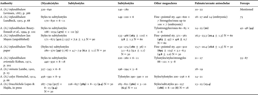
Table 2 lists previous descriptions of A. (A.) infundibulum as well as closely related species sharing the same type of palmate/arcuate anisochela, a peduncle and a cup-shaped body. Forceps and smaller curved strongyles and oxea in A. (A.) infundibulum are, as reported by Lundbeck (Reference Lundbeck1905), associated with embryonic structures, which would explain their absence in some of the descriptions. Slender, fine-pointed styles are found only in the upper body of A. (A.) infundibulum, which was not recovered in the case of Boury-Esnault et al., Reference Boury-Esnault, Pansini and Uriz1994. The species A. (A.) minuta Lambe, Reference Lambe1900 (north-west Atlantic) and A. (A.) calyx Hentschel, Reference Hentschel and von Drygalski1914 (Antarctic) are very similar to each other, though geographically separated. The species A. (A.) bracchichela Lopes & Hajdu, in press is closely related as well, and is described partly (and named) on the basis of the serrations of the lower lateral alae, which however are present in A. (A.) infundibulum (and possibly other species) as well (Figure 5 and comparative materials examined). Given the close relationship among the species listed and the geographical range in which they appear, their precise relationship should be regarded as uncertain.
Previous reports of Asbestopluma (A.) infundibulum (Levinsen, Reference Levinsen and Lütken1887) as well as A. (A.) infundibulum orientalis Koltun, 1970, A. (A.) minuta Lambe, Reference Lambe1900, A. (A.) calyx Hentschel, Reference Hentschel and von Drygalski1914 and A. (A.) bracchichela Lopes & Hajdu, in press. Spicule dimensions (µm). When available, minimum, maximum and average dimensions are given, as well as the number of spicules measured (N).
Asbestopluma (Asbestopluma) lycopodium (Levinsen, Reference Levinsen and Lütken1887)
(Table 3; Figure 6A–F)
ORIGINAL DESCRIPTION
Esperella cupressiformis var. lycopodium Levinsen, Reference Levinsen and Lütken1887: 365, pls XXIX, XXXI.
EXAMINED MATERIAL
Caracole K3 (2001-08-01, Station T, 51°25.00′N, 011°45.00′W, 1010 m) and R132d (2001-08-13, Station R2, 55°32.00′N015°40.00′W, 884 m).
The specimens were recovered from the Porcupine and Rockall Banks using box corer during the Caracole cruise.
COMPARATIVE MATERIAL EXAMINED
Asbestopluma (A.) lycopodium: unpublished specimens from Jan Mayen (SEM).
DESCRIPTION
Two small, incomplete specimens of an erect sponge made of a single main axis carrying radiating processes. The specimens are 5 and 30 mm long and 0.3 and 1 mm in diameter. In the larger specimen, the axis is divided into a light brown basal part and a white upper part, while the smaller specimen is all white. Tiny filaments or filament relicts radiate in all directions from the central axis, except for the light-brown, lower part of the larger specimen. The smaller specimen has the remains of a basal plate with which the sponge is attached to the substrate (Figure 6A).
Asbestopluma (A.) lycopodium (Levinsen, Reference Levinsen and Lütken1887): (A) habitus of smaller specimen; (B) detail; (C) large mycalostyle and smaller subtylostyle; (D) forceps; (E) mycalostyle detail; (F) palmate anisochelae.
SKELETON
The axis and filaments are composed of bundles of the larger mycalostyles, while the smaller mycalostyles/subtylostyles have a more diffuse placement within the main supporting skeleton. Microscleres line the surface of both axis and filaments (Figure 6B).
SPICULE TYPES
1. Mycalostyles. In the main axis and filaments. Slender, slightly fusiform, straight or slightly curved. 500–1090 (761 ± 158) µm long and 4.7–12.6 (8.7 ± 2.1) µm wide (N = 60) (Table 3; Figure 6C, E).
2. Mycalostyles/subtylostyles. In the main axis and filaments. Slender, slightly fusiform, straight or slightly curved. 200–427 (292 ± 55) µm long and 4.7–9.4 (7.2 ± 1.1) µm wide (N = 60) (Table 3; Figure 6C).
3. Palmate anisochelae. Covering the filaments. Shaft curved. With upper lateral alae large (4/5 of total length) curving and completely fused to the shaft and a large (1/2 of total length) upper frontal tooth. Small, denticulate lower lateral alae and small, plate-like lower frontal tooth. 7.9–11.0 (9.3 ± 0.9) µm long (N = 60). (Table 3; Figure 6F).
4. Forceps. A small circular swelling at both ends. The ends are parallel and close together near the middle, then curve somewhat outwards closer to the ends of the spicule. 36.1–44.0 (39.6 ± 2.1) µm long (N = 30). (Table 3; Figure 6D).
Asbestopluma (A.) lycopodium (Levinsen, Reference Levinsen and Lütken1887). Spicule dimensions (µm). Minimum, maximum and average dimensions are given, as well as the number of spicules measured (N). Some selected earlier descriptions are also given (Levinsen, Reference Levinsen and Lütken1887; Lundbeck, Reference Lundbeck1905; van Soest & Baker, Reference van Soest and Baker2011).
PREVIOUSLY REPORTED DISTRIBUTION
The north-eastern Atlantic and Arctic: Kara Sea (Levinsen, Reference Levinsen and Lütken1887; Koltun, Reference Koltun1959), the east coast of Greenland (Burton, Reference Burton1934; Koltun, Reference Koltun1964a), Norwegian Sea (Hansen, Reference Hansen1885), the Faroe Islands (Lundbeck, Reference Lundbeck1905), the Rockall Bank (van Soest et al., Reference van Soest, Cleary, de Kluijver, Lavaleye, Maier and van Duyl2007).
REMARKS
The spicule sizes are generally smaller than those given in earlier A. (A.) lycopodium descriptions (e.g. Levinsen, Reference Levinsen and Lütken1887; Lundbeck, Reference Lundbeck1905), but correspond very well to the sizes given for a specimen previously found at the Rockall Bank (van Soest et al., Reference van Soest, Cleary, de Kluijver, Lavaleye, Maier and van Duyl2007; van Soest & Baker, Reference van Soest and Baker2011).
Asbestopluma (Asbestopluma) parvula sp. nov.
(Table 4; Figure 7A–E)
TYPE MATERIAL
Holotype: MNHN DJV159—Bengal 5 Be5prél.39 (1998-03-15, Station KGS 39, 48°50′N016°30′W, 4845 m).
Paratypes: MNHN DJV160—Bengal 2 Be2prél.59; MNHN DJV161—Bengal 2 Be2prél.63; MNHN DJV162—Bengal 3 Be3prél.47; MNHN DJV163—Bengal 5 Be5prél.25; MNHN DJV164—Biozaïre 3 BZ3_R7_KGS_76_2; MNHN DJV165 – Biozaïre 3 BZ3_R7_KGS_76_a; MNHN DJV166—Biozaïre BZ3_R7_KGS_76_b; MNHN DJV167—Biozaïre 3 BZ3_R7_KGS_76_c; MNHN DJV168—Biozaïre 3 BZ3_ZB-D_KGS_51. The paratypes are in the form of spicule preparations only.
The specimens were recovered using box corer from the Porcupine Abyssal Plain, adjacent to the Irish continental margin (Bengal 2, 3 and 5 samples; 48°49′–48°50′N, 016°29′–016°30′W, 4842–4845 m), and from outside the western coast of Africa at Gabon–Congo (Biozaïre 3 samples; 05°48′–05°51′S009°43′–008°2′E, 3160–3964 m).
COMPARATIVE MATERIAL EXAMINED
Asbestopluma (A.) hydra: Syntype, Ingolf Expedition Station 113 (69°31′N007°06′W, 2394 m), SEM.
ETYMOLOGY
From parvula Latin, adj. fem. form, meaning tiny, referring to the very small size of the sponge.
DIAGNOSIS
Small, stipitate sponge composed of a basal peduncle and a spherical body with a skeleton of radiating spicules, and numerous short, protruding processes. Megascleres are two types of subtylostyle. Microscleres are palmate anisochelae.
DESCRIPTION
A white, small (specimens range from under 1 to 3–4 mm including the peduncle) sponge consisting of a spherical or oblong body (0.5–1.5 mm ) with radiating spicules, connected to the substrate with a peduncle (Figure 7A, C). The attachment base is missing in all specimens. There is no discernible pattern in variation between specimens from the two collection areas.
Asbestopluma (A.) parvula sp. nov.: (A, C) whole specimens and fragments; (B) subtylostyles; (D) palmate anisochela detail; (E) palmate anisochelae including growth form of small anisochela.
SKELETON
The peduncle consists of bundles of slender subtylostyles (typically 700–900 µm) arranged in parallel. In the body shorter subtylostyles (typically 300–400 µm) are arranged radially. While longer, the peduncle subtylostyles are equal in thickness to the radiating subtylostyles. The palmate anisochelae are found at the surface of the sponge.
SPICULE TYPES
1. Subtylostyles. In the peduncle. Straight and fusiform. 600–1030 (806 ± 106) µm long and 4.7–7.9 (7.3 ± 0.9) µm wide (N = 94) (Table 4; Figure 7B).
2. Subtylostyles. Radiating outwards in the body. Straight and fusiform. 220–580 (358 ± 59) µm long and 4.7–12.6 (7.7 ± 0.5) µm wide (N = 276) (in specimen Be2prél.59 9.4–12.6 µm wide) (Table 4; Figure 7B).
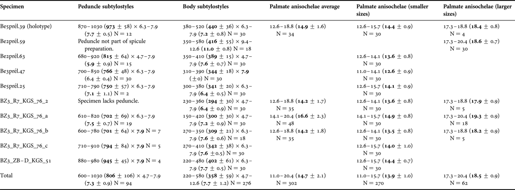
3. Palmate anisochelae. Shaft curved. With upper lateral alae large (3/4 of total length) curving and completely fused to the shaft and a large (3/5 of total length) upper frontal tooth. Smaller, denticulate lower lateral alae and plate-like lower frontal tooth. 11.0–20.4 (14.7 ± 2.1) µm long (N = 302). These may in some cases be divided into two different size-classes, with a common smaller type, 11.0–15.7 (13.9 ± 1.0) µm long (N = 270), and a less common larger type, 17.3–20.4 (18.5 ± 0.9) µm long (N = 62) (Table 4; Figure 7D, E).
Asbestopluma (A.) parvula sp. nov. Spicule dimensions (µm). Minimum, maximum and average dimensions are given, as well as the number of spicules measured (N). Large palmate anisochelae measurements were discontinued after 30 small ones were found, in order to give a more realistic average size.
Both types of subtylostyle are typically straight, but slightly curved forms are also present. There is some intraspecific variation in average subtylostyle length, with some specimens having on average longer subtylostyles than others (Table 4).
In many specimens, their minute size combined with the fact that many megascleres were broken made it impossible to get the preferred number of measurements for the peduncle subtylostyles.
REMARKS
In addition to Asbestopluma s. str., (pinnate, with two or more rows of filaments, containing one or two characteristic type of palmate anisochelae and sigmas), Lundbeck proposed the subgenera Cotylina, (stipitate, with a cup-shaped body and characteristic palmate anisochelae with upper alae and frontal tooth about half the length of the shaft, large foot and curved shaft, no sigmas) and Lycopodina (central stem, with lateral filaments issuing in all directions and characteristic palmate anisochelae with upper alae 2/3 or 3/4 of total spicule length and completely fused with shaft, smaller foot and less curved shaft, no sigmas) (Lundbeck, Reference Lundbeck1905) (compare Figure 5B and 7E in this paper for the two different anisochela types). The Lycopodina and Cotylina subgenera were questioned by Hentschel when describing Asbestopluma. (A.) callithrix Hentschel, Reference Hentschel and von Drygalski1914, given the stipitate form together with anisochelae of the Lycopodina type of that species (Hentschel, Reference Hentschel and von Drygalski1914), and they have fallen into disuse (Vacelet & Boury-Esnault, Reference Vacelet and Boury-Esnault1996; Hajdu & Vacelet, Reference Hajdu, Vacelet, Hooper and van Soest2002).
Like A. (A.) hydra Lundbeck, Reference Lundbeck1905, A. (A.) callithrix and more recently A. (A.) hypogea Vacelet & Boury-Esnault, Reference Vacelet and Boury-Esnault1996 and A. (A.) communis Lopes & Hajdu, in press, A. (A.) parvula sp. nov. is stipitate, with a thin peduncle connected to an oblong or spherical body, together with Lycopodina-type anisochelae. With the exception of A. (A.) hydra, these species have distinct radial skeletal body architectures, with filaments or shorter protrusions issuing in all directions from the centre. On the other hand, species containing Cotylina-type anisochelae typically have a cup-shaped body, with a skeletal architecture with spicules oriented mostly towards the rim of the cup (in the case of A. (A.) flabellum Koltun, 1970 giving the body a fan-like appearance) (Koltun, Reference Koltun and Bogorov1972).
Asbestopluma (A.) parvula sp. nov. seems to be a close relative of A. (A.) hydra, but differs from it by being generally smaller (the largest specimen of A. (A.) parvula sp. nov. examined is nearly the size of the smallest of Lundbeck's A. (A.) hydra specimens however, but this specimen is equally different from A. (A.) hydra in morphology and spiculation than the smaller specimens), by having protrusions issuing in all directions from the body rather than from the top only, in the much thinner peduncle subtylostyles, and in the presence of larger palmate anisochelae than those of A. (A.) hydra. It can be distinguished from A. (A.) hypogea most easily by the larger upper teeth of the palmate anisochelae, and from A. (A.) communis by differences in megasclere types and sizes, and anisochela size. Given that the distinguishing characters of A. (A.) parvula sp. nov. are almost completely uniform across all the specimens examined, it is well characterized as a new species.
Asbestopluma (Asbestopluma) pennatula (Schmidt, Reference Schmidt1875)
(Table 5; Figure 8A–G)
ORIGINAL DESCRIPTION
Cladorhiza pennatula Schmidt, Reference Schmidt1875: 119.
EXAMINED MATERIAL
Caracole K7b, K7c, K7d and K8a (2001-08-06, Station P1, 52°09.00′N012°46.00′W, 670 m, box corer).
The specimens were recovered from Propeller Mound, the Porcupine Bank, during the Caracole cruise.
DESCRIPTION
Each sample consists of an erect, pinnate sponge or sponge fragments, each of which is composed of a thin axis ranging from 22 to 60 mm long and 0.5 to 1 mm in diameter, attached to the substrate with rhizoids in specimens where the basal part was recovered. The axis is clearly divided into a light-brown lower part bearing rhizoids towards the base, and a much shorter (~10–15 mm ) white feather-like top part with numerous, up to 5 mm long very thin filaments. The filaments are regularly arranged perpendicularly to the axis in two alternating lateral rows, with a spacing of approximately 0.5 mm (Figure 8A, B).
Asbestopluma (A.) pennatula (Schmidt, Reference Schmidt1875): (A) habitus of specimen K8a; (B) detail from filaments; (C) subtylostyle; (D, F) acanthotylostyle; (E) palmate anisochelae; (G) sigma.
SKELETON
The axis is composed of a dense core of large, longitudinally arranged subtylostyles. Acanthotylostyles are abundant in the lower part of the axis but rare or absent in the filament-bearing top part. The microscleres are two types of palmate anisochelae and sigmas, and are found covering the top part of the axis and the filaments (Figure 8C–G).
SPICULE TYPES
1. Subtylostyles. In the main axis and filaments. Straight and fusiform. 330–800 (532 ± 79) µm long and 6.3–18.8 (10.8 ± 2.3) µm wide (N = 120) (Table 5; Figure 8C).
2. Acanthotylostyles. In the basal part of the axis and the rhizoids. 49–249 (104 ± 44) µm long and about 2 µm wide (N = 108) (Table 5; Figure 8D, F).
3. Palmate anisochelae. Shaft curved. With upper lateral alae large (4/5 of total length), curving and completely fused to the shaft, small lower alae and a central lower tooth ending in a point. 7.9–11.0 (9.3 ± 0.5) µm long (N = 120) (Table 5; Figure 8E).
4. Palmate anisochelae. Shaft straight. With upper lateral alae and frontal tooth around half of total length, upper lateral alae completely fused to the shaft. Lower lateral alae small. One or two small, lower frontal teeth. 36.1–47.1 (41.4 ± 2.2) µm long (N = 120) (Table 5).
5. Sigmas. 18.8–23.6 (20.5 ± 1.2) µm long (N = 120) (Table 5; Figure 8G).
Asbestopluma (A.) pennatula (Schmidt, Reference Schmidt1875). Spicule dimensions (µm). Minimum, maximum and average dimensions are given, as well as the number of spicules measured (N). With earlier description of holotype (Topsent, Reference Topsent1909).
PREVIOUSLY REPORTED DISTRIBUTION
The North Atlantic and Arctic (Schmidt, Reference Schmidt1875; Carter, Reference Carter1876; Vosmaer, 1882; Hansen, Reference Hansen1885; Fristedt, Reference Fristedt and Nordenskiöld1887; Lambe, Reference Lambe1896; Topsent, Reference Topsent1901b; Arnesen, Reference Arnesen1903; Lundbeck, Reference Lundbeck1905; Topsent, Reference Topsent and Richard1913; Arnesen, Reference Arnesen1932; Koltun, Reference Koltun1959).
Asbestopluma (Asbestopluma) rastrichela sp. nov.
(Table 6; Figure 9A–E)
TYPE MATERIAL
Holotype: MNHN DJV169—Epicea 2 Ep2KG 19b (1988-03, Station 19, 46°N017°W, ~470o m), in the form of spicule preparation.
Paratype: MNHN DJV170—Bengal 2 Be2prél.19 (1997-03-17, Station KGS 19, 48°50′N016°30′W, 4846 m), in the form of spicule preparation.
The specimens were recovered from the Porcupine Abyssal Plain during the Epicea 2 and Bengal 2 cruises.
ETYMOLOGY
From Latin raster, meaning rake, and chela, referring to the peculiar shape of the anisochelae.
DIAGNOSIS
Asbestopluma (Asbestopluma) with megascleres one type of subtylostrongyle and one type of subtylostyle. Microscleres are palmate anisochelae with lower lateral alae fused into a plate with a row of four teeth.
DESCRIPTION
Specimens are several fragments of two sponges, white in colour. Specimens examined include a part of an axis (2.2 mm long) and a couple of amorphous fragments, the largest being almost 5 mm long (Figure 9A).
Asbestopluma (A.) rastrichela sp. nov.: (A) specimen fragments; (B) substrongyle; (C) subtylostyle; (D) subtylostrongyle tylote end; (E) palmate anisochelae.
SKELETON
The axis is made up of bundles of megascleres. The structure of the other fragments is difficult to ascertain.
SPICULE TYPES
1. Subtylostrongyles. Straight and fusiform. 710–990 (890 ± 57) µm long and 11.0–17.3 (13.8 ± 1.6) µm wide (N = 50) (Table 6; Figure 9B, D).
2. Subtylostyles. Straight and fusiform. 340–450 (397 ± 27) µm long and 6.3–9.4 (7.9 ± 1.0) µm wide (N = 60) (Table 6; Figure 9C).
3. Palmate anisochelae. Ovoid and stout with the shaft slightly curved. With upper lateral alae large (3/4 of total length) slightly curving and completely fused to the shaft, and an equally large upper central tooth. Lower tooth plate-like, with one long central and two lateral processes, and lower lateral alae fused into a comb-like plate with a row of four long processes. 11.0–12.6 µm long (12.0 ± 0.8 µm) (N = 14) and 14.1–18.8 µm long (16.0 ± 1.4 µm) (N = 30) in specimen Be2prél.19; 14.1–15.7 µm long (15.2 ± 0.7 µm) (N = 30) in specimen Ep2KG 19b (Table 6; Figure 9E).
Asbestopluma (A.) rastrichela sp. nov. Spicule dimensions (µm). Minimum, maximum and average dimensions are given, as well as the number of spicules measured (N).
In specimen Be2prél.19 there is a certain separation into small and large forms of the palmate anisochelae, but there are overlapping sizes, so that they could perhaps be seen as simply size variation of the anisochelae. There is no corresponding variation in the specimen Ep2KG 19b.
REMARKS
The designation of the new species is given based on the presence of subtylostrongyles rather than styles or subtylostyles and the peculiar form of the palmate anisochelae, which is unlike any present in known species of Asbestopluma. The anisochelae seem related to the Lycopodina-type of palmate anisochela (see remarks for A. parvula sp. nov.), but with the addition of the comb-like lower plate. The unusual shape of the palmate anisochelae is clearly visible using a light microscope, which makes it unlikely that the specimens represent a previously described species.
Asbestopluma (Asbestopluma) sp.
(Figure 10A–F)
MATERIAL EXAMINED
MNHN DJV171—Epicea 2 Ep2KG 19a, (1988-03, Station 19, 46°N, 017°W, ~4700 m), used for making spicule preparation. The specimen was recovered from the Porcupine Abyssal Plain.
DESCRIPTION
The fragment examined is a mass of filaments covered with palmate anisochelae, ~2.5 mm long and 1.5 mm wide (Figure 10A).
Asbestopluma (A.) sp.: (A) specimen fragment; (B) the different straight and bent (sub)tylostyles; (C) detail from large type of subtylostyle/tylostyle; (D) detail from large type of bent subtylostyle/tylostyle; (E) tyles; (F) palmate anisochela.
SKELETON
The poor state of the specimen makes it difficult to observe the detailed structure of the skeleton of the sponge. There are several megasclere types: Straight/curving and bent tylostyles/subtylostyles, each in two size-classes, either smooth or with numerous (5–10) tyles or swellings, as well as large tylostyles/subtylostyles. The palmate anisochelae cover the surface of the fragment.
SPICULE TYPES
1. Tylostyles/subtylostyles. Fusiform, straight or slightly curving, some with irregularities or rudimentary tyles along length of the spicule. 570–850 (741 ± 73) µm long and 6.3–7.9 (7.5 ± 0.7) µm wide (N = 12) (Figure 10B, C).
2. Tylostyles/subtylostyles. Fusiform, straight or slightly curving, without additional tyles. 440–530 (479 ± 22) µm long and 7.9–9.4 (7.0 ± 0.8) µm wide (N = 30) (Figure 10B).
3. Tylostyles/subtylostyles. Non-fusiform, straight or slightly curving, without additional tyles. 190–320 (252 ± 34) µm long and 3.1–4.7 (4.5 ± 0.5) µm wide (N = 30) (Figure 10B).
4. Tylostyles/subtylostyles. Bent, with 5–10 tyles on the half closest to the blunt end, the bend being between half and two-thirds towards the point. 265–332 (304 ± 17) µm long and 3.1–6.3 (4.6 ± 0.6) µm wide (N = 30) (Figure 10B, D, E).
5. Tylostyles/subtylostyles. Bent, with 5–10 tyles on the half closest to the blunt end, the bend being between half and two thirds towards the point. 169–196 (183 ± 9) µm long and 3.1 µm wide (N = 13) (Figure 10B).
6. Palmate anisochelae. 17.3–20.4 (19.2 ± 1.0) µm long (N = 30) (Figure 10F).
7. Palmate anisochelae. 11.0–14.1 (12.1 ± 1.1) µm long (N = 30).
REMARKS
While the material available is insufficient for a proper species description, the megasclere complement, especially the presence of the curious bent tylostyles/subtylostyles, is unlike any form present in known species of Asbestopluma. The specimen was used in toto for making a slide for optical measurements, meaning no SEM images are available, and thus the figures are based on optical microscope observations.
Genus Chondrocladia Thomson, Reference Thomson1873
TYPE SPECIES
Chondrocladia virgata Thomson, Reference Thomson1873.
DIAGNOSIS
Cladorhizidae with a slender stalk and spherical body with filamentous processes that may be inflated and turgid in life. The base of attachment may be root-like or solid. Axial skeleton composed of monactinal megascleres (mycalostyles), from which radiating extra-axial tracts diverge to lateral processes. Microscleres include unguiferous anchorate isochelae (Vacelet et al., Reference Vacelet, Kelly and Schlacher-Hoenlinger2009).
Subgenus Chondrocladia Thomson, Reference Thomson1873
TYPE SPECIES
Chondrocladia virgata Thomson, Reference Thomson1873.
DIAGNOSIS
Chondrocladia without a layer of special spicules (spear-like tylostyles or trochirhabds) (Lopes et al., Reference Lopes, Bravo and Hajdu2011).
Chondrocladia (Chondrocladia) vaceleti Cristobo, Urgorri & Rios, Reference Cristobo, Urgorri and Rios2005
(Figure 11A–G)
ORIGINAL DESCRIPTION
Chondrocladia vaceleti Cristobo, Urgorri & Rios, Reference Cristobo, Urgorri and Rios2005: 203–213; figure 3.
EXAMINED MATERIAL
ABY_CP 18 (1981-05-01, Station CP 18, 42°52.30′N, 015°53.10′W, 4330 m).
The specimen was recovered from the north-east Atlantic Ocean some distance west from Galicia using a beam trawl during the Abyplaine cruise.
DESCRIPTION
The sponge is pedunculate, with a white to light-beige and roughly spherical body with a slightly elongated, conical distal part. The basal part was not recovered. The body is about 15 mm long and 10 mm across. Numerous long fistules project from the body, up to 10 mm long and approximately 0.8 mm in diameter. The body surface is firm and slightly hispid (Figure 11A, B).
Chondrocladia (C.) vaceleti Cristobo, Urgorri & Rios, Reference Cristobo, Urgorri and Rios2005: (A) habitus, (B) stalk and fistules; (C) cross-section; (D) anchorate isochelae; (E) sigmas; (F) reproductive structures; (G) section of the outer layer of the sponge.
SKELETON
The peduncle is composed of bundles of large mycalostyles, and reaches up to the centre of the spherical body. This centre is also the focal point for most of the radiating mycalostyle bundles forming the projecting fistules, and together with other diffusely arranged mycalostyles forms the core of the sponge (Figure 11C).
Surrounding the core there is an interstitial cavity nearly devoid of spicules but containing a number of spherical, probably reproductive, structures (Figure 11F). The outer layers and surface of the sponge is made up of smaller mycalostyles arranged confusedly closest to the inner cavity, then forming a single layer palisade at the surface of the sponge, points directed outwards (Figure 11G). Anchorate isochelae and sigmas are found in great number throughout the body, and the isochelae cover the surface of the main body as well as the fistules.
SPICULE TYPES
1. Mycalostyles. Forming the main skeleton of the peduncle and fistules. Straight and fusiform. 1550–4617 (2608 ± 600) µm long and 23.6–44.0 (35.9 ± 4.6) µm wide (N = 30).
2. Mycalostyles. Forming the outer skeleton of the spherical body. Straight and fusiform. 630–1260 (948 ± 155) µm long and 12.6–23.6 (16.5 ± 2.4) µm wide (N = 30).
3. Anchorate isochelae. Three teeth in each end in addition to lateral fimbriae. 56.5–72.2 (63.8 ± 3.7) µm long (N = 30) (Figure 11D).
4. Anchorate isochelae. Three teeth in each end in addition to lateral fimbriae. 28.3–50.2 (38.5 ± 5.6) µm long (N = 30) (Figure 11D).
5. Sigmas. Slender, contorted about 90 degrees. 72.2–86.4 (79.9 ± 4.5) µm long (N = 30) (Figure 11E).
PREVIOUSLY REPORTED DISTRIBUTION
Previously described from the Angola Basin at 5460 m (Cristobo et al., Reference Cristobo, Urgorri and Rios2005).
Genus Cladorhiza Sars, Reference Sars1872
TYPE SPECIES
Cladorhiza abyssicola Sars, Reference Sars1872.
DIAGNOSIS
Cladorhizidae with anchorate/unguiferate anisochelae (Hajdu & Vacelet, Reference Hajdu, Vacelet, Hooper and van Soest2002).
Cladorhiza abyssicola Sars, Reference Sars1872
(Table 7; Figure 12A–G)
ORIGINAL DESCRIPTION
Cladorhiza abyssicola Sars, Reference Sars1872: 65, pl. IV.
EXAMINED MATERIAL
Caracole K5b (2001-08-02, Station T, 51°25.00′N011°45.00′W, 875 m) and K16b (2001-08-12, Station R2, 55°32.00′N015°40.00′W, 702 m).
The specimens were recovered using box corer from the Porcupine and Rockall Banks during the Caracole cruise.
DESCRIPTION
One sponge from each of the two stations. The K5b specimen is a small fragment of a sponge central axis, 15 mm long. The K16b specimen is a nearly complete specimen, 45 mm long, with additional fragments, lacking the basal part. This specimen consists of a central axis (less than 1 mm in diameter) with branches which in many cases carry branches themselves. The central axis and branches carry numerous clearly separated thin filaments, ~2 mm long, in all directions from the sponge. The colour of the living sponge is white, but it turns whitish-yellow in ethanol (Figure 12A).
Cladorhiza abyssicola Sars, Reference Sars1872: (A) habitus; (B) stem detail; (C) cross-section; (D) mycalostyles/strongyles; (E) mycalostyle point and blunt end; (F) anchorate anisochela; (G) large sigma.
SKELETON
The axis, branches and filaments all consist of a dense core of longitudinally arranged mycalostyles/strongyles, covered by a thin lining. The microscleres are anchorate anisochelae and two sizes of sigmas. The anisochelae are very numerous both covering the axis/branch and filament surface. The large sigmas are less common, and are found in the axis/branch lining and at the base of the filaments. The small sigmas are rare (Figure 12B, C).
SPICULE TYPES
1. Mycalostyles/strongyles. In the main axis, branches and filaments. Thicker megascleres are strongyles, thinner ones mycalostyles. Straight or slightly curved. Slightly fusiform or non-fusiform. 270–430 (357 ± 51) µm long and 7.9–14.1 (11.0 ± 1.7) µm wide (N = 60) (Table 7; Figure 12D, E).
2. Anchorate anisochelae. Five teeth in each end. Fimbriae are upper end only, and are about twice the length of the upper teeth. 15.7–22.0 (17.4 ± 1.4) µm long (N = 60) (Table 7; Figure 12F).
3. Sigmas. Non-contort. 56.5–81.4 (67.4 ± 5.8) µm long (N = 50) (Table 7; Figure 12G).
4. Sigmas. 31.4–44.0 (38.7 ± 4.8) µm long (N = 6) (Table 7).
Cladorhiza abyssicola Sars, Reference Sars1872. Spicule dimensions (µm). Minimum, maximum and average dimensions are given, as well as the number of spicules measured (N). With earlier description of holotype (Sars, Reference Sars1872).
PREVIOUSLY REPORTED DISTRIBUTION
The Mid-Atlantic (Topsent, Reference Topsent1909; Burton, Reference Burton1956), North Atlantic, North Sea and Arctic (Sars, Reference Sars1872; Schmidt, Reference Schmidt1875; Marenzeller, Reference Marenzeller1878; Vosmaer, Reference Vosmaer1882; Lambe, Reference Lambe1896; Lundbeck, Reference Lundbeck1905). Possibly the Mediterranean (Babiç, Reference Babiç1922).
REMARKS
The specimens are finer, and the megasclere and large sigma sizes are smaller than most earlier descriptions (e.g. Sars, Reference Sars1872; Lundbeck, Reference Lundbeck1905), which is also to varying degrees true for the two other species (Asbestopluma (A.) lycopodium and A. (A.) pennatula) described in this paper recovered from the Rockall Bank area.
Cladorhiza acanthoxea sp. nov.
(Table 8; Figure 13A–I)
TYPE MATERIAL
Holotype: MNHN DJV172—Biozaïre 2 BZ2_DIA_138-1_ASPI_1a (2001-11-17, Dive 138-1. loc. DIAPIR, 06°35.57′S, 010°24.23′E, 2627 m).
Paratype: MNHN DJV173—Biozaïre 2 BZ2_DIA_138-1_ASPI_2 (2001-11-17, Dive 138-1. loc. DIAPIR, 06°35.57′S 010°24.23′E, 2627 m).
The specimens were recovered from outside the western coast of Africa at Gabon–Congo during the Biozaïre 2 cruise by the Victor 6000 ROV.
ETYMOLOGY
From Greek, acanthos, ‘thorny’, and oxea, referring to the heavily spined acanthoxea megascleres.
DIAGNOSIS
Elongate sponge composed of a main axis bearing numerous thin, unevenly placed, radiating filaments. Megascleres are two size-classes of mycalostyles to strongyles and stout, heavily spined acanthoxea. Microscleres are anchorate anisochelae.
DESCRIPTION
The samples examined are several fragments of two specimens collected during the same dive. The sponge is yellowish-light brown, erect, and consists of a main axis attached to the substrate with a small base. The axis is bare closest to the base, but carries numerous filaments along most of its length. The filaments of the examined specimens are very short, and possibly damaged. Spherical elements are present in the axis of both specimens and are probably reproductive structures (Figure 13A, B).
Cladorhiza acanthoxea sp. nov. (A) habitus of BZ2_DIA_138-1_ASPI_1a; (B) detail of reproductive structures in axis; (C) large mycalostyle; (D) smaller mycalostyle/strongyle; (E) acanthoxea; (F) cross-section of axis with lining; (G) anchorate anisochela; (H) small sigma; (I) large sigma.
SKELETON
The axis is made up of a central stem composed of bundles of long, straight, fusiform mycalostyles/strongyles. The stem is lined with a thick tissue layer containing numerous anchorate anisochelae and a ring of acanthoxea. From the central axis, bundles of mycalostyles/substrongyles emerge forming the stem of the projecting filaments (Figure 13F). The lining is somewhat thicker than in most species of Cladorhiza.
SPICULE TYPES
1. Blunt mycalostyles or strongyles. Forming the skeleton of the main axis. Long, straight and fusiform. Varying between being blunt styles to having no discernible difference between both ends. 710–1780 (1041 ± 240) µm long and 14.1–31.4 (21.4 ± 3.4) µm wide (N = 60) (Table 8; Figure 13C).
2. Blunt mycalostyles or strongyles. Straight or curved, fusiform. Generally thinner and straighter in the filamentous part of the sponge. 260–780 (470 ± 127) µm long and 6.3–20.4 (12.7 ± 2.4) µm wide (N = 65) (Table 8; Figure 13D).
3. Acanthoxea. Associated with the tissue cover lining the main axis. Stout, with large spines. 162–233 (204 ± 14) µm long and 7.9–17.3 (13.2 ± 2.0) µm wide (N = 60) (Table 8; Figure 13E).
4. Anchorate anisochelae. Associated with the tissue cover lining the main axis and the filaments. Five teeth in each end. Fimbriae are upper end only, and are about twice the length of the upper teeth. 20.4–29.8 (24.4 ± 1.5) µm long (N = 60) (Table 8; Figure 13G).
5. Sigmas. Contort. 37.7–45.5 (41.4 ± 1.9) µm long (N = 60) (Table 8; Figure 13H).
Cladorhiza acanthoxea sp. nov. Spicule dimensions (µm). Minimum, maximum and average dimensions are given, as well as the number of spicules measured (N).
*, only a few sigmas of this type were found.
The size and shape of the mycalostyles/strongyles is rather variable, and overlapping forms appear between the two sizes listed. The shorter sizes may be slightly curved, and the relative amount of curved and straight mycalostyles/strongyles as well as the number of spicules that appear as styles rather than strongyles varies, with curved, thicker and more strongyle-like spicules being more common in the basal part of the sponge.
A few sigmas were found of a second, larger type (three in each specimen: 76.9–164.9, 114.9 ± 39.6 µm). These might be contamination, but as two types of sigmas are present in many of the species of the genus, and as these sometimes are quite rare, they might also belong to the specimens examined (Figure 13I).
REMARKS
Acanthoxea of roughly similar length are found only in Cladorhiza arctica Koltun, Reference Koltun1959, but much finer, with shorter spines. Non-spined stout oxea are found in Cladorhiza corona Lehnert, Watling & Stone, Reference Lehnert, Watling and Stone2005. The presence of these heavily spined acanthoxea is therefore highly unusual in the genus.
Cladorhiza aff. gelida Lundbeck, Reference Lundbeck1905
(Figure 14A–D)
EXAMINED MATERIAL
FR 19 no.4_6a (1992-02, Dive 19, 15°35′N046°43′E, 3436 m).
The specimen was recovered during the Faranaut cruise, at the intersection of the Mid-Atlantic Ridge with the fracture zone at 15°20′N using the ‘Nautile’ submersible.
DESCRIPTION
The sponge is white and stipitate, consisting of a 3 mm long peduncle connecting a hispid 7 mm long basal part to a body from which single or partially fused fine up to 5 mm long filaments project, giving it a brush-like appearance. The whole sponge is about 17 mm long (Figure 14A).
Cladorhiza aff. gelida Lundbeck, Reference Lundbeck1905: (A) habitus; (B) palmate anisochela; (C) sigma; (D) mycalostyle.
SKELETON
The structural skeleton is made up by bundles of subtylostyles which form the central stem of the root, axis and head, and the filaments. Thicker megascleres are less tylote, and may be described as mycalostyles. There is no discernible spicular differentiation between the different parts of the sponge, though the anchorate anisochelae are more numerous in the filaments.
SPICULE TYPES
1. Subtylostyles or mycalostyles. Forming the main skeleton. Straight or slightly curved, fusiform. 630–1000 (770 ± 80) µm long and 7.9–18.8 (13.4 ± 2.6) µm wide (N = 30) (Figure 14D).
2. Anchorate anisochelae. Five teeth in each end. Fimbriae are upper end only, and are about 1.5 times the length of the upper teeth. 31.4–37.7 (35.1 ± 1.4) µm long (N = 30) (Figure 14B).
3. Sigmas. Non-contort. 123–147 (137 ± 7) µm long (N = 30) (Figure 14C).
REMARKS
This specimen is very close to Cladorhiza gelida Lundbeck, Reference Lundbeck1905 in spiculation, but differs in one respect: the megascleres are mainly subtylostyles rather than mycalostyles only, as for C. gelida, and are somewhat longer. The smaller size-category of sigma is absent, but is very rare or absent in several previously examined C. gelida as well (Lundbeck, Reference Lundbeck1905). The shape of the specimen is one that could be expected of any newly settled Cladorhiza species. It is, however, found far to the south of the currently reported geographical range of C. gelida. Based on the material available, a certain identification of the specimen cannot be made.
Cladorhiza methanophila Vacelet & Boury-Esnault, Reference Vacelet and Boury-Esnault2002
(Table 9; Figure 15A–E)
ORIGINAL DESCRIPTION
Cladorhiza methanophila Vacelet & Boury-Esnault, Reference Vacelet and Boury-Esnault2002: 141–148.
EXAMINED MATERIAL
FR 03 n°6 (Dive 3, 15°05′N044°57′W, 2932 m), FR 04a (Dive 4, 15°05′N044°59′W, 2600 m), FR 13 n°3 and FR 13 n°7a (Dive 13, 15°32′N046°33′W, 3283–3578 m).
The specimens were recovered from three separate dives during the 1992 Faranaut cruise to the area around the axis of the Mid-Atlantic Ridge and the fracture zone at 15°20′N and 043°W.
DESCRIPTION
The sponge is white, erect, symmetrical, with a central axis covered with numerous small, unbranched 1–4 mm long filaments. The stem of one of the specimens ends in a cluster of partially fused filaments pointing upwards, giving the impression of the tip of a fine brush. In all, four specimens were examined (Figure 15A).
Cladorhiza methanophila Vacelet & Boury-Esnault, Reference Vacelet and Boury-Esnault2002: (A) habitus; (B) large and small type of mycalostyle; (C) small style; (D) palmate anisochela; (E) sigma.
SKELETON
The main branches are made up of a central stem lined with a cover of living tissue. The stem consists of bundles of mycalostyles from which the stems of the filaments emerge. Anchorate anisochelae are abundant both in the tissue covering the axis and in the filaments. Sigmas were less abundant and varied somewhat in size between specimens (Table 9; Figure 15B–E).
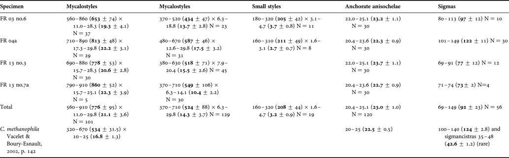
Cladorhiza methanophila Vacelet & Boury-Esnault, Reference Vacelet and Boury-Esnault2002. Spicule dimensions (µm). Minimum, maximum and average dimensions are given, as well as the number of spicules measured (N). With earlier description of holotype (Vacelet & Boury-Esnault, Reference Vacelet and Boury-Esnault2002).
SPICULE TYPES
1. Mycalostyles. Forming the skeleton of the main axis and filaments. Some may have minor subtylote swellings. Straight and fusiform. 560–910 (776 ± 95) µm long and 11.0–29.8 (21.1 ± 3.6) µm wide (N = 101) (Table 9; Figure 15B).
2. Mycalostyles. Forming the skeleton of the main axis and filaments. Some may have minor subtylote swellings Straight and fusiform. 370–710 (524 ± 89) µm long and 6.3–29.8 (14.3 ± 3.7) µm wide (N = 129) (Table 9; Figure 15B).
3. Small styles. Present in small numbers in two specimens. Straight, thin and non-fusiform. 160–320 (208 ± 44) µm long and 1.6–4.7 (3.2 ± 0.9) µm wide (N = 19) (Table 9; Figure 15C).
4. Anchorate anisochelae. Associated with the lining of the main branches and the filaments. Five teeth in each end. Fimbriae are upper end only, and are about the same length as the upper teeth. 20.4–25.1 (23.0 ± 1.0) µm long (N = 120) (Table 9; Figure 15D).
5. Sigmas. Non-contort. 69–149 (92 ± 23) µm long (N = 56) (Table 9; Figure 15E).
The two types of main mycalostyle are quite similar, but a histogram of length–frequencies (not included) showed a two-topped distribution, though with somewhat different length maxima in the different specimens. Some of the mycalostyles have minor swellings at the blunt end, but not to a sufficient degree as to be considered true subtylostyles. Smaller styles were found in small number in the axis of some specimens. One small sigma (50 µm long) was found, and might possibly indicate a second type of sigma or sigmancistra.
PREVIOUS REPORTED DISTRIBUTION
This species has previously been reported from mud volcano fields in the Barbados Trench, from 4700–4900 m depth (Vacelet & Boury-Esnault, Reference Vacelet and Boury-Esnault2002).
REMARKS
While the main mycalostyles are separated into two groups in this description, a case might also be made that they are similar enough that they should constitute one type, as in the earlier description (Vacelet & Boury-Esnault, Reference Vacelet and Boury-Esnault2002). The specimen examined differs in a few properties from those previously described: in particular, the small styles are not included in the previous description, and no sigmancistras were found in these specimens. Given the geographical proximity, the fact that the specimens were collected from near an active methane source, and the close match of the rest of the spicule complement and general morphology, a total assessment still favours assigning the specimen to Cladorhiza methanophila.
Cladorhiza thomsoni Topsent, Reference Topsent1909
(Figure 16A–F)
ORIGINAL DESCRIPTION
Cladorhiza thomsoni Topsent, Reference Topsent1909: 15–18; pls I–II.
EXAMINED MATERIAL
Specimen KN 20_01 (1992-12, Dive 20, 23°N045°W, ~4000 m).
The specimen was recovered at the Kane Fracture Zone at the Mid-Atlantic Ridge as part of the 1992 Kanaut cruise by the ‘Nautile’ submersible.
DESCRIPTION
The specimen examined consists of the dried out debris of a single sponge and consists of a main axis carrying secondary filaments, which in most cases are damaged (Figure 16A, B).
Cladorhiza thomsoni Topsent, Reference Topsent1909: (A) species fragments; (B) stem detail; (C) mycalostyles; (D) anchorate anisochela; (E) large sigma; (F) small sigma.
SKELETON
The skeleton in the main axis is composed of bundles of mycalostyles, which also form the projecting filaments. Anchorate anisochelae and sigmas are found at the surface of the main axis and filaments. The smaller type of sigma seems to be confined to the filaments (Figure 16B).
SPICULE TYPES
1. Mycalostyles. Forming the skeleton of the main axis and filaments. Some may have minor subtylote swellings. Straight and fusiform. 940–1230 (1055 ± 88) µm long and 15.7–29.8 (27.0 ± 3.1) µm wide (N = 30). (Figure 16C).
2. Mycalostyles. Forming the skeleton of the main axis and filaments. Some may have minor subtylote swellings. Straight and fusiform. 580–840 (700 ± 65) µm long and 12.6–28.3 (20.5 ± 2.9) µm wide (N = 30), (Figure 16C).
3. Anchorate anisochelae. Lining the main branches and the filaments. Five teeth in each end. Fimbriae are upper end only, and are a little bit longer than the upper teeth. 26.7–29.8 (28.1 ± 1.0) µm long (N = 30) (Figure 16D).
4. Sigmas. Non-contort. 137–174 (158 ± 9) µm long (N = 30) (Figure 16E).
5. Sigmas. Present in filaments. Non-contort. 41–61 (52 ± 4) µm long (N = 30) (Figure 16F).
PREVIOUS REPORTED DISTRIBUTION
Previously described from 3300–5300 m depth between Gough Island and Cape Town (Topsent, Reference Topsent1909).
DISCUSSION
The Ifremer material examined in this study includes five previously undescribed and ten previously known cladorhizids collected from a series of Atlantic cruises ranging from the better studied north-eastern Rockall Rise to the Central Mid-Atlantic and the continental slope of Central Africa, at depths ranging from 670 to 4800 m.
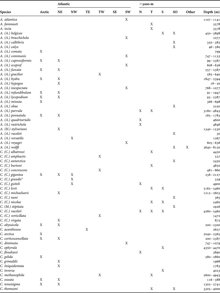
Table 10 lists the distribution of currently described cladorhizids from different regions of the Arctic, the Atlantic Ocean and the Atlantic parts of the Antarctic (Southern Ocean). The regions are based on borders between biogeographic realms as outlined in the Marine Ecoregions of the World (MEOWs) scheme (Spalding et al., Reference Spalding, Fox, Allen, Davidson, Ferdaña, Finlayson, Halpern, Jorge, Lombana, Lourie, Martin, McManus, Molnar, Recchia and Robertson2007), but dividing the northern, tropical and southern Atlantic realms each into an eastern and a western part, as well as extending the scope of the regions into the deep-sea parts of the Atlantic Ocean. Three lower bathyal/abyssal areas were thus designated for parts of the Atlantic Ocean deeper than 3000 m (see also Lopes & Hajdu, in press, for maps of approximate collection locations for Cladorhizidae globally).
Checklist of known Arctic, Atlantic and Southern Ocean species of family Cladorhizidae (Abyssocladia, Asbestopluma, Chondrocladia, Cladorhiza) divided by region and depth.
*, C. (C.) grandis is described with no spicule information, and is possibly a synonym of C. (C.) gigantea (Verrill, Reference Verrill1879).
A couple of attempts have been made to analyse global sponge biogeography patterns at different levels of systematic resolution (van Soest, Reference van Soest, van Soest, Van Kempen and Braekman1994; van Soest et al., Reference van Soest, Boury-Esnault, Vacelet, Dohrmann, Erpenbeck, de Voogd, Santodomingo, Vanhoorne, Kelly and Hooper2012). For sponges in general, the number of Atlantic species is highest in European waters and the Caribbean, with a lower number of species recorded in other tropical areas as well as the southern Atlantic. Certainly, the higher European numbers in part reflect the high collection effort in these waters. For the deep-sea Cladorhizidae globally, van Soest (Reference van Soest, van Soest, Van Kempen and Braekman1994) found a bipolar cold-water distribution pattern, in contrast to a global distribution pattern of increased species richness in warmer regions for sponges in general. In the Atlantic, this is reflected most strikingly in the relatively few Cladorhizidae known from the otherwise very sponge-rich Caribbean.
The data in Table 10 confirms the high number of reported cladorhizid species from the north-east Atlantic and Arctic, with the exception of the genera Chondrocladia and Abyssocladia. These regions are comparatively well-sampled, with multiple reports of most species, showing that a few species have an exclusively Arctic distribution while some species are reported only from the north-east Atlantic. Most species in these areas are reported from both regions however. The Caracole samples included in this paper represent previously described species from this area, from shallower (670–1010 m) waters at the Rockall Bank at the European Shelf.
To the south and down onto the abyssal plains, the picture becomes more complex: in the case of Asbestopluma (A.) parvula, numerous samples were collected both from the Porcupine Abyssal Plain (~48°N, 4800 m), outside the Irish continental margin, and much farther south, on the lower continental slope outside Congo–Gabon (~05°S, 3150–3900 m). Similarly, Chondrocladia (C.) vaceleti, here described from the abyssal plain outside the Iberian Peninsula (~42°N, 4200 m), has previously been described from the Angola Basin (~17°S, 5450 m) (Cristobo et al., Reference Cristobo, Urgorri and Rios2005). Additionally, the species A. (A.) belgicae, previously reported from the shallower waters in the Antarctic (370–570 m) (Topsent, Reference Topsent1901b; Koltun, Reference Koltun, Pavlovskii, Andriyashev and Ushakov1964b), is here described from the lower continental slope outside Congo–Gabon (3150 m), and the species Cladorhiza thomsoni, previously described from the abyssal plain west of South Africa (between Gough Island and Cape Town, 3300–5500 m) (Topsent, Reference Topsent1909), is described from the Mid-Atlantic Ridge at 23°N (~4000 m).
In general, Cladorhizidae in the tropical and southern Atlantic are known only from one or a couple of records. In their paper describing cladorhizids from coral mounds at 605–1135 m in the Campos Basin (south-west coast of Brazil) Lopes & Hajdu (Reference Lopes and Hajdu2013) found seven species, all previously undescribed. While a couple of species have previously been described from the abyssal plain in the south-west Atlantic, this increased the number of known cladorhizids from the Southern Atlantic/South American shelf from zero to seven (see also Lopes et al. (Reference Lopes, Bravo and Hajdu2011) for similar results off the Diego Ramírez Archipelago, not included in the biogeographical scope of this paper). Similarly, the only currently known cladorhizid from the tropical and southern Atlantic shelf and upper slope of Africa is Cladorhiza acanthoxea sp. nov. described in this paper.
Small cladorhizids are easily damaged and may be overlooked in general benthic surveys. Higher concentrations and diversity of cladorhizid species are often found at particular bathymetric features where the general fauna diversity is also higher, such as cold-water coral mounds like those of Campos Basin (e.g. Lopes & Hajdu, in press) or in the vicinity of vent- and seep areas (Vacelet, Reference Vacelet, Desbruyères, Segonzac and Bright2006b). Table 10 shows that with the exception of the north-east Atlantic/Arctic cladorhizids, most species are known only from a single region.
Sponge larvae have limited dispersal capabilities, and overall, sponges are typically endemic to regional or local areas (van Soest et al., Reference van Soest, Boury-Esnault, Vacelet, Dohrmann, Erpenbeck, de Voogd, Santodomingo, Vanhoorne, Kelly and Hooper2012). While the Cladorhizidae in general is described as a deep-sea family, the reported depth ranges of known cladorhizids (although admittedly in many cases based on limited data) are different between species in the family. Table 11 lists the depth distribution of cladorhizid species recorded from the Atlantic, showing that while a roughly similar number of species are recorded from the deepest parts of the ocean in all regions, the number of shallower recorded cladorhizids is currently dominated by Arctic and North Atlantic species. Of special interest is the low level of overlap in cladorhizid species composition between different depths, though in many cases specific distributions are poorly known since species might only be recorded from one or a few sites.
Number of cladorhizid species for different depth zones for different parts of the Atlantic, Arctic and Atlantic-facing parts of the Antarctic, as well as total number of species for the whole extent of the analysis. Species overlap refers to the percentage of species that the particular depth zone shares with the zone(s) above it.
The large biogeographical range of several abyssal species shown in this study compared to previous reports indicates that Cladorhizidae at abyssal depths have a much wider geographical distribution than their shallower counterparts. The case of A. (A.) belgicae reported here is a detail of special interest. Antarctic cladorhizid sponges are to a great extent endemic. The high degree of endemism has also been reported for other sponges in the Antarctic (e.g. Janussen & Tendal, Reference Janussen and Tendal2007; Rapp et al., Reference Rapp, Göcke, Tendal and Janussen2013) and extends to Antarctic deep-sea sponge communities, which instead are more closed elated to shallower Antarctic habitats (Göcke & Janussen, 2013). Asbestopluma (A.) belgical might then represent a more unusual case of a more widely distributed species that has also spread into shallower Antarctic waters.
The two Abyssocladia species described here were collected during the Faranaut cruise to the geologically active area at the intersection of the 15°20′N fracture zone and the Mid-Atlantic Ridge. The affinity of cladorhizids to the general enrichment zones around vent and seep sites is well documented (e.g. Vacelet, Reference Vacelet, Desbruyères, Segonzac and Bright2006b; Reference Vacelet, Custódio, Lôbo-Hajdu, Hajdu and Muricy2007). In general, the greater presence of cladorhizids at such sites is explained as deep-sea fauna benefitting from the greater abundance of prey close to the nutrient-rich vent and seep areas. Interestingly, specimens belonging to Cladorhiza methanophila were also recovered from the Faranaut site, a species known to harbour symbiotic bacteria feeding directly on methane discharge, which has previously been described from mud volcano fields in the Barbados Trench (Vacelet et al., Reference Vacelet, Boury-Esnault, Fiala-Medioni and Fisher1995, Reference Vacelet, Fiala-Medioni, Fisher and Boury-Esnault1996; Vacelet & Boury-Esnault, Reference Vacelet and Boury-Esnault2002). While the extent of such symbiosis within the family is not known, the presence of C. methanophila together with CH4 plumes generated by serpentinization of ultramafic rocks at the 15°20′N site (Charlou et al., Reference Charlou, Fouquet, Bougault, Donval, Etoubleau, Jean-Baptiste, Dapoigny, Appriou and Rona1998) indicates that symbiotic interactions are present.
In conclusion, it can be shown that there are large areas of the Atlantic Ocean where cladorhizid diversity is poorly known and that both new records of known species and new species can be expected to be found. More specifically, regionally mostly endemic cladorhizid faunas can be expected in shelf and upper slope areas in previously poorly sampled areas. The lower bathyal and abyssal may on the other hand prove a distinct habitat where species can be encountered over wide distances. The current biogeographical knowledge of the Cladorhizidae is for most parts of the Atlantic Ocean as yet insufficient, but proper examination of existing museum collections as well as future sampling efforts can be expected to improve the situation.
ACKNOWLEDGEMENTS
We gratefully acknowledge the sorting centre, Centob, of the Laboratoire Environnement Profond (Ifremer, Brest, France) for entrusting us with the study of the specimens, especially Michel Segonzac. We would also like to thank the Ifremer and Genavir personnel, crew, captains and scientists of the cruises during which the specimens were collected. Finally, we would like to thank the Editor and two anonymous referees whose comments helped improve the quality of the manuscript.
FINANCIAL SUPPORT
This research received no specific grant from any funding agency, commercial or not-for-profit sectors.