The material contained in this work was presented at the symposium on Heterotaxy and Isomerism on 25 February, 2007 in Orlando, Florida, USA at Cardiology 2007: Tenth Annual Update on Paediatric Cardiovascular Disease. The symposium was dedicated to the memory of Stella Van Praagh, one of the most accomplished morphologists, teachers and paediatric cardiologists to study the topic.
The word heterotaxy stems from Greek origin, with “heteros” meaning other and “taxis” meaning arrangement. This term is, therefore, quite appropriate to describe a group of disorders that involves abnormal lateralization of the abdominal and thoracic organs, and also the atrial appendages. In heterotaxy syndrome, structures that typically manifest asymmetry on the right and left sides of the body develop as mirror-images of each other. In addition to these anomalies, it is usual to find complex malformations within the heart, often requiring cardiac surgery in the neonatal period. The syndrome itself can be stratified into two identifiable subtypes, namely the subset characterized by isomerism of the right atrial appendages, found most frequently with absence of the spleen, and that associated with isomerism of the left atrial appendages, usually found with multiple spleens, although not all patients show the expected combinations.Reference Van Praagh and Bergsma1 The outcome for patients falling within these two subtypes may differ, and depends on the complexity of the cardiac disease, and the severity of extra-cardiac anomalies. In this review, we describe some of the controversies surrounding how best to categorize and describe heterotaxy syndrome, and address the genetic and developmental background to the disorder. We also discuss non-invasive and invasive assessment, long-term outcome and the impact of prenatal diagnosis on outcome.
What’s in a name?
From the stance of the school of nomenclature developed using the teachings of Van PraaghReference Van Praagh and Bergsma1, visceral arrangement can be stratified into three types. Situs solitus is the usual arrangement, while situs inversus is the mirror-imaged variant of solitus. The third type is situs ambiguus, a combination of situs solitus and situs inversus in the same individual. Examples of situs ambiguus include the presence of atrial appendages and bronchuses of the same morphology on both sides of the body, the liver and stomach both being on the same side of the body, or the abdominal organs mostly being mirror-imaged with the thoracic organs in their usual arrangement, or vice-versa. Heterotaxy syndrome is considered to be present whenever there is situs ambiguus, in other words when the structures are not identifiably in their usual or mirror-imaged positions as defined above. Most patients with heterotaxy syndrome have splenic abnormalities. In the postmortem collection at The Children’s Hospital of Philadelphia, nearly half of patients have asplenia, and onethird have multiple small spleens. In the opinion of those using this system of analysis, the concept of bilateral right-sidedness and bilateral left-sidedness, while useful in helping to remember which abnormalities are likely to occur in asplenia or polysplenia, should not be granted the status of a specific “situs”, since there are numerous examples of exceptions to these patterns.
Heterotaxy syndrome is the Murphy’s Law of congenital heart disease, in that almost anything that can go wrong, will. Virtually all patients with asplenia syndrome have a common atrioventricular junction, as do almost two-thirds of those with polysplenia syndrome. Most patients with asplenia syndrome have either double outlet right ventricle, or right ventricular aorta with pulmonary atresia, whereas more than half of those with polysplenia syndrome have normal anatomy of the ventricular outflow tracts. Nearly all patients with asplenia syndrome have both a common atrioventricular junction and abnormal ventricular outflow tracts. In contrast, about half of those with polysplenia syndrome have either a common atrioventricular junction or an abnormal ventricular outflow tract, another one-third have both lesions, and less than one-sixth have neither defect. Most patients with asplenia syndrome have subpulmonary stenosis or atresia, while those with polysplenia syndrome are roughly evenly distributed among subpulmonary, subaortic, or no obstruction.
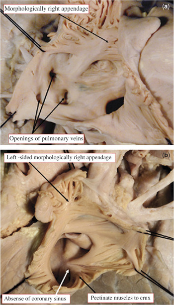
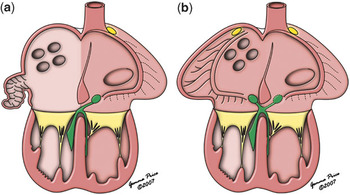
Those who developed the European Paediatric Cardiac Code, in contrast, took a different approach to analysis, To them, the key to analysis of patients with heterotaxy syndrome is the approach taken to the structure of the heart. Thus, the European Paediatric Cardiac Code takes as the starting point for diagnosis of complex cardiac malformations the establishment of the arrangement of the atrial chambers. All then depends on the rules used to distinguish between morphologically right and left atriums. As discussed above, the school of nomenclature that developed using the tenets of Van Praagh argues that, in the setting of heterotaxy syndrome, the atrial arrangement is ambiguous. In the past,Reference Van Praagh and Bergsma1 however, it had been suggested that atrial “situs” could be determined on the basis of the venoatrial connections. Is it justified to use this feature as the arbiter of morphologically rightness and leftness in the atrial segment of the heart? Another important principle established by Van Praagh and his colleagues,Reference Van Praagh, David, Wright and Van Praagh2 over and above segmental analysis,Reference Van Praagh and Bergsma1 was the so-called “Morphological method”. Initially put forward to show that an atrioventricular valve should not be used as the determinant of whether a chamber within the ventricular mass was a true ventricle, or a so-called rudimentary chamber,Reference Keeton, Macartney and Hunter3 Van Praagh and colleaguesReference Van Praagh, David, Wright and Van Praagh2 stated that variable structures within the heart should not be distinguished on the basis of other components that were themselves variable. Instead, they argued that structures should be distinguished on the basis of their own intrinsic morphology. The principle of the “morphological method”,Reference Van Praagh, David, Wright and Van Praagh2 therefore, is crucial when one is faced with the challenge of defining atrial arrangement in the setting of heterotaxy. One of the paramount features of patients with the splenic syndromes is their anomalous venoatrial connections. It has also been shown, nonetheless, that the atrial chambers in patients with heterotaxy syndrome can be distinguished on the basis of one feature that is independent of venoatrial connections, as well as size or shape, namely the extent of the pectinate muscles in the appendages relative to the vestibules of the atrioventricular valves.Reference Uemura, Ho, Devine, Kilpatrick and Anderson4 In the morphologically right appendage, these pectinate muscles extend all round the vestibule of the atrioventricular valve, reaching to the cardiac crux, and separate the vestibule from the venous component. In the morphologically left appendage, in contrast, the pectinate muscles are confined within the appendage, the smooth vestibule being in continuity with the smooth atrial venous component. On the basis of these anatomical features, the study of Uemura and colleaguesReference Uemura, Ho, Devine, Kilpatrick and Anderson4 showed that it was possible always to distinguish morphologically right from left appendages, and that the appendages in those with heterotaxy were either both of right morphology, or else both of left morphology. In other words, they showed that heterotaxy could be stratified on the basis of isomerism of either the morphologically left or morphologically right appendages.Reference Uemura, Ho, Devine, Kilpatrick and Anderson4 The lungs and bronchuses in patients with heterotaxy also typically show evidence of isomerism,Reference Van Mierop, Gessner and Schiebler5 albeit that the type of isomerism is not always in harmony with splenic morphology.Reference Uemura, Ho, Devine and Anderson6 The approach taken by the European school to analysis of patients with heterotaxy, therefore, is to analyse each system or organs independently, and to describe specifically the arrangement of each system. Within the heart, analysis starts with establishing the arrangement of the appendages, and only then defining the specific venoatrial connections. It may well be that the venoatrial connections are relatively normal, or akin to the mirror-imaged arrangement. This does not mean that the atrial arrangement is “solitus” or “inversus”. On the contrary, if the appendages are isomeric, then as dictated by the “morphological method”, it is their structure that determines “situs”. It is also the arrangement of the appendages that provides the key to interpretation of the electrocardiogram, since the sinus node is positioned adjacent to the morphologically right appendage, and establishes the likely arrangement of the atrioventricular conduction axis.Reference Smith, Ho, Anderson, Connell, Arnold and Wilkinsion7 If, for example, in the setting of absence of the spleen, all the pulmonary veins return to a right-sided atrium with a right atrial appendage, and all the systemic veins to a left-sided atrium, also with a right atrial appendage, then the arrangement is not situs inversus, nor even situs ambiguus. The arrangement is one of isomerism of the right atrial appendages, with pulmonary venous return to the right-sided atrium, systemic venous return to the left-sided atrium, and with bilateral sinus nodes (Fig. 1). Furthermore, if the ventricular topology is left-handed, then the atrioventricular connections will be biventricular and mixed (Fig. 2), and we should anticipate paired atrioventricular nodes. In patients with visceral, therefore, it is not so much as “what’s in a name”, but rather “how do we determine atrial arrangement”? Those who developed the European Paediatric Cardiac Code took care to follow the precepts of Van Praagh and his colleagues,Reference Van Praagh, David, Wright and Van Praagh2 and used the “morphological method” to distinguish the arrangement of the atrial chambers, basing this on the morphology of the atrial appendages.
Photographs showing a heart with isomerism of the right atrial appendages, with a) all the pulmonary veins returning to right-sided atrium, and b) all the systemic veins to left-sided atrium. Despite the mirror-imaged arrangement of the venous return, the starting point for cardiac analysis is the recognition of the bilateral right atrial appendages, this being based on the extent of the pectinate muscles relative to the atrioventricular vestibules.

Cartoon comparing the disposition of the sinus node (yellow commas) and atrioventricular conduction axis (green) in the setting a) of mirror-imaged arrangement and left-hand topology, and b) isomerism of the right atrial appendages with mirror-imaged venous return, again with left-hand ventricular topology. Unless the fundamental difference is taken into account with regard to the morphology of the atrial appendages, those diagnosing the situation as shown in b) as “situs inversus” would make major mistakes in predicting the location of the conduction tissues.

Genetic aetiology
Heterotaxy is an abnormality of formation of the left-right axis of the body. It is not yet clear whether differences in the classification of defects of laterality are primarily due to dogma, or imperfect description. What is clear is that heterotaxy is clinically and genetically heterogeneous, suggesting that normal laterality, in other words left-right asymmetry, develops by a complex mechanism.
Cardiac development has seen a shift in experimental approach. From the 18th century to the present, it was the descriptive approach that formed the backbone of developmental biology. Descriptive approaches, though precise, are also inherently limited, in that there can be no proof of mechanism, solely associations. In fact, most of our anatomic and developmental tenets in paediatric cardiac disease are associations, with few instances of experimentally proven descriptive hypotheses. From the 1970s to the present, a more experimental approach has been taken, using animal systems to model congenital cardiac defects.Reference Harh, Paul, Gallen, Friedberg and Kaplan8 Since the 1980s, this research has been extended to the use of gene-targeted mice, providing a powerful tool to unravel single gene defects.Reference Gruber and Epstein9 Contiguous gene syndromes, and complex genetic traits, are proving more challenging. In order to change the course of a disease, nonetheless, we need to understand the mechanism by which it occurs.
What, therefore, are the molecules that provide instructions for cardiac assembly? One way to identify these is to look at natural mutations that result in defects of laterality. One of the first indications of a molecular basis for such decisions has been the identification of the iv, or inversum viscerum, mouse.Reference Pehlivan, Pober and Brueckner10–Reference Icardo and Sanchez de Vega13 In this spontaneous mouse mutant, there is a randomization of ventricular looping, shown to reflect a mutation in the Lrd geneReference Hummel and Chapman11, Reference De Tommasi, Daliento, Ho, Macartney and Anderson45 Another mouse with related defects is the inv mouse, showing “inversion of embryonic turning”, in which the ventricular loop always turns to the left rather than right.Reference Watanabe, Saijoh and Nonaka16 Both of these mutants also have abnormalities in nodal cilia,Reference Afzelius17 reminiscent of Kartagener’s syndrome.Reference Marszalek, Ruiz-Lozano, Roberts, Chien and Goldstein18 Since these discoveries, more mutants have been discovered that harbour similar defects, such as Dnahc11/Lrd/iv and Kif3A.Reference Hummel and Chapman11, Reference Azakie, Merklinger, Williams, Van Arsdell, Coles and Adatia69
A number of molecular investigations have identified asymmetric expression of signaling molecules, including Sonic hedgehog, cNR1, Lefty-2, and Pitx2.Reference Heymer, Kuehn and Ruther20–Reference Afzelius24 Many of these have had direct mechanistic implications for the development of asymmetric structures. The question still remains concerning the mechanism responsible for setting up initial sidedness. The focus has now shifted back to nodal cilia. The node, a transient midline structure that forms during gastrulation, is critical for decisions concerning laterality. It forms after the establishment of the anteroposterior and dorsoventral axes. On the ventral surface of the node is a ciliated pit covered with specialized monocilia. These monocilia are distinct from the arrangement of conventional cilia in that they lack central microtubules. Patients lacking cilia display disorders of laterality. But why does lack of cilia result in symmetry abnormalities? Nodal monocilia in fish, rabbits, and mice rotate rapidly to produce flow from right to left, a mechanism now identified as the initial event in breaking left-right symmetry.Reference McGrath, Somlo, Makova, Tian and Brueckner25–Reference Hirokawa, Tanaka, Okada and Takeda27 Unlike conventional cilia, the monocilia demonstrate a clockwise rotation, and most importantly, are tilted 40° posterior such that the rightward sweep is close to the surface and the leftward sweep is away from the surface. Thus, due to the high turbulence, the net flow is leftward. Although many details need to be filled in, there is a clear mechanistic explanation for the net leftward flow that uniquely integrates the biochemical, biophysical, genetic, and clinical information.
There are two alternative models that have been proposed to explain how asymmetric gene expression comes from nodal flow. First, there is a model that predicts that directional flow produces concentration gradients of secreted morphogens. Perhaps complementary, perhaps contradictory, is the model that predicts that flow itself is sensed through motile cilia in the central portion of the node, producing the leftward flow as described above, while immotile cilia on the border of the node sense the flow. This sensing of flow is translated by the cells harbouring the immotile border cilia into activation of genes in an asymmetric fashion. Supporting the first chemical gradient model was the discovery of nodal particles that are released and transported to the tips of motile cilia. After reaching the left side of the node, they collide with border cells and increase calcium, resulting in changes in gene expression. All of the above mechanisms have strong experimental support, suggesting that the true mechanism may in fact be a combination of these models.Reference Ryan, Blumberg and Rodriguez-Esteban22, Reference Gaynor, Collins, Rychik, Gaughan and Spray67
These discoveries have had a profound impact on one of the most basic biological decisions an organism must make, namely what is morphologically left and what is morphologically right? Despite this, there remain a number of considerable challenges. The clinical obstacles are apparent as this is a perhaps the most difficult set of patients for whom to care. The scientific questions left to answer are also prodigious. They include a heterogeneous data set where the delineation of phenotype is essential. Moreover, there is a need to identify more completely determinants of laterality in normal development, and a further identification of genes and locuses that are mutated in abnormal phenotypes. We must now integrate these clinical and scientific discoveries in the aim to develop rational strategies in tackling this disorder.
Impact of prenatal diagnosis
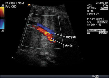
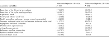
Recognition of cardiac disease in the fetus, generally at 18–20 weeks gestation, occurs well after the embryonic development of heterotaxy described above. Accurate fetal diagnosis of heterotaxy syndrome is one of the greatest challenges for the echocardiographer. The segmental approach to diagnosis is essential to allow for systematic description of fetal congenital cardiac disease. Thus, it is essential to distinguish the right and left sides of the fetus, and then note the position of the organs, so as to establish an accurate diagnosis. Discrepancy between the position of the stomach and the cardiac apex, particularly if associated with a midline liver, is often the first clue that heterotaxy syndrome may be present in the fetus. The heart is frequently positioned abnormally in heterotaxy syndrome, and this feature is easily identified once the fetal lie has been established. Approximately one-third of fetuses with heterotaxy syndrome have right-sided hearts.Reference Cohen28 The atrial appendages can sometimes be identified in the fetus, but not consistently. Identification of the subtype of heterotaxy syndrome, therefore, is often inferred from the associated cardiac defects seen. For example, interruption of the inferior caval vein, with azygos continuation to the superior caval vein, seen almost exclusively in those with isomerism of the left atrial appendages or polysplenia syndrome, is easy to recognize on fetal echocardiography (Fig. 3).Reference Gilljam, McCrindle, Smallhorn, Williams and Freedom29–Reference Jacobs and Mayer31 In contrast, anomalous pulmonary venous connections, frequently associated with isomerism of the right atrial appendages or asplenia syndrome, can be difficult to detect during fetal life because of the minimal flow of blood to the lungs, especially in the presence of subpulmonary obstruction.Reference Cohen, Schultz and Tian32 Extracardiac totally anomalous pulmonary venous connection occurs in up to two-thirds of fetuses with isomerism of the right atrial appendages, but is rare in those with isomeric left atrial appendages.Reference Anderson, Webb and Brown30, Reference Cohen, Schultz and Tian32 A diagnosis of isomerism of the right atrial appendages in fetal life, nonetheless, should alert the practitioner to the likely presence of the abnormal pulmonary venous connections. Intracardiac lesions associated with heterotaxy syndrome, such as unbalanced ventricles with common atrioventricular junction, functionally univentricular arrangements, malformations of the ventricular outflow tracts, and pulmonary obstruction are typical findings in those with isomeric right atrial appendages or asplenia syndrome. In those with isomeric left atrial appendages or polysplenia syndrome, a common atrioventricular junction is frequent, but some patients have normal intracardiac anatomy (Table 1). In a recent study assessing accuracy of fetal diagnosis in heterotaxy syndrome, assessment of atrioventricular and ventriculoarterial connections was 100% sensitive and specific. Prediction of ductal dependency after birth was also correct in all patients.Reference Cohen, Schultz and Tian32
Fetal echocardiographic color Doppler longitudinal view demonstrating demonstrating the descending aorta in parallel with the azygos vein, which is enlarged secondary to interruption of the inferior caval vein. This finding is commonly seen in left isomerism.

Prevalence of anatomic cardiac variables in 81 patients with prenatal and postnatal diagnosis of heterotaxy syndrome.

Fetal diagnosis may have a beneficial impact on outcome. Some studies suggest, for example, that infants with either hypoplastic left heart syndrome, or discordant ventriculo-arterial connections, have improved survival with prenatal diagnosis,Reference Tworetzky, McElhinney, Reddy, Brook, Hanley and Silverman33, Reference Bonnet, Coltri and Butera34 albeit that others have shown no difference in outcome.Reference Tworetzky, McElhinney, Reddy, Brook, Hanley and Silverman33, Reference Garcia, Mehta and Pickoff56 Significant morbidity, such as metabolic acidosis, may be avoided by prenatal diagnosis, which may reflect on better long-term neurodevelopmental outcome.Reference Verheijen, Lisowski and Stoutenbeek37 The impact for heterotaxy syndrome was assessed by Lim and colleagues,Reference Lim, McCrindle and Smallhorn38 who showed that prenatal diagnosis did not improve outcome for either form even if immediate and aggressive care was given after birth, a finding also shown for those with functionally univentricular hearts.Reference Cohen, Schultz and Tian32 If deaths during fetal life were included, those diagnosed prenatally had greater mortality than those diagnosed after birth, indicating that heterotaxy syndrome, when identified during fetal life, is more severe than the same disease presenting after birth. Risk factors for poor outcome include a diagnosis of complete heart block, or totally anomalous pulmonary venous connection, which were mutually exclusive in the series.Reference Cohen, Schultz and Tian32 Indeed, the association of congenitally complete heart block with isomeric left atrial appendages or polysplenia syndrome is almost always uniformly lethal, despite aggressive management after birth. Complete heart block is readily identified in the fetus because of the slow heart rate, and is likely to lead to referral for fetal echocardiography. Accurate prenatal diagnosis allows for consultation with families faced with this very complex problem regarding potential therapies. Though progress has been made in the treatment of patients with heterotaxy syndrome, this remains a condition with one of the highest mortalities. In some cases, neonatal cardiac transplantation may be the most viable option for long-term survival.Reference Cohen, Schultz and Tian32
Postnatal echocardiographic assessment
Since heterotaxy syndrome is associated with a wide variety of complex cardiac malformations, echocardiographic evaluation requires strict adherence to an organized protocol, which starts with localizing the heart within the chest, and is followed by sequential segmental analysis. The heart is right-sided in up to half of patients, and in the middle in up to one tenth.Reference Uemura, Ho, Devine, Kilpatrick and Anderson4, Reference Rose, Izukawa and Moes39 When the heart is in the right chest, the echocardiographer should also distinguish between a rightward and a leftward apex. The latter can be seen with hypoplasia of the right lung or diaphragmatic hernia. Subcostal views generally provide the best echocardiographic assessment of the right-sided heart, though care should be taken to maintain true right-left orientation in apical views, as well as accurate long-axis and short-axis displays in parasternal views.
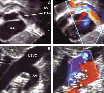
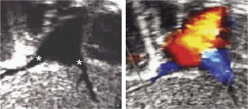
Abnormal venoatrial connections are the rule. Bilateral superior caval veins are seen in up to seven-tenths of these patients.Reference Uemura, Ho, Devine, Kilpatrick and Anderson4, 40, Reference Rubino, Van Praagh, Kadoba, Pessotto and Van Praagh41 In those with isomerism of the right atrial appendages, the caval veins enter the atrial roof, with the coronary sinus being universally absent.Reference Rubino, Van Praagh, Kadoba, Pessotto and Van Praagh41, Reference Webber, Uemura, Anderson, Anderson, Baker, Macartney, Rigby, Shinebourne and Tynan42 The inferior caval vein is intact in almost all those with isomeric right appendages or asplenia syndrome,Reference Rubino, Van Praagh, Kadoba, Pessotto and Van Praagh41, Reference Ruscazio, Van Praagh, Marrass, Catani, Iliceto and Van Praagh43 but is interrupted with azygos continuation in most of the patients with isomeric left appendages or polysplenia syndrome (Fig. 4).40, Reference Webber, Uemura, Anderson, Anderson, Baker, Macartney, Rigby, Shinebourne and Tynan42 Anomalous hepatic venous drainage occurs in up to onethird of these patients.Reference Uemura, Ho, Devine, Kilpatrick and Anderson4, Reference Webber, Uemura, Anderson, Anderson, Baker, Macartney, Rigby, Shinebourne and Tynan42 Most children will ultimately require a Fontan procedure in their staged surgical management, so careful delineation of the individual hepatic venous connections to the atrial mass or inferior caval vein should be undertaken, using posterior sweeps in subcostal and apical views (Fig. 5).
Interrupted inferior caval vein with left azygos (or hemiazygos) continuation into the left superior caval vein. A: Subcostal short-axis view. B: Modified high left parasagittal view. AV, azygos vein; Dao, descending aorta; RA, right atrium; LSVC, left superior caval vein.

Subcostal long-axis view of separate hepatic veins (stars) entering the floor of the atrial chamber.

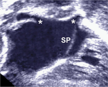
The pulmonary veins should be evaluated individually, because of the high likelihood of abnormal pulmonary venous connections.Reference Uemura, Ho, Devine, Kilpatrick and Anderson4, 40 Totally anomalous return to an extra-cardiac site is seen commonly in those with isomeric right atrial appendages, but rarely in the setting of isomerism of the left appendages. In contrast, abnormal drainage of all or some of the pulmonary veins to the right side of the atrial mass occurs more frequently in those with isomerism of the left atrial appendages. Closer inspection of such patients may reveal displacement of the primary atrial septum to the left side of the common atrial chamber (Fig. 6).Reference Van Praagh, Carrera, Sanders, Mayer and Van Praagh44
Subcostal long-axis view of a nearly common atrium with leftward displacement of the primary atrial septum and anomalous drainage of the pulmonary veins (stars). SP, primary atrial septum.

Echocardiographic assessment of atrial morphology, including the atrial appendages and pectinate muscles, is not always straightforward. A common atrioventricular junction is frequently seen, and modified subcostal imaging is usually the best approach to evaluate this area. This view is also useful for ventricular hypoplasia, with left ventricular hypoplasia seen in up to two-fifths of those with isomeric right appendages.40
Assessment of the ventricular outflow tracts, taken in association with the venoatrial connections, can help to distinguish between the two categorical subtypes of heterotaxy, and should therefore be carefully assessed by echocardiography. Bilateral infundibulums, usually associated with a double outlet right ventricle and subpulmonary obstruction, occur most frequently in patients with isomeric right appendages.40, Reference De Tommasi, Daliento, Ho, Macartney and Anderson45 In contrast, many with isomerism of the left atrial appendages have normal outflow tracts.40 Moreover, up to one-quarter of such patients have some type of aortic obstruction. A right aortic arch, with mirror-imaged branching, occurs in approximately one-third of patients with either subtype of heterotaxy syndrome.Reference Cohen28 This can have important implications if an aorto-pulmonary shunt is required to provide a reliable source of flow of blood to the lungs.
Cardiac magnetic resonance imaging
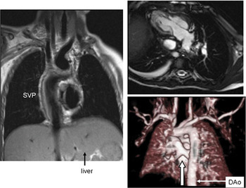
Heterotaxy, with the myriad of visceral and cardiovascular malformations, is well suited to comprehensive imaging using a modality such as cardiac magnetic resonance, which is especially advantageous because of its wide field of view, permitting establishment of the arrangement of the abdominal and thoracic organs in one scan. The technique has been used to demonstrate the abnormal cardiac morphology (Fig. 7) for a number of years.Reference Masui, Seelos, Kersting-Sommerhoff and Higgins46 Prospective studiesReference Geva, Vick, Wendt and Rockey47, Reference Hong, Park and Ryu48 have shown that the diagnostic yield is superior to echocardiography, and often to cardiac catheterization, in defining pulmonary and systemic venous anatomy, and their relationship to mediastinal structures.
Examples of the use of magnetic resonance imaging in the setting of heterotaxy. The image on the left is a double inversion dark blood image of a patient with a functionally single ventricle after conversion to the Fontan circulation. Note the midline liver and the systemic venous pathway (SVP). The upper right panel is an example of steady state free precession bright blood imaging of a “2-chamber view” of another patient with a functionally single ventricle who has a right-sided heart with the apex pointing to the right. The patient also has a small right pleural effusion. The lower right panel is a three-dimensional reconstruction utilizing gadolinium viewed posteriorly of a patient with totally anomalous venous connection to the superior caval vein. Note the ascending vertical vein (arrow) receiving all four pulmonary veins. DAo = descending aorta.

The protocol begins with a set of bright blood axial images of the chest to survey the thoracic anatomy, followed by sets of targeted static or cine bright blood images further to define the cardiovascular anatomy. Specialized sequences can be used to assess the coronary arterial system if needed. The technique also shows the presence of a spleen, and the sidedness of the liver, stomach, and other abdominal organs. In the thorax, the tracheobronchial tree can be visualized to determine lateralization of the lungs, along with the bronchial-arterial relationships. Gadolinium is used with specialized sequences to create three-dimensional images of the cardiovascular system to aid in comprehending the anatomy and in surgical reconstruction.
Utilizing cine cardiac magnetic resonance, ventricular function is quantified by measuring ejection phase parameters, cardiac index, mass and ventricular volumes, which can be especially important in lesions where variable degrees of ventricular hypoplasia may be present or in patients undergoing the Fontan operation. Phase encoded velocity mapping can quantify flow, and is generally used to quantify cardiac output, measure pulmonary and systemic blood flow, regional blood flow to each lung, and the relative contributions of specific pulmonary or systemic veins to venous return. Viability sequences identify areas of myocardial scarring which may have resulted from perinatal insults or injuries during cardiac surgery.
Angiography
In the current era, noninvasive imaging has replaced angiography as the primary modality for anatomic evaluation, and this is as true for patients with heterotaxy syndromes as any others. As the majority of these patients will require repeated surgical treatment for physiologic correction or palliation, nonetheless, catheterization is required at different stages for haemodynamic evaluation, intervention, and even angiographic assessment. Angiography and echocardiography are complementary imaging tools, and should be used as such. The principles guiding angiographic assessment in heterotaxy are no different from standard approaches to any other structural heart defects. Because the complexity of these disorders may require multiple angiographic series, there is the potential for toxicity from excess contrast and radiation. To minimize risk, a focused approach is required to obtain the needed information for treatment. Echocardiographic evaluation will generally provide excellent information about atrial, ventricular, and atrioventricular valvar anatomy. Angiographic assessment should be focused on delineation of venous anatomy, pulmonary arterial architecture and supply, and delineation of ventricular anatomy and function.
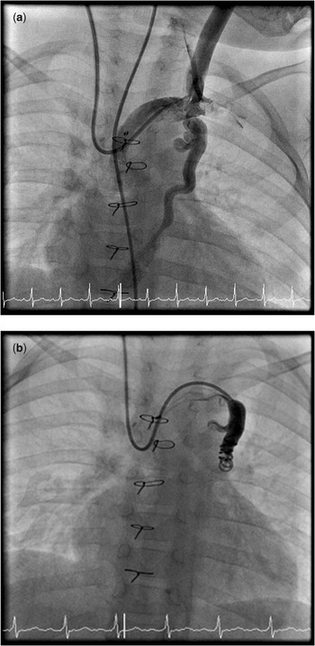
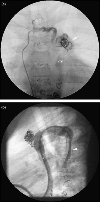
The best method for establishing the venous return is balloon-occlusion angiography. When bilateral superior caval veins are present with a large brachiocephalic vein, embolization of the non-dominant vein is indicated prior to superior cavopulmonary anastomosis (Fig. 8). It is also important to recognize that variations in azygos drainage are common, and can have important implications. Embolization or ligation of a superior caval vein proximal to an azygos vein can result in a large right-to-left shunt after creation of the Fontan circulation (Fig. 9). From the stance of pulmonary venous drainage, the angiographer should define the drainage from all parts of each lung in every patient. The initial image should be a laevophase after opacification of the pulmonary trunk, either with direct injection or balloon-occlusion aortography if there is pulmonary atresia. Subsequent selective injections are often required in the pulmonary arteries to further define drainage. In the presence of obstructed venous return, wedge angiography may be helpful, although direct injection in the pulmonary veins may be required.
Angiography of the brachiocephalic vein in a patient who has undergone a bidirectional Glenn procedure. (a) Balloon-occlusion injection via an endhole catheter demonstrates the large brachiocephalic vein with persistence of a left superior caval vein, which drains to the coronary sinus. (b) After coil embolization of the vessel, there is no residual flow to the atrium and all contrast drains the brachiocephalic vein to the pulmonary arteries via the right superior caval vein.

Angiograms in a patient who has become cyanotic several years after a modified Fontan operation. (a) Injection of contrast via a catheter which has been advanced into a small venous branch arising from the posterior aspect of the right superior caval vein fills a plexus of veins across the midline, and then opacifies a persistent left azygos which drains to the left superior caval vein (LSVC) below where it had been previously coil embolized (arrow) in the past. Contrast then flows via the coronary sinus (CS) to the atrium. (b) A transseptal puncture of the Fontan baffle was performed and a catheter advanced through the coronary sinus into the left azygos vein for embolization. Lateral projection of an injection demonstrates the left azygos (arrow) draining via the left superior caval vein to the coronary sinus (asterisk).

Non-invasive imaging may be suboptimal for delineation of the pulmonary arterial supply and anatomy. The most commonly missed features are clinically important stenoses of the pulmonary arteries, and accessory sources of pulmonary flow. Standard angiographic techniques suffice to identify these features. The most common situation where such imaging is required is in pulmonary atresia, where a balloon-occlusion image of the descending aorta using a Berman angiographic catheter advanced in an antegrade manner will demonstrate the features of importance.
Ventriculography can often add useful information, particularly when questions arise about the adequacy of ventricular size for biventricular repair, or as an additional modality to rule out multiple ventricular septal defects. Standard axial imaging approaches should be used optimally to demonstrate the ventricular septum.
The conduction system and arrhythmias
Electrophysiologic abnormalities are pervasive in patients with heterotaxy syndrome, and equally challenging to the paediatric cardiologist and cardiothoracic surgeon. Aside from the readily identifiable anatomic features unique to normal atriums, there are well-described properties of the specialized conduction tissue in the normal heart. The sinus node resides within the terminal groove at the junction between the morphologically right atrial appendage and the right atrium. In hearts with isomeric right atrial appendages, the sinus nodes are always bilateral, and it is incumbent for the surgeon to respect the terminal grooves in both atriums, as each likely harbours a sinus node.Reference Smith, Ho, Anderson, Connell, Arnold and Wilkinsion7 In contrast, solitary sinus nodes are identified in less than half of hearts with isomeric left atrial appendages. They tend to be hypoplastic and malpositioned.Reference Smith, Ho, Anderson, Connell, Arnold and Wilkinsion7 Caution should be exercised with incisions near the atrioventricular vestibule.
The orifice of the coronary sinus, septal leaflet of the tricuspid valve, and the tendon of Todaro form the triangle of Koch, which is the landmark for the compact atrioventricular node. Neither the atrial septum nor the coronary sinus, however, should be used as a definitive landmark. While generalities abound with regards to the location of the atrioventricular node in hearts with isomeric atrial appendages, the associated structural malformations seen in heterotaxy syndrome play a large part in the displacement of the normal conduction tissue. For example, in some hearts with common atrioventricular junction, and in those with straddling tricuspid valve, the node is displaced posteroinferiorly. In those with left handed ventricular topology, the node sits anterolaterally, often with a “sling” of conduction tissue connecting to a postero-inferior node. The disposition of the atrioventricular conduction axis reflects both the atrioventricular connection and the ventricular topology. When there are biventricular atrioventricular connections, the dominant feature is ventricular topology. When there are functionally univentricular connections, the dominant feature is ventricular morphology.Reference Smith, Ho, Anderson, Connell, Arnold and Wilkinsion7, Reference Webber, Uemura, Anderson, Anderson, Baker, Macartney and Rigby49 Discontinuity of the ventricular conduction axis has been reported in more than four-fifths of patients with isomeric left atrial appendages.Reference Ho, Fagg, Anderson, Cook and Allan50
Multiple morphologies of the P wave, owing to the frequent occurrence of dual sinus nodes, is seen in the surface electrocardiogram of about one-sixth of patients with isomeric right atrial appendages.Reference Cheung, Yan-wah, Yung and Chau51, Reference Wren, Macartney and Deanfield52 Supraventricular tachycardia has been reported in up to one-quarter of patients, and is more prevalent in hearts with biventricular and mixed atrioventricular connections.Reference Wren, Macartney and Deanfield52, Reference Wu, Wang and Lin53 The tachycardia is presumed to be due to nodal macro-reentry, with antegrade conduction through a posteriorly located node, and retrograde via the sling of conduction tissue to the other node. Medical management with vagal maneuvers, or nodal blocking drugs, is a reasonable first-line approach to treatment. The discernible electrophysiology features unique to twin nodes and supraventricular tachycardia include two distinct non-preexcited QRS morphologies, with each QRS dependent on the site of spontaneous activation or atrial pacing, separate discernible His-bundle electrograms, antegrade, decremental conduction over at least one node and possibly both, retrograde conduction over one node, and inducible supraventricular tachycardia. Successful treatment with catheter ablation, modifying one of the two nodes, is an effective option, and should be considered in patients with intolerance to antiarrhythmic medications or frequent reoccurrences.Reference Epstein, Saul, Weindling, Triedman and Walsh54
Sinus nodal dysfunction in those with isomeric left atrial appendages, attributable to the hypoplastic and malpositioned sinus node, may manifest as sinus bradycardia, junctional rhythm, or some combination of the two abnormal rhythms.Reference Wu, Wang, Lin, Lai, Lue and Hsieh55 Variations may result in a variety of conduction abnormalities, ranging from first degree to complete atrioventricular block. Complete atrioventricular block has been reported in up to one-third of patients with isomeric left atrial appendages.Reference Wren, Macartney and Deanfield52, Reference Wu, Wang, Lin, Lai, Lue and Hsieh55, Reference Gaynor, Collins, Rychik, Gaughan and Spray67 In addition, abnormal automaticity of the atrioventricular node may explain the relatively high incidence of junctional ectopic tachycardia occurring after congenital heart surgery.Reference Wu, Wang, Lin, Lai, Lue and Hsieh55 In patients with either sinus nodal dysfunction or atrioventricular block requiring implantation of a pacemaker, it is necessary to have a thorough understanding of the anatomy and previous operative procedures before embarking on delivery of a transvenous pacing system. Abnormalities of the superior caval veins may preclude successful implantation of endocardial leads. The presence of complete atrioventricular block, heterotaxy syndrome, and functionally univentricular arrangements is an ominous sign, and families should be counseled appropriately.Reference Cohen, Schultz and Tian32
Non-cardiac anomalies
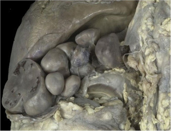
Isomerism of the bronchial tree is an integral part of the phenotype of most patients with heterotaxy, and although helpful in determining the subtype, this feature has few clinical implications with regard to respiratory function. In contrast, gastrointestinal abnormalities are frequently associated with heterotaxy syndrome, and may have significant impact on outcome. The spleen is typically affected. In those with isomeric right atrial appendages, most frequently the spleen is absent, while multiple, small, poorly functioning spleens are the rule in those with isomeric left atrial appendages (Fig. 10).40 Assessment of splenic function, therefore, is important in the neonate presenting with heterotaxy syndrome, because antibiotic prophylaxis and vaccination against pneumococcus may be required to protect against bacterial organisms. In the era prior to vaccination, up to one-quarter of patients with congenital asplenia developed sepsis.Reference Waldman, Rosenthal, Smith, Shurin and Nadas58
Pathologic specimen of the abdomen demonstrating polysplenia; there are multiple, small, variable-sized spleens seen inferior to the liver edge.

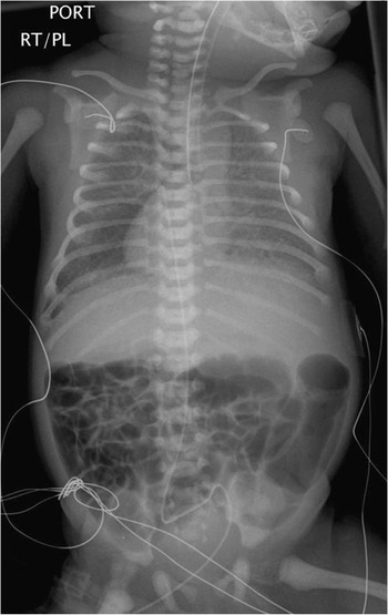
Malrotation of the gut is also very common in both subtypes of heterotaxy syndromeReference Choi, Borenstein, Hornberger and Langer59 (Fig. 11). Some have recommended screening of all individuals with heterotaxy syndrome using barium contrast studies.Reference Ferdman, States, Gaynor, Hedrick and Rychik60, Reference Applegate, Goske, Pierce and Murphy61 Once malrotation is diagnosed, a prophylactic Ladd procedure is generally recommended to prevent volvulus of the midgut, a potentially life-threatening complication. Other associated gastrointestinal anomalies include microgastria, hiatal hernia, absence of the gall bladder, omphalocele, and horseshoe adrenal gland.Reference Ticho, Goldstein and Van Praagh62–Reference Strouse, Haller, Berdon, Rosovsky, Bellah and Barr64 Anal atresia occurs in those with isomeric right atrial appendages, and up to one-tenth of children with isomeric left atrial appendages may have biliary atresia, another potentially lifethreatening disorder.Reference Ticho, Goldstein and Van Praagh62, Reference Chandra65 As expected, the combination of complex cardiac disease with biliary atresia has a very poor outcome.
This total body roentgenogram of an infant with heterotaxy syndrome demonstrates many of its clinical features, including right-sided heart, abnormal lung fields, this being the result of obstructed totally anomalous pulmonary venous connection, midline liver, and malrotation, with the majority of the small intestines on the right and the large intestines on the left.

Other midline defects have been observed in association with heterotaxy syndrome.Reference Ticho, Goldstein and Van Praagh62 Abnormalities of the central nervous system include meningomyelocele, porencephalic cyst and cerebellar agenesis. Cranio-facial anomalies seen include cleft lip and palate, micrognathia and choanal atresia. Absence or hypoplasia of the kidney can be seen. Moreover, musculoskeletal abnormalities such as kyphoscoliosis, fused or hemivertebrae or pectus deformities are seen in one-eighth of patients.Reference Ticho, Goldstein and Van Praagh62 Ultimately, the infant with heterotaxy syndrome requires a complete and thorough multi-system assessment to identify non-cardiac anomalies, with particular emphasis on splenic anatomy and function, hepatic function and assessment for malrotation.
Outcomes following cardiac surgery
The natural history for patients with heterotaxy and complex cardiac involvement is poor. The types of associated cardiac abnormalities preclude a biventricular repair in the majority of patients. Initial surgical palliations for these patients have been characterized by high mortality and unfavorable midterm survival. This pertains to those with isomeric left atrial appendages,Reference Gilljam, McCrindle, Smallhorn, Williams and Freedom29 and to a greater degree those with isomeric right atrial appendages, who frequently have totally anomalous pulmonary venous drainage with obstruction. Half of those patients who undergo initial palliation do not survive.Reference Hashmi, Abu-Sulaiman, McCrindle, Smallhorn, Williams and Freedom66, Reference Gaynor, Collins, Rychik, Gaughan and Spray67 Recent reports, however, suggest that the presence of heterotaxy does not independently increase the risk of mortality. The rate of re-operation for those with totally anomalous pulmonary venous connections, nonetheless, is higher than in those with normal pulmonary venous connections.Reference Morales, Braud and Booth68
Survivors of functionally univentricular palliation are subsequent candidates for the Fontan procedure. Factors associated with increased operative risk for construction of this circulation have historically included abnormalities of the systemic or pulmonary venous connections, an incompetent common atrioventricular valve, a morphologically dominant right ventricle, and disturbances of cardiac rhythm, all frequently present in patients with heterotaxy. A variety of surgical and medical innovations have developed over the last decade to improve outcomes in such patients. Contemporary studies suggest that this improvement in surgical outcome has also translated to those with heterotaxy.Reference Azakie, Merklinger, Williams, Van Arsdell, Coles and Adatia69
Surgical modifications of the Fontan procedure have now been compared across surgical eras, revealing improved early survival in those with heterotaxy.Reference Bartz, Driscoll and Dearani70 Analysis of a recent cohort revealed the presence of heterotaxy no longer to be an independent risk factor for early or late death after completion of the Fontan circuit, as it had been in previous eras.Reference Gentles, Gauvreau and Mayer71, Reference Stamm, Friehs and Duebener72 Although mortality has improved, morbidity compared with patients not having heterotaxy remains significant, and includes early and late arrhythmias, as well as perioperative prolonged pleural effusions.Reference Stamm, Friehs and Duebener72 Continued reassessment and close follow up are essential in order to optimize the long-term outcome of these complicated patients.
Conclusions
In 2007, the heterotaxy syndrome remains one of the most challenging and complex anomalies for the paediatric cardiologist and cardiothoracic surgeon. It even continues to stir controversy with regard to its nomenclature! The key to the genetic aetiology is not beyond reach, now that genes that encode laterality are being discovered and investigated. Prenatal diagnosis early in gestation allows for appropriate counselling for families faced with such a difficult diagnosis, and instigates prompt treatment after birth. The diagnosis of totally anomalous pulmonary venous connection or complete heart block in association with heterotaxy should raise concern regarding the long-term outcome. Practitioners need to identify and treat non-cardiac anomalies, such as functional asplenia and malrotation of the gut. Arrhythmias may occur during the lifetime of the patients, and insertion of pacemakers may be required. Despite all of these adversities, the patient with heterotaxy who achieves survival after conversion to the Fontan circulation now has similar outcomes to the patient without the disorder. Cardiac transplantation as an initial therapy may become a viable option for those with the most complex aspects of the disease.