Introduction
The monogeneric dytiscid subfamily Coptotominae (Coleoptera: Dytiscidae) is a Nearctic endemic group of diving beetles that includes five medium-sized (6.0–8.0 mm long) species generally inhabiting weedy areas of permanent ponds, lakes, and slowly flowing streams (Larson et al. Reference Larson, Alarie and Roughley2000; Nilsson Reference Nilsson2001). The genus Coptotomus Say has a broad range of distribution in North America being found from southern Canada to northern Mexico (Larson et al. Reference Larson, Alarie and Roughley2000).
The taxonomic placement of Coptotomus within the Dytiscidae has been the matter of much debate. Traditionally, Coptotomus has been treated as a monotypic tribe in the subfamily Colymbetinae (e.g., Brinck Reference Brinck1948; Burmeister Reference Burmeister1976; Ruhnau and Brancucci Reference Ruhnau and Brancucci1984). In a comparative study of the Hydradephaga ovipositor, Burmeister (Reference Burmeister1976) found a close relationship between Coptotomus and the colymbetine genera Colymbetes Clairville, Rhantus Dejean, and Meladema Laporte. More recently, Miller (Reference Miller2001) provided convincing arguments in support of the elevation of Coptotomus to subfamily level based mainly on female genitalia and postulated a sister-group relationship between Coptotomus and a clade comprised the subfamilies Copelatinae, Hydrodytinae, and Hydroporinae. Barman (Reference Barman2004), based on larval characters, postulated a sister-group relationship between Coptotominae and Laccophilinae, and Ribera et al. (Reference Ribera, Vogler and Balke2008), based on molecular data, found Coptotomus nested within Copelatinae, as sister to the genus Agaporomorphus Zimmermann.
The study of larval morphology provides another line of evidence to test phylogenetic hypotheses. As different expressions of the same genotype, larval characters help to complement adult characters, which have been traditionally the primary basis for classification. Recent studies have demonstrated the taxonomic and phylogenetic value of chaetotaxy in studying larval Dytiscidae (e.g., Alarie et al. Reference Alarie, Nilsson, Hendrich, Watts and Balke2000, Reference Alarie, Michat, Nilsson, Archangelsky and Hendrich2009, Reference Alarie, Michat and Miller2011; Alarie and Michat Reference Alarie and Michat2007; Michat et al. Reference Michat, Alarie, Torres and Megna2007; Michat and Alarie Reference Michat and Alarie2009; Michat and Torres Reference Michat and Torres2009). There is an overall pattern of setae and pores, which is widespread among taxa, though it is modified in a variety of groups. This generalised pattern is consistent enough to be used for phylogenetic analysis and yet sufficiently variable to allow for taxonomic distinction.
Knowledge of the larval morphology of Coptotomus is scanty. Larvae of two species have been described: Coptotomus interrogatus (Fabricius) (Wilson Reference Wilson1923; De Marzo Reference De Marzo1976; Barman Reference Barman2004) and Coptotomus longulus lenticus Hilsenhoff (Bacon et al. Reference Bacon, Barman and White2000, only the mature larva). Descriptions at the genus level were also provided by Ruhnau and Brancucci (Reference Ruhnau and Brancucci1984), Nilsson (Reference Nilsson1988) (only leg primary chaetotaxy), and Larson et al. (Reference Larson, Alarie and Roughley2000). Most of these treatments, however, lack comparative precision and detail and can hardly be used in the context of the recent system developed for the Dytiscidae (e.g., Alarie Reference Alarie1995, Reference Alarie1998; Alarie et al. Reference Alarie, Archangelsky, Nilsson and Watts2002, Reference Alarie, Michat, Nilsson, Archangelsky and Hendrich2009, Reference Alarie, Michat and Miller2011; Michat and Torres Reference Michat and Torres2009). The aims of this paper therefore are: (i) to describe and illustrate all larval instars of C. longulus lenticus including detailed morphometric and chaetotaxic analyses of the cephalic capsule, head appendages, legs, last abdominal segment, and urogomphus; (ii) to propose the ground plan pattern of primary setae and pores for the subfamily Coptotominae; and (iii) to revisit the relative phylogenetic placement of the Coptotominae within Dytiscidae based on an extensive data set of larval characters.
Materials and methods
Material examined
Eight specimens of instar I, five of instar II, and three of instar III of C. longulus lenticus were used for the descriptions. Larvae were collected in association with adults at the following locality: Canada, Trois-Rivières area (46°22′N, 72°31′W), which is the southern border of the region (41 794 km2) drained by the St-Maurice River, mid-way between the cities of Québec and Montréal. Sampling sites included open pools surrounded by willow shrubs (Salix bebbiana Sargent and Salix discolor Mühlenberg (Salicaceae)) and cattails (Typha angustifolia Linnaeus and Typha latifolia Linnaeus (Typhaceae)). The identification is firm as C. longulus lenticus is the only species of Coptotomus collected at that locality. Coptotomus loticus Hilsenhoff, the only other species of the genus present in Canada, was never collected in the region where the larvae and adults were collected. Also, that species has been most frequently found in flowing waters.
Methods
Specimens were cleared in lactic acid, dissected and mounted on glass slides with polyvinyl-lacto-glycerol. Observation (at magnifications up to 1000×) and drawings were made using an Olympus CX31 (Olympus Corporation, Tokyo, Japan) compound microscope equipped with a camera lucida. Drawings were scanned and digitally inked using a Genius PenSketch tablet (KYE Corporation, Taipei, Taiwan). The material is held in the larval collection of Y. Alarie (Laurentian University, Sudbury, Ontario, Canada).
Morphometric analysis
We employed the terms used in previous papers dealing with the larval morphology of Agabinae and Colymbetinae (Michat Reference Michat2005; Alarie et al. Reference Alarie, Michat, Nilsson, Archangelsky and Hendrich2009; Michat and Archangelsky Reference Michat and Archangelsky2009). Paired structures of each individual were considered independently. The following measurements were taken (with abbreviations shown in parentheses). Head length (HL) (total HL including the frontoclypeus, measured medially along the epicranial stem); maximum head width (HW); length of frontoclypeus (FRL) (from apex of nasale to the joint of frontal and coronal sutures); occipital foramen width (OCW) (maximum width measured along dorsal margin); coronal line length (COL); length of mandible (MNL) (measured from laterobasal angle to apex); width of mandible (MNW) (maximum width measured at base); length of maxillary palpifer (PPF); length of galea (GA). Length of antenna (A), maxillary (MP) and labial (LP) palpi were derived by adding the lengths of the individual segments; each segment is denoted by the corresponding letter(s) followed by a number (e.g., A1, first antennomere). A3′ is used as an abbreviation for the apical lateroventral process of the third antennomere. Length of leg, including the longest claw (CL), was derived by adding the lengths of the individual segments; each leg is denoted by the letter L followed by a number (e.g., L1, prothoracic leg). The length of trochanter includes only the proximal portion, considered from the base to the beginning of the femur. The legs were considered as being composed of six articles following Lawrence (Reference Lawrence1991). Length of last abdominal segment (LAS) (measured dorsally along midline from anterior to posterior margin). Length of urogomphus (U) (total length from base to apex). These measurements were used to calculate several ratios that characterise body shape.
Chaetotaxic analysis
Primary (present in first instar) and secondary (added in later instars) setae and pores were distinguished in the cephalic capsule, head appendages, legs, LAS, and urogomphus. Sensilla were coded by two capital letters, in most cases corresponding to the first two letters of the name of the structure on which are located, and a number (setae) or a lower case letter (pores). The following abbreviations were used: AB, abdominal segment VIII; AN, antenna; CO, coxa; FE, femur; FR, frontoclypeus; LA, labium; MN, mandible; MX, maxilla; PA, parietal; PT, pretarsus; TA, tarsus; TI, tibia; TR, trochanter; UR, urogomphus. Setae and pores present in first instars were labelled by comparison with the groundplan of chaetotaxy of the subfamilies Agabinae and Colymbetinae (Alarie Reference Alarie1995, Reference Alarie1998; Michat Reference Michat2005; Alarie et al. Reference Alarie, Michat, Nilsson, Archangelsky and Hendrich2009; Michat and Archangelsky Reference Michat and Archangelsky2009). Homologies were recognised using the criterion of similarity of position (Wiley Reference Wiley1981). Setae located at the apices of the antenna and maxillary and labial palpi were extremely difficult to distinguish due to their position and small size. Accordingly, they are not well represented in the drawings.
Phylogenetic analysis
The phylogenetic relationships of Coptotomus (Coptotominae) were analysed cladistically using the program TNT (Goloboff et al. Reference Goloboff, Farris and Nixon2008) and considering the character set provided by the larval chaetotaxy and morphology. A broad sampling of taxa was included comprising members of eight of the remaining nine dytiscid subfamilies. The subfamily Hydrodytinae was not included because their larvae are unknown. Data were scored directly from the observation of the specimens except for C. interrogatus; data for this species were scored from Barman (Reference Barman2004). In order to avoid the problem of comparing different semaphoronts (see Meier and Lim Reference Meier and Lim2009), in the codification of each character the same instars were compared. The tree was rooted in Aspidytidae (another family of aquatic Adephaga) to allow the dytiscid taxa to vary freely, thus testing the relationships of Coptotominae with the other dytiscid subfamilies. All characters were treated as unordered and equally weighted. Multistate characters were treated as nonadditive. A heuristic search was implemented using “tree bisection reconnection” as algorithm, with 200 replicates and saving 100 trees per replication (previously setting “hold 20000”). Bremer support values were calculated using the commands “hold 20000”, “sub n” and “bsupport”, where “n” is the number of extra steps allowed. The process was repeated increasing the length of the suboptimal cladograms by one step, until all Bremer values were obtained (Kitching et al. Reference Kitching, Forey, Humphries and Williams1998).
Results
Description of the larvae of C. longulus lenticus Hilsenhoff
Diagnosis
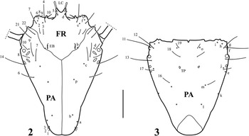
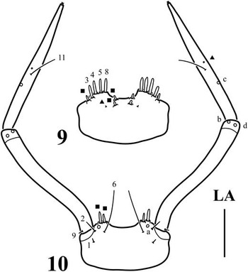
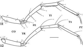
The larvae of Coptotomus are characterised by the following combination of characters: occipital suture absent (Figs. 2, 16); egg bursters elongate (Fig. 2); anterior margin of FR projected forward in a horn (nasale) (Figs. 2, 16); instar I bearing two lamellae clypeales (Fig. 2); A3 with a ventroapical spinula (Fig. 5); cleaning device present on proTI and proTA (Figs. 17–18); abdominal segments I–VI bearing tracheal gills (Fig. 1); U long, one-segmented, bearing rows of elongate spinulae (instar I, Fig. 15) or setae (instars II–III, Fig. 19) on lateral margins; seta AN3 located basally (Fig. 5); A3 without additional pores (Figs. 4–5); stipes with one additional seta contiguous to MX5 and MX6 (Fig. 7); prementum with 5–7 additional spine-like setae (Figs. 9–10); setae LA10 and LA12 absent (Figs. 9–10); seta CO7 inserted proximally on all legs (Fig. 11); seta FE1 inserted submedially (Fig. 11); setae FE5 and TI6 short, spine-like (Fig. 12); seta TI7 elongate, hair-like (Fig. 12); seta AB13 and pore ABc absent (Figs. 13–14); TI and TA with natatory setae (instars II–III) (Fig. 18); LAS with a row of natatory setae on each lateral margin (instars II–III) (Fig. 19).
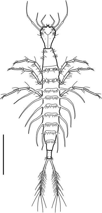
Coptotomus longulus lenticus, first instar, habitus, dorsal aspect. Scale bar = 1.50 mm.

Coptotomus longulus lenticus, first instar. (2) Cephalic capsule, dorsal aspect; (3) Cephalic capsule, ventral aspect. FR, frontoclypeus; PA, parietal; EB, egg bursters; LC, lamellae clypeales; TP, tentorial pits. Scale bar = 0.20 mm.

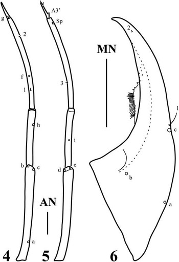
Coptotomus longulus lenticus, first instar. (4) Right antenna, dorsal aspect; (5) left antenna, ventral aspect; (6) right mandible, dorsal aspect. MN, mandible; AN, antenna; SP, spinula. Scale bars = 0.10 mm.

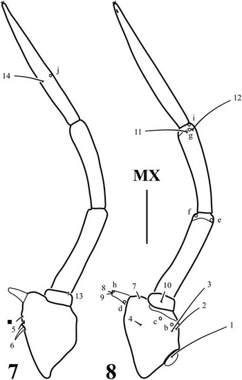
Coptotomus longulus lenticus, first instar. (7) Right maxilla, dorsal aspect; (8) left maxilla, ventral aspect. Solid square refers to additional seta. MX, maxilla. Scale bar = 0.10 mm.

Coptotomus longulus lenticus, first instar. (9) Prementum, dorsal aspect; (10) labium, ventral aspect. Solid squares refer to additional setae; solid triangles refer to additional pores. LA, labium. Scale bar = 0.10 mm.

Coptotomus longulus lenticus, first instar. (11) Left metathoracic leg, anterior aspect; (12) right metathoracic leg, posterior aspect. CO, coxa; TR, trochanter; FE, femur; TI, tibia; TA, tarsus; PT, pretarsus. Scale bar = 0.15 mm.

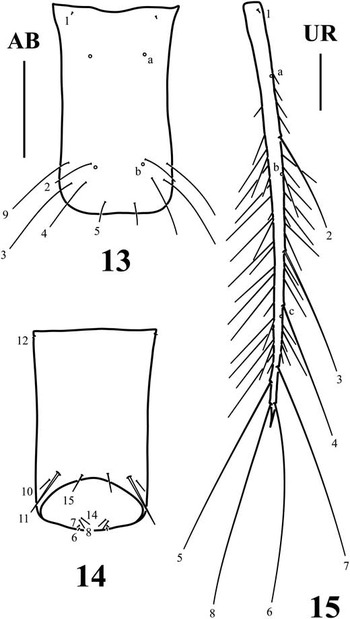
Coptotomus longulus lenticus, first instar. (13) Abdominal segment VIII, dorsal aspect; (14) abdominal segment VIII, ventral aspect; (15) right urogomphus, dorsal aspect. AB, abdominal segment VIII; UR, urogomphus. Scale bars = 0.20 mm.

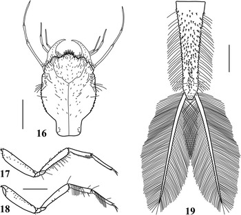
Coptotomus longulus lenticus, third instar. (16) Head, dorsal aspect; (17) left prothoracic leg, anterior aspect; (c) right prothoracic leg, posterior aspect; (19) abdominal segment VIII and urogomphi, dorsal aspect. Scale bars = 0.70 mm.

First instar (Figs. 1–15)
Colour (alcohol-preserved specimens). Larva uniformly light brown to brown; cephalic capsule, thorax and abdomen sometimes darker than head appendages, legs, and urogomphi.
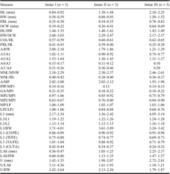
Body. Subcylindrical, widest at metathorax, narrowing anteriorly and posteriorly (Fig. 1). Measurements and ratios that characterise the body shape are shown in Table 1.
Measurements and ratios for the three larval instars of Coptotomus longulus lenticus.

HL, Head length; HW, head width; FRL, frontoclypeus length; OCW, occipital foramen width; COL, coronal line length; MNL, length of mandible; MNW, width of mandible; MP, maxillary palpi; PPF, length of maxillary palpifer; GA, length of galea; LP, labial palpi; LAS, last abdominal segment.
Head. Cephalic capsule (Figs. 2–3). Subtriangular, longer than broad; posterodorsal and lateral surfaces covered with minute spinulae; maximum width at level of stemmata, narrowing towards occipital foramen; neck constriction present, moderately marked; occipital suture absent; ecdysial line visible, coronal line long; occipital foramen deeply emarginate ventrally; posterior tentorial pits visible ventrally; FR subtriangular, with one elongate, slightly prominent egg burster on each posterolateral margin, anterior margin projected forward in a V-shaped horn (nasale) bearing two well-developed lamellae clypeales; anterolateral lobes well developed, not projected beyond nasale; six stemmata at each side, arranged in two vertical rows. Antenna (Figs. 4–5). Very long, slender, much longer than HW, composed of four antennomeres; A4 the shortest, A1 and A3 the longest, subequal, A3 with a ventroapical spinula; A3’ short. Mandible (Fig. 6). Prominent, broad basally, distal half projected inwards, apex sharp, basoexternal margin with minute spinulae, ventrointernal margin with denticles; mandibular channel present. Maxilla (Figs. 7–8). Cardo small, subovate; stipes short, broad, with minute spinulae on dorsointernal and ventrointernal surfaces, anterointernal angle prominent; galea well developed, subconical, lacinia absent; PPF short, palpomere-like; MP elongate, composed of three palpomeres, MP1 and MP2 the shortest, subequal, MP3 the longest. Labium (Figs. 9–10). Prementum well developed, subrectangular, broader than long, with minute spinulae dorsally; LP long, composed of two palpomeres subequal in length.
Thorax. Terga convex, pronotum about as long as mesonotum and metanotum combined, mesonotum and metanotum subequal, wider than pronotum; protergite short, subrectangular, not covering anterior and posterior parts of pronotum, more developed than mesotergite and metatergite; mesotergite and metatergite small, transverse, not covering the whole dorsal surface; all sclerites without anterior transverse carina, with sagittal line; thoracic sterna membranous; spiracles absent. Legs (Figs. 11–12). Long, composed of six segments, L1 the shortest, L3 the longest; CO robust, elongate, TR transversely divided into two parts by an annulus, FE, TI, and TA slender, subcylindrical, PT with two long, slender, slightly curved claws, posterior claw shorter than anterior one; surface of CO (dorsoproximal), FE, TI, and TA covered with minute slender spinulae; ventral surface of TI and TA with a row of long spinulae; spinulae on anteroventrodistal portion of proTI and anteroventroproximal portion of proTA dense, forming weakly delimited patches (cleaning device).
Abdomen. Eight-segmented; segments I–VI sclerotised dorsally, membranous ventrally, bearing a long tracheal gill on each lateral; segment VII narrower, sclerotised dorsally, membranous ventrally; tergites I–VII small, narrow, transverse, rounded laterally, without sagittal line; all sclerites without anterior transverse carina, covered with minute slender spinulae; spiracles absent on segments I–VII; LAS (Figs. 13–14) the narrowest, truncate posteriorly, lacking a siphon; completely sclerotised, ring-like, covered with minute slender spinulae. Urogomphus (Fig. 15). Very long, composed of one urogomphomere, much longer than LAS, surface covered with minute slender spinulae, lateral margins with rows of elongate spinulae.
Chaetotaxy (Figs. 1–15). Similar to that of generalised Agabinae and Colymbetinae larvae (Alarie Reference Alarie1995, Reference Alarie1998; Michat Reference Michat2005; Alarie et al. Reference Alarie, Michat, Nilsson, Archangelsky and Hendrich2009; Michat and Archangelsky Reference Michat and Archangelsky2009) except for the following features: pore FRe absent; seta AN3 located basally; pore ANg proximal; A3 without additional pores; seta MN1 close to pore MNc; stipes with one additional seta contiguous to MX5 and MX6; MP3 with a minute structure (possibly a spinula) on ventrodistal margin; prementum with 4–6 additional spine-like setae on anterodorsal and anteroventral surfaces, and one additional pore on dorsal surface; setae LA10 and LA12 absent; LP2 with a minute structure (possibly a pore) on ventral surface, near seta LA11; seta CO7 inserted proximally on all legs; seta FE1 inserted submedially; seta FE5 short, spine-like; additional setae occasionally present on dorsal and ventral surfaces of mesoFE and metaFE; seta TI6 short, spine-like; seta TI7 long, hair-like; seta TA7 inserted apically on proTA and mesoTA; TI and TA without additional setae; seta AB13 and pore ABc absent; seta AB15 inserted medially to seta AB11; seta UR7 long, hair-like.
Second instar
As for first instar except for the following features:
Body. Measurements and ratios that characterise the body shape are shown in Table 1.
Head. Cephalic capsule. Subovate, with well-developed neck (neck constriction well marked); egg bursters absent; anterior margin of nasale rounded, bearing 11–13 lamellae clypeales. Antenna. A1 the longest, A3 somewhat shorter than A1. Maxilla. MP2 the shortest, MP1 slightly longer than MP2, MP3 slightly longer than MP1. Labium. LP1 somewhat longer than LP2.
Thorax. Protergite, mesotergite, and metatergite covering the whole pronota; mesotergite and metatergite with anterior transverse carina. Legs. Surface spinulae of FE, TI, and TA less abundant; spinular patches on anteroventrodistal portion of proTI and anteroventroproximal portion of proTA (cleaning device) well developed.
Abdomen. Siphon present, very short, rounded apically. Urogomphus. Long spinulae on lateral margins absent.
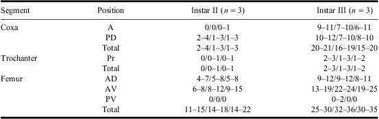
Chaetotaxy. Cephalic capsule with some minute or hair-like secondary setae scarcely distributed on dorsal and ventral surfaces; PA with 9–10 spine-like secondary setae on each lateral margin and 1–2 spine-like secondary setae on each side of ventral surface; MN with one hair-like, secondary seta on basoexternal margin, proximal to pore MNa; stipes with one minute secondary seta on ventral surface, near setae MX2 and MX3; prementum with one secondary pore on ventral surface, near seta LA1; thoracic tergites with several hair-like secondary setae; secondary leg setation detailed in Table 2; TI and TA with a row of natatory setae on posterodorsal margin; CO with one secondary pore on posterior surface; TR with one secondary pore on proximal portion; abdominal sclerites I–VII with some secondary setae mainly on lateral and posterior margins; LAS with several spine-like secondary setae on dorsal surface, 0–1 spine-like secondary setae on ventral surface, and a row of natatory setae on each lateral margin; U with a row of natatory setae on each lateral margin.
Number and position of secondary setae on the legs of larvae of Coptotomus longulus lenticus.
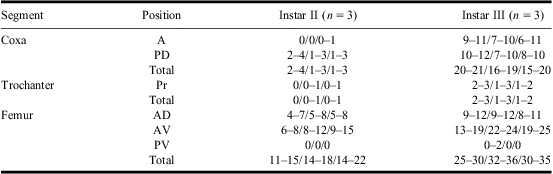
Numbers between slash marks refer to prothoracic, mesothoracic, and metathoracic leg, respectively. Total, total number of secondary setae on the segment (excluding primary and natatory setae).
A, anterior; D, dorsal; P, posterior; Pr, proximal; V, ventral.
Third instar (Figs. 16–19)
As for second instar except for the following features:
Body. Measurements and ratios that characterise the body shape are shown in Table 1.
Head (Fig. 16). Cephalic capsule. Maximum width posterior to stemmata; anterior margin of nasale bearing 21 lamellae clypeales. Mandible. Without spinulae. Maxilla. MP3 slightly longer than MP2, MP1 slightly longer than MP3.
Thorax. Spiracles present on mesothorax. Legs (Figs. 17–18). Surface spinulae of FE, TI, and TA less abundant.
Abdomen. Tergites I–VII more developed, sclerites I and II with poorly delimited anterior transverse carina; spiracles present on segments I–VII. Urogomphus (Fig. 19). Somewhat longer than LAS.
Chaetotaxy. Secondary setae on cephalic capsule somewhat more abundant; PA with 7–9 spine-like secondary setae on each lateral margin and one spine-like secondary seta on each side of ventral surface; MN with some minute secondary setae on external margin; stipes with 1–2 minute secondary setae on ventral surface, near setae MX2 and MX3; prementum with two secondary pores on ventral surface, near seta LA1; secondary leg setation detailed in Table 2 and Figs. 17–18; CO with 1–2 secondary pores on posterior surface; LAS with numerous spine-like secondary setae on dorsal and ventral surfaces (Fig. 19).
Remarks
With the exception of C. longulus lenticus whose larval stages are described in this article, C. interrogatus represents the only other species of Coptotomus for which the first larval stage is known (Barman Reference Barman2004). Interestingly, Barman (Reference Barman2004) postulated that the urogomphus of the first instar of C. interrogatus characterised by the presence of additional setae, which is contrary to the condition observed in C. longulus lenticus. Indeed the urogomphus of C. longulus lenticus has eight primary setae and three primary pores along with several elongate and seta-like spinulae. Species of Coptotomus have long been recognised for their overall morphological similarity (Hilsenhoff Reference Hilsenhoff1980; Larson et al. Reference Larson, Alarie and Roughley2000). It is therefore postulated that the additional setae reported in C. interrogatus are in fact elongate hair-like spinulae similar to the condition observed in the closely related species C. longulus lenticus. These spinulae can easily be confounded with setae when observed at low magnifications. Similarly, Bacon et al. (Reference Bacon, Barman and White2000) reported the presence of two prothoracic sternites and of a completely sclerotised abdominal segment VII in the third instar of C. longulus lenticus. Both these character traits are not corroborated in this study.
Character analysis
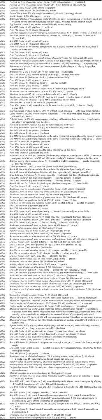
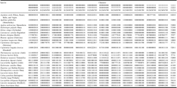
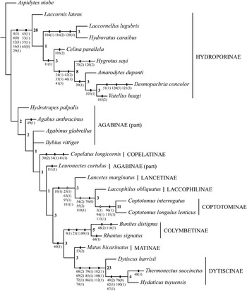
The final data matrix included 25 taxa and 125 characters (94 binary and 31 multistate) (Tables 3–4). The parsimony analysis with TNT yielded two most parsimonious cladograms of 374 steps (consistency index = 0.45; retention index = 0.69). Both trees differed in the relative position of the agabine genera Agabinus Crotch and Ilybius Erichson only. The strict consensus tree was therefore calculated (Fig. 20) in which Coptotominae was recovered as part of the clade Lancetinae + (Coptotominae + Laccophilinae). The derived nodes were generally better supported than the most basal ones. Character states were mapped for each clade (Fig. 20).
Characters and states used for the cladistic analysis.

Data matrix used for the cladistic analysis.

Missing data coded “?”.
Strict consensus cladogram with unique or interesting character changes mapped. Solid rectangles indicate unique character state transformations; open rectangles indicate homoplastic character state transformations. Bremer support values are indicated on each branch.

Discussion
The larvae of C. longulus lenticus described in this paper turn out to be characterised by several unique character states: (1) the presence of well-developed tracheal gills on the lateral margins of the abdominal segments I–VI (character 96.1); (2) the presence of elongate seta-like spinulae on the urogomphus of the first instar (character 115.1); (3) the presence of a short nasale (character 5.1); (4) the presence of a row of natatory setae on the lateral margin of abdominal segment VIII (character 113.1); and (5) the presence of a basoventral patch of dense slender spinulae on the protarsus (character 94.1), postulated to represent either a cleaning or a device for spreading a microbial growth inhibitor (De Marzo and Nilsson Reference De Marzo and Nilsson1986), which is less evident in the first instar (Fig. 20).
The presence of abdominal tracheal gills is noteworthy as this trait resembles those observed in the larvae of Gyrinidae (Michat et al. Reference Michat, Archangelsky and Fernández2010). Indeed all gyrinid larvae share the presence of tracheal gills on the abdominal segments I–IX. Larvae of Coptotomus, however, differ from those of Gyrinidae in that the gills are devoid of spinulae along the lateral edges (Fig. 1). This condition in combination with the fact that larvae of Coptotominae are clearly Dytiscidae allows us to conclude that the presence of abdominal gills in both Gyrinidae and Coptotominae is the result of convergent evolution.
The presence of elongate seta-like spinulae on the urogomphus of the first instar (Fig. 15) represents another putative synapomorphy for the Coptotominae. Whereas such a character state could not be corroborated for C. interrogatus (Barman Reference Barman2004), we do believe that the trait is generalised within the subfamily and that the additional setae described by Barman (Reference Barman2004) refer to these elongate spinulae. Interestingly, the second and third instars of C. longulus lenticus do not have spinulae on the urogomphus but bear a row of natatory setae on both the internal and external margins (Fig. 19). Such setal fringes resemble those of Dytiscus Linnaeus and Hyderodes Hope (Alarie et al. Reference Alarie, Michat and Miller2011), although in those dytiscine genera the natatory setae are restricted to the external margin only.
The presence of a frontoclypeal projection (=nasale) in both the Coptotominae and Hydroporinae is also noteworthy. Compared with the Hydroporinae, which are characterised by an extensive nasale reaching approximately the tip of the mandibles, the nasale of Coptotomus barely gets beyond the level of the lateral lobes (Figs. 2, 16). Moreover, as it arises post marginally from within the frontoclypeal interior in the Coptotominae (Barman Reference Barman2004), compared with being strictly an elongation of the distal margin within the Hydroporinae, it is reasonable to postulate that both conditions evolved independently. It is worth mentioning that the structure sometimes described as a “clypeal horn” in the Coptotominae reflects the typical bifid shape of the first instar nasale, which varies through the ontogenetic development of the larva (compare Figs. 2 and 16).
The presence of a basoventral patch of spinulae on the ventroproximal portion of the protarsus (which is accompanied by another patch on the anteroventrodistal portion of protibia (Fig. 17) to form the device mentioned above) and the presence of a row of natatory setae on the lateral margin of abdominal segment VIII are other interesting features of Coptotominae larvae. No other dytiscid has these features except members of the subfamily Dytiscinae (Alarie et al. Reference Alarie, Michat and Miller2011). Whereas these character states support a closer relationship between both subfamilies, our results suggest that the conditions found in Coptotominae and Dytiscinae are the result of convergent evolution.
Several different hypotheses have been proposed as to where to place the Coptotominae within Dytiscidae (Brinck Reference Brinck1948; Burmeister Reference Burmeister1976; Ruhnau and Brancucci Reference Ruhnau and Brancucci1984; Miller Reference Miller2001; Barman Reference Barman2004; Ribera et al. Reference Ribera, Vogler and Balke2008). Although not strongly supported (Bremer value = 3), our study suggests a monophyletic origin of Lancetinae + Laccophilinae + Coptotominae. This relationship is interesting from an evolutionary point of view, as the larvae of these subfamilies share a unique feature within Dytiscidae, the presence of an unusually low number of lamellae clypeales in the first instar (character 10.1) (Fig. 2). Such a character trait is somewhat related to the “four-peg-pattern” suggested by Ruhnau and Brancucci (Reference Ruhnau and Brancucci1984), which combines the two anteromedial lamellae of the frontoclypeus with the two sublateral spines labelled as FR10 within the Dytiscidae. The “four-peg-pattern” is also observed in the Carabidae and Gyrinidae, where the two anteromedial lamellae are labelled FR11 (Bousquet and Goulet Reference Bousquet and Goulet1984; Michat et al. Reference Michat, Archangelsky and Fernández2010). Ruhnau and Brancucci (Reference Ruhnau and Brancucci1984) postulated that the “four-peg-pattern” has arisen independently in Laccophilinae, Coptotominae, and Lancetinae, and that it represents a reversal to the ancestral condition found in Carabidae and Gyrinidae. This reinforces the idea that the presence of only two lamellae clypeales within the Dytiscidae evolved secondarily from patterns composed of a larger number of lamellae. Our results, however, do not support an independent acquisition of this pattern in each of the three subfamilies as long as they form a distinct clade in our analysis. See De Marzo and Nilsson (Reference De Marzo and Nilsson1986) and Friis et al. (Reference Friis, Bauer and Betz2003) for hypotheses on the possible function of the lamellae clypeales.
The monophyletic origin of Lancetinae + Laccophilinae + Coptotominae proposed in our study is also supported by: (1) the submedial insertion of seta AN3 on the third antennomere (character 23.1); (2) the proximal insertion of seta CO7 on the meso- and metacoxa (character 63.1); and (3) the absence of an anterior transverse carina on abdominal tergites I–VII in the first instar (characters 97.1 and 101.1, shared with Copelatinae). These characters must be treated with caution knowing that they are also present in some taxa of Hydroporinae and Dytiscinae. However, as both subfamilies are strongly supported, monophyletic groups (Fig. 20) they most probably represent independent evolutionary processes.
Our study gives moderate support (Bremer value = 3) to a monophyletic origin of Laccophilinae + Coptotominae, which reinforces the hypothesis formulated by Barman (Reference Barman2004) based on a smaller data set. This grouping is supported by the absence of setae LA10 and LA12 on the second labial palpomere (characters 54.2 and 55.2, homoplastic in Vatellus Aubé), and the absence of pore ABc on the LAS (character 110.1, homoplastic in Amarodytes Régimbart and Thermonectus Dejean) (Fig. 20). Both character states, however, must be seen as less meaningful evolutionarily because they are present in other dytiscid taxa. The primary setae LA10 and LA12 are well developed in the Hydroporinae except Vatellini (see Michat and Torres Reference Michat and Torres2005, Reference Michat and Torres2011) compared with very small to minute in the Colymbetinae, Copelatinae, Dytiscinae, and Lancetinae (Michat et al. Reference Michat, Archangelsky and Torres2005; Alarie et al. Reference Alarie, Michat, Nilsson, Archangelsky and Hendrich2009, Reference Alarie, Michat and Miller2011; Michat and Torres Reference Michat and Torres2009). On the other hand, the pore ABc is also absent in the hydroporine tribe Bidessini (Michat and Alarie Reference Michat and Alarie2008) and in the dytiscine tribes Aciliini and Eretini (Alarie et al. Reference Alarie, Michat and Miller2011).
Acknowledgements
We thank the anonymous referees for their useful comments on the manuscript. Laboratory work by M. C. Michat was supported by an external scholarship from the Consejo Nacional de Investigaciones Científicas y Técnicas (CONICET) and projects PIP 112-200801-02759 from CONICET and PICT-2007-01438 from the Agencia Nacional de Promoción Científica y Tecnológica. Financial support was also provided by the Natural Sciences and Engineering Research Council of Canada in the form of an operating research grant to Y. Alarie.