Introduction
Steinernematid nematodes (Steinernematidae: Rhabditida) are obligate lethal pathogens of insects with a worldwide distribution (Poinar, Reference Poinar1979; Adams et al., Reference Adams, Fodor, Koppenhöfer, Stackebrandt, Stock and Klein2006). So far around 90 steinernematid species have been described from all continents except Antarctica. Despite its large area and high diversity of habitats, only 15 steinernematid nematodes have been reported so far from the African continent.
The only well-explored country on the continent is South Africa, where several surveys have been performed (Malan et al., Reference Malan, Nguyen and Addison2006, Reference Malan, Knoetze and Moore2011; Hatting et al., Reference Hatting, Stock and Hazir2009) resulting in the recovery of seven entomopathogenic nematode (EPN) species, including six new species. The rest of the continent is still largely unexplored and data on the occurrence of EPNs are available only from a few countries, including Egypt (Shamseldean & Abd-Elgawad, Reference Shamseldean and Abd-Elgawad1994; Abd-Elbary et al., Reference Abd-Elbary, Shamseldean, Stock and Abu-Shady2012), Ethiopia (Mekete et al., Reference Mekete, Gaugler, Nguyen, Mandefro and Tessera2005), Kenya (Mwaniki et al., Reference Mwaniki, Nderitu, Olubayo, Kimenju and Nguyen2008), Tanzania (Mwaitulo et al., Reference Mwaitulo, Haukeland, Sæthre, Laudisoit and Maerere2011), Cameroon (Kanga et al., Reference Kanga, Waeyenberge, Hauser and Moens2012), Nigeria (Akyazi et al., Reference Akyazi, Ansari, Ahmed, Crow and Mekete2012), Benin (Zadji et al., Reference Zadji, Baimey, Afouda, Houssou, Waeyenberge, De Sutter, Moens and Decraemer2013) and DR Congo (Haukeland et al., unpublished). These data mainly represent the results of single surveys, often reporting unidentified steinernematids and heterorhabditids.
A survey in Tanzania revealed 4.4% EPN-positive out of a total of 90 samples (Mwaitulo et al., Reference Mwaitulo, Haukeland, Sæthre, Laudisoit and Maerere2011). Among the isolated nematodes a strain belonging to the ‘glaseri’ group has been recovered. Morphological and molecular examination revealed that this nematode differs from other African species from the ‘glaseri’ group: S. karii Waturu, Hunt & Reid, 1997; S. khoisanae Nguyen, Malan & Gozel, 2006; S. ethiopiense Tamiru, Waeyenberge, Hailu, Ehlers, Půža & Mráček, 2012; S. innovationi Cimen, Lee, Hatting, Hazir & Stock, 2014; S. tophus Cimen, Lee, Hatting, Hazir & Stock, 2014; and S. jeffreyense Malan, Knoetze & Tiedt 2015. The new species is described herein as Steinernema pwaniensis n. sp.
Materials and methods
Collection and examination of nematodes
The nematode was isolated within a survey of biocontrol agents for the control of the banana weevil (Cosmopolites sordidus) (Mwaitulo et al., Reference Mwaitulo, Haukeland, Sæthre, Laudisoit and Maerere2011) in Mwalusembe Village, Pwani region, Tanzania (07°16’46.9”S, 39°08’35.9”E). The soil type in the locality was loamy sand.
Steinernema pwaniensis n. sp. was cultured on last-instar Galleria mellonella as described by Poinar (Reference Poinar1979). First-generation and second-generation adults were obtained by dissecting infected insects approximately 2 days and 4 days, respectively, after the death of the host. The specimens of different stages were heat killed, fixed in TAF (triethanolamine, formalin and water) (Courtney et al., Reference Courtney, Polley and Miller1955) and subsequently processed to glycerine by the Seinhorst method (Seinhorst, Reference Seinhorst1959). Nematodes were transferred to the fresh drop of glycerine on a microscope slide and covered with a coverglass supported by stainless steel wires slightly larger in diameter than the nematodes. Morphometric analysis of the nematode specimens was done for 20 individuals of the adult stages of both generations and infective juveniles (IJs), using light microscopy and the image analysing software Cell D (Olympus Soft Imaging Solutions GMBH, Germany).
Scanning electron microscopy (SEM)
Adults of the first generation and IJs were fixed in 4% formalin buffered with 0.1 m sodium cacodylate at pH 7.2 for 24 h at 4–6°C. They were post-fixed with a 2% osmium tetroxide solution for 12 h at 25°C, dehydrated in a graded ethanol series, critical point dried with liquid CO2, mounted on SEM stubs, and coated with gold (Nguyen & Smart, Reference Nguyen and Smart1995, Reference Nguyen and Smart1997). The mounts were examined with a JEOL 7401 FE scanning electron microscope (JEOL, Eching, Germany).
Molecular analysis
DNA was extracted from single females. Each female was transferred into a sterile Eppendorf tube (500 μl) with 20 μl of extraction buffer (17.7 μl of double-distilled water (ddH2O), 2 μl of 10 × polymerase chain reaction (PCR) buffer, 0.2 μl of 1% Tween and 0.1 μl of proteinase K). Buffer and nematode were frozen at − 20°C for 20 min and then immediately incubated at 65°C for 1 h, followed by 10 min at 95°C. The lysates were cooled on ice, then centrifuged (2 min, 9000 g) and 1 μl of supernatant was used for PCR. A fragment of rDNA containing the internal transcribed spacer regions (ITS1, 5.8S, ITS2) was amplified using primers 18S: 5′-TTGATTACGTCCCTGCCCTTT-3′ (forward), and 28S: 5′-TTTCACTCGCCGTTACTAAGG-3′ (reverse) (Vrain et al., Reference Vrain, Wakarchuk, Levesque and Hamilton1992). The other fragment containing D2–D3 expansion segments of the 28S rDNA gene was amplified using primers D2F: 5′-CCTTAGTAACGGCGAGTGAAA-3′ (forward) and 536: 5′-CAGCTATCCTGAGGAAAC-3′ (reverse) (Nguyen, Reference Nguyen, Nguyen and Hunt2007). The PCR master mix consisted of: ddH2O, 7.25 μl; 10 × PCR buffer, 1.25 μl; deoxynucleoside triphosphates (dNTPs), 1 μl; 0.75 μl of each forward and reverse primer; polymerase, 0.1 μl; and 1 μl of DNA extract. The PCR profiles were used as follows for ITS: 1 cycle of 94°C for 7 min; followed by 35 cycles of 94°C for 60 s, 50°C for 60 s and 72°C for 60 s; and a final extension at 72°C for 7 min (Nguyen, Reference Nguyen, Nguyen and Hunt2007); and for 28S rDNA: 1 cycle of 94°C for 7 min; followed by 35 cycles of 94°C for 60 s, 55°C for 60 s and 72°C for 60 s; and a final extension at 72°C for 10 min (Mráček et al., Reference Mráček, Půža and Nermut'2014). PCR was followed by electrophoresis (45 min, 120 V) of 2 μl of PCR product in a 1% Tris–acetate–EDTA (TAE)-buffered agarose gel stained with ethidium bromide (ETB, 20 μl per 100 ml of gel). The PCR products were sequenced by Macrogen Inc. (Korea). The presence of a double peak in the otherwise flawless ITS sequence chromatogram suggested the presence of intra-individual variability (Půža et al., Reference Půža, Chundelová, Nermut', Žurovcová and Mráček2015). Thus, the PCR products of two single females were cloned into the pGEM®-T Easy vector (Promega, Madison, Wisconsin, USA) following the manufacturer's protocol. For each female, DNA from five recombinant clones was re-amplified and purified with Exo-SAP-IT (USB Corporation, Cleveland, Ohio, USA) according to the manufacturer's instructions and sequenced using the universal primers T7 (5′-GATAATACGACTCACTATAGGGCTTCTTGCATTCTG-3′) and SP6 (5′-TCATTTAGGTGACACTATAGGACTTCAGGTGGCTGGA-3′).
The PCR products were sequenced and deposited in GenBank under accession numbers KT358811 and KT358812 (ITS sequences) and KT358813 (28S sequence). The sequences were edited and compared with those present in GenBank by means of a Basic Local Alignment Search Tool (BLAST) of the National Center for Biotechnology Information (NCBI). An alignment of our samples together with sequences of the species of the ‘glaseri’ group was produced for each amplified DNA region using default ClustalW parameters in MEGA 6.0 (Tamura et al., Reference Tamura, Stecher, Peterson, Filipski and Kumar2013) and optimized manually in BioEdit (Hall, Reference Hall1999). Pairwise distances were computed using MEGA 6.0 (Tamura et al., Reference Tamura, Stecher, Peterson, Filipski and Kumar2013). Codon positions included were first + second + third + non-coding.
The phylogenetic trees were obtained by the Minimum Evolution method (Rzhetsky & Nei, Reference Rzhetsky and Nei1992) in MEGA 6.0 (Tamura et al., Reference Tamura, Stecher, Peterson, Filipski and Kumar2013). Steinernema akhursti was used as an outgroup. The Minimum Evolution tree was searched using the Close-Neighbour-Interchange (CNI) algorithm (Nei & Kumar, Reference Nei and Kumar2000). The neighbour-joining algorithm (Saitou & Nei, Reference Saitou and Nei1987) was used to generate the initial tree. The evolutionary distances were computed using the p-distance method (Nei & Kumar, Reference Nei and Kumar2000) and are expressed as the number of base differences per site.
Results and discussion
Description of Steinernema pwaniensis n. sp.
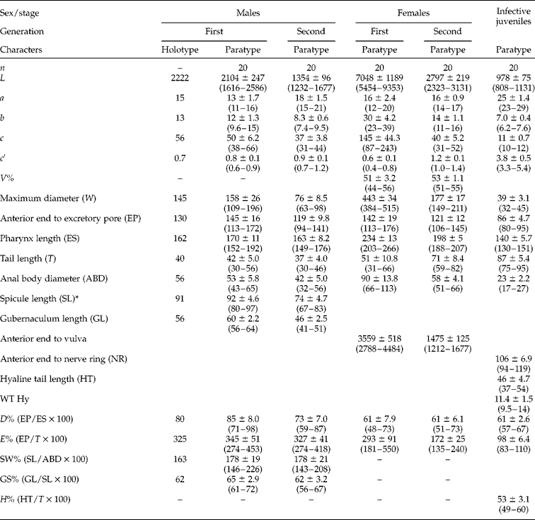
Morphometrics of the holotype (the first-generation male) and paratypes (other stages) are summarized in table 1. Figures 1–3 illustrate the description.
Morphometric characters (in μm) of Steinernema pwaniensis n. sp. based on the holotype and 20 paratypes of each generation with mean ± SD and ranges given in brackets.

* Measured along the chord. L= body length; a= L/W; b= L/ES; c= L/T; c′ = T/ABD; V% = distance from anterior end to vulva/L; WT Hy = tail width at beginning of hyaline layer.
Steinernema pwaniensis n. sp.: (A) first-generation male, tail, lateral; (B) second-generation male, tail with mucron, lateral; (C) second-generation male, tail with arrangement of genital papillae; (D) first-generation male, anterior region with excretory pore; (E) first-generation male, spicule; (F) first-generation female, tail with terminal peg; (G) second-generation female, tail with mucron; (H) first-generation female, vulval region; (I–L) infective juvenile: (I) anterior region with excretory pore; (J) tail with anus and hyaline region; (K) lateral field with ridges; (L) adult arrangement of labial (lp), cephalic (cp) papillae and amphidial apertures (a). Scale bars: (A–C, E, G, I–L) 50 μm; (D, F, H) 100 μm.

Steinernema pwaniensis n. sp. infective juvenile, male and female. (A–C) Infective juvenile: (A) head region with four papillae and amphid openings; (B) lateral field in mid-body (ridges numbered 1–8); (C) lateral field in tail region with phasmid opening (arrow). (D) First-generation female tail with terminal peg and projections. (E) Second-generation female tail with mucron. (F) First-generation male tail, lateral. (G–H) Second-generation male: (G) tail with genital papillae, ventro-lateral; (H) tail tip, dorso-lateral, showing two sublateral post-cloacal pairs and one subdorsal post-cloacal pair of genital papillae and mucron (arrow).

Steinernema pwaniensis n. sp. infective juvenile, male and female. (A, C) First-generation female: (A) tail region; (C) vulval region. (B, D) Second-generation female: (B) tail with spine-like mucron; (D) vulval region. (E) First-generation male, tail with spicules and gubernaculum. (F) Second-generation male, tail with mucron, spicules and gubernaculum. (G–H) Infective juvenile: (G) anterior portion showing rounded head and excretory pore (arrow); (H) tail with anus and hyaline region.

Infective juvenile
Body elongate, on average 978 μm long, almost straight or slightly C-shaped when heat-killed, often enclosed in second-stage cuticle sheath. Cephalic region rounded, not offset from body contour. Lip region bearing four cephalic papillae and a pair of pore-like amphidial apertures laterally. Cuticle with prominent striations (distinct under SEM) c. 2 μm wide at mid-body. Lateral fields consisting of eight equally spaced and developed ridges in mid-body region (i.e. nine lines). Lateral field beginning anteriorly with a cuticular depression (line) on the third annulus; at 18th annulus, two ridges appearing and changing to seven ridges (eight lines) at about excretory pore level. Posteriorly, one additional ridge appears, creating a total of eight ridges. In posterior part of body, number of ridges gradually reducing. At the level of 71st annulus, two central ridges fuse, making seven ridges in total. Close to anus, the central ridge disappears (six ridges). Around phasmid, four inner ridges fuse to two, making four ridges in total. Few annuli posteriorly, two marginal ridges disappear, and the last two ridges continue to three-quarters the length of the tail. Formula for lateral field = 2, 7, 8, 7, 6, 4, 2.
Deirids not observed. Hemizonid visible, located at the level of the middle of basal bulb. Pharynx corpus slender, cylindrical, isthmus distinct, surrounded by nerve ring. Excretory pore located posterior to mid-pharynx (D% = 61). Cardia present. Bacterial vesicle usually well developed, ellipsoid, c. 10 μm wide and 20 μm long. Tail conoid, tapering gradually, contour slightly depressed both dorsally and ventrally at about commencement of hyaline region. Hyaline layer occupying c. half of tail length. Anus c. 10 μm wide, sickle-shaped. Phasmids distinct, located c. 35% of tail length posterior to anus, with aperture anterior to the commencement of the hyaline portion.
Male, first generation
Body J-shaped in heat-killed specimens, curved ventrally posteriorly, tapering toward head region. Cuticle with a faint transverse striation. Stoma c. 4 μm deep and 7–9 μm wide. Excretory pore located posterior to nerve ring (c. 85% of distance from anterior body end to base of pharynx). Spicules paired, curved, dark golden-brown in colour, c. 90 μm long, spicule tip pointed. Spicule manubrium elongated, almost twice as long as wide with the ratio manubrium length/manubrium width of 1.7:1. Calomus distinct, but short. Lamina with two internal ribs, well curved. Velum extending from calomus almost to the end of lamina. Gubernaculum boat-shaped, manubrium of gubernaculum curved ventrally, usually bearing one or two protuberances proximally. Tail short, dorsally convex conoid, with a bluntly rounded terminus, mucron absent. Genital papillae totalling 23, comprising 11 pairs and a single midventral papilla located anterior to anus. Paired papillae arranged as follows: six pairs subventral pre-cloacal, one sublateral pre-cloacal, one sublateral ad-cloacal, two sublateral post-cloacal pairs and one subdorsal post-cloacal pair.
Second-generation male
Similar to first-generation male, but smaller. Spine-like tail mucron developed as a short, c. 2 μm long, projection.
First-generation female
Body variable in length, C-shaped when heat relaxed. Head bluntly rounded, slightly tapering anteriorly, continuous with body, bearing six labial and four cephalic papillae. Stoma c. 6–8 μm long and 11–13 μm broad. Excretory pore located anterior to the basal bulb. Vulva indistinct, slightly asymmetrical and in the form of a transverse slit, protruding from body contour. Vulval opening at mid-body. Small epiptygma rarely observed. Vagina short, leading into paired uteri. Rectum narrow, anal opening distinct, without post-anal swelling in mature females. Tail of mature females obese with a peg-like tip bearing three spine-like projections, the more prominent of them being positioned dorsocentrally, corresponding to the arrangements described for the species in the ‘glaseri’ group (Mráček et al., Reference Mráček, Půža and Nermut'2015). Endotokia matricida observed in c. 30% of the females.
Second-generation female
Similar to first generation in general morphology, but smaller. Vulval opening slightly posterior to mid-body, slightly to moderately protruding. Tail conical, longer than anal body diameter, with a pointed tip, extending to form a mucron in some specimens. Post-anal swelling slightly developed.
Life cycle
The life cycle of Steinernema pwaniensis n. sp. is very short. In an experimental infection of G. mellonella with a dose of 50 IJs/insect at a temperature of 22°C, the majority of insects were dead after 24 h, and fourth-stage juveniles were present in the hosts. After 48 h, males and young females could be observed. First IJs appeared 10 days after inoculation.
Type material
Holotype first-generation male, paratype males (11 slides with 31 specimens), paratype females (8 slides with 17 specimens) and third-stage juveniles, (5 slides with 70 specimens) were deposited at the EPN collection in the Laboratory of Entomopathogenic Nematodes, Institute of Entomology, BC CAS, Česke Budějovice, Czech Republic. Three slides with paratype IJs, males and females were deposited at the USDA Nematode Collection, Beltsville, Maryland, USA under accession numbers T-6678p, T-6679p and T-6680p.
Etymology
The name is derived from the regional name Pwani. In Swahili, ‘pwani’ means coast or coastal.
Bacterial symbiont
The symbiotic bacterium of S. pwaniensis n. sp. (bacterial strain TZ01) was determined via its phenotypic characters, fatty-acid profiles, and a molecular and multigene approach by Tailliez et al. (unpublished data). The data show that isolate TZ01 shares a common ancestor with Xenorhabdus griffiniae ID10 and X. ehlersii DSMZ16337, the symbiotic bacteria associated with Steinernema hermaphroditum and Steinernema longicaudum, respectively. Sequences of the genes 16S, recA, gyrB, dnaN, gltX and infB are available under accession numbers JQ687358 and JQ687369–JQ687373. Isolate TZ01 demonstrated high pathogenicity for G. mellonella and Spodoptera littoralis (Tailliez et al., unpublished data).
Diagnosis
Infective juveniles possess a body length of, on average, 978 μm. Lateral fields have eight equally spaced and developed ridges in mid-body region forming the formula 2, 7, 8, 7, 6, 4, 2. Excretory pore located posterior to mid-pharynx (D% = 61). Hyaline layer occupies c. half of tail length. Male spicules curved, dark golden-brown in colour, manubrium elongate with ratio of length to width 1.7:1. The tail of the first-generation male is non-mucronate, whereas a small spine-like mucron is developed in the second generation. Genital papillae: 11 pairs and one unpaired pre-anal papilla. First-generation females possess a slightly protruding vulva, non-protruding post-anal swelling and peg-like tail tip with three minute spine-like protuberances. Second-generation females possess a slightly to moderately protruding vulva and slightly protruding post-anal swelling.
Relationships
Steinernema pwaniensis n. sp. can be distinguished from other Steinernema species by means of a combination of morphological and morphometric characteristics of males and infective juveniles. A relatively high IJ body length, eight ridges in the lateral fields and the shape of the male spicules indicate that S. pwaniensis n. sp. belongs to the ‘glaseri’ group (sensu Spiridonov et al., Reference Spiridonov, Reid, Podrucka, Subbotin and Moens2004).
At present, this group consists of the following species: S. aciari Qiu, Yan, Zhou, Nguyen & Pang, 2004; S. apuliae Triggiani, Mráček & Reid, 2004; S. arenarium (Artyukhovsky, 1967) Wouts, Mráček, Gerdin & Bedding, 1982; S. australe Edgington, Buddie, Tymo, Hunt, Nguyen, France, Merino & Moore, 2009; S. boemarei Lee, Sicard, Skeie & Stock, 2009; S. brazilense Nguyen, Ginarte, Leite, Santos & Harakava, 2010; S. caudatum Xu, Wang & Li, 1991; S. cubanum Mráček, Hernández & Boemare, 2004; S. diaprepesi Nguyen & Duncan, 2002; S. ethiopiense; S. glaseri (Steiner, 1929) Wouts, Mráček, Gerdin & Bedding, 1982; S. guangdongense Qiu, Fang, Zhou, Pang & Nguyen, 2004; S. hermaphroditum Stock, Griffin & Chaerani, 2004; S. innovationi; S. jeffreyense; S. karii; S. khoisanae; S. leizhouense Nguyen, Qiu, Zhou & Pang, 2006; S. loci Phan, Nguyen & Moens, 2001; S. longicaudum Shen & Wang, 1992; S. puertoricense Román & Figueroa, 1994; and S. tophus.
Of these, six species – S. karii, S. khoisanae, S. ethiopiense, S. innovationi, S. tophus and S. jeffreyense – are native to the African continent, and thus the diagnosis focuses mainly on these species, with a special focus on S. ethiopiense and S. karii, which are phylogenetically most closely related to S. pwaniensis n. sp.
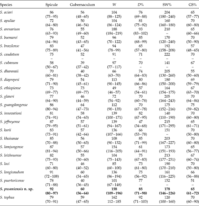
Males of S. pwaniensis n. sp. can be distinguished from the males of S. ethiopiense and S. karii by their spicule length of 92 μm (80–97) vs. 73 μm (69–77) and 83 μm (73–91), respectively. The length of spicules is greater than that of all members of the ‘glaseri’ group, with the sole exception of S. longicaudum (table 2). Further, S. pwaniensis n. sp. differs from S. karii in the presence of a mucron on the tail of second-generation males. The mucron in second-generation males is also absent in S. jeffreyense, S. tophus and S. khoisanae, while it is present in S. innovationi and S. ethiopiense.
Comparative morphometrics (in μm) of first-generation males of Steinernema pwaniensis n. sp. and other species of the ‘glaseri’ group; mean with ranges given in brackets, see table 1 for abbreviations. Data for Steinernema pwaniensis n. sp. in bold.

–, Data not available.
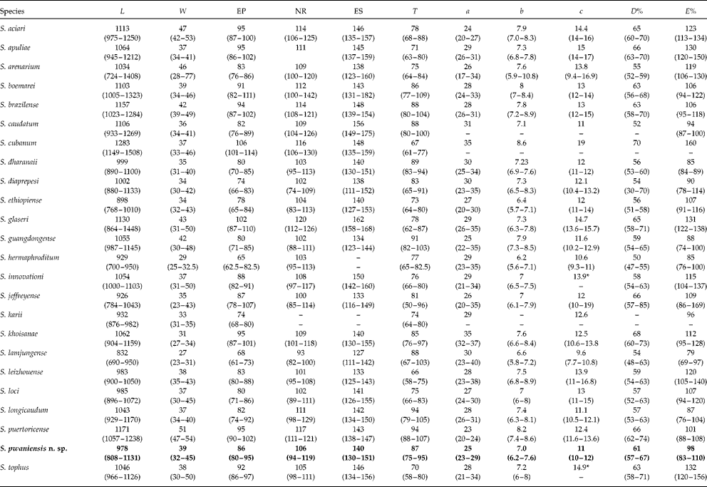
The IJs of S. pwaniensis are longer than those of S. ethiopiense and S. karii (978 μm (808–1131) vs. 898 μm (768–1010) and 932 μm (876–982), respectively). The body length of other African ‘glaseri’ group nematodes is similar to that of S. pwaniensis. Furthermore, S. pwaniensis can also be distinguished from S. ethiopiense and S. karii based on the distance from anterior end to excretory pore and the length of the tail (86 μm (80–95) vs. 78 μm (65–84) and 74 μm (68–80), respectively, and 87 μm (75–95) vs. 73 μm (64–80) and 74 μm (64–80), respectively). The ratios a, c and E% of S. pwaniensis are substantially lower than all other African ‘glaseri’ group members, with the exception of the E% ratio of S. karii which is the same (table 3).
Comparative morphometrics (in μm) of infective juveniles of Steinernema pwaniensis n. sp. and other species of the ‘glaseri’ group; mean with ranges given in brackets, see table 1 for abbreviations. Data for Steinernema pwaniensis n. sp. in bold.

–, Data not available. *, Calculated from the original description.
Molecular characterization and phylogenetic analysis
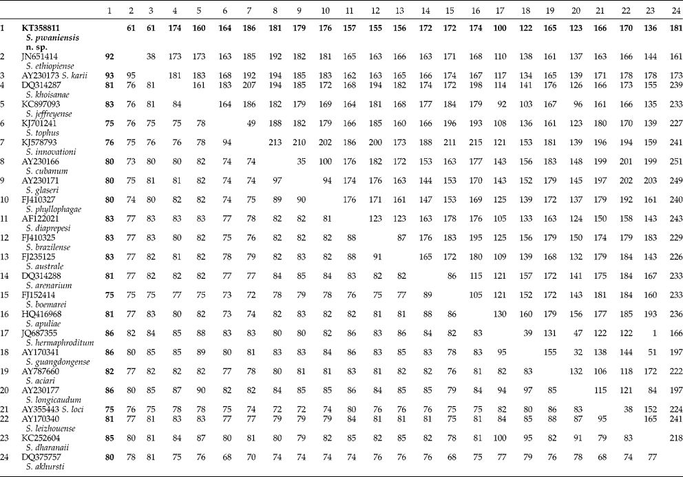
Cloning of the PCR products of the ITS region showed the presence of two haplotypes with a variation in the position 225 bp of the ITS1, with either nucleotide A (KT358811) or G (KT358812). This variation is minimal in comparison to that observed in other species in the ‘glaseri'group (Půža et al., Reference Půža, Chundelová, Nermut', Žurovcová and Mráček2015). Steinernema pwaniensis n. sp. is separated from other species in the ‘glaseri’ group by 61–186 bp (table 4).
Pairwise distances of the ITS region between species of the ‘glaseri’ group. Below diagonal: percentage similarity; above diagonal: total character differences. Data for Steinernema pwaniensis n. sp. in bold.

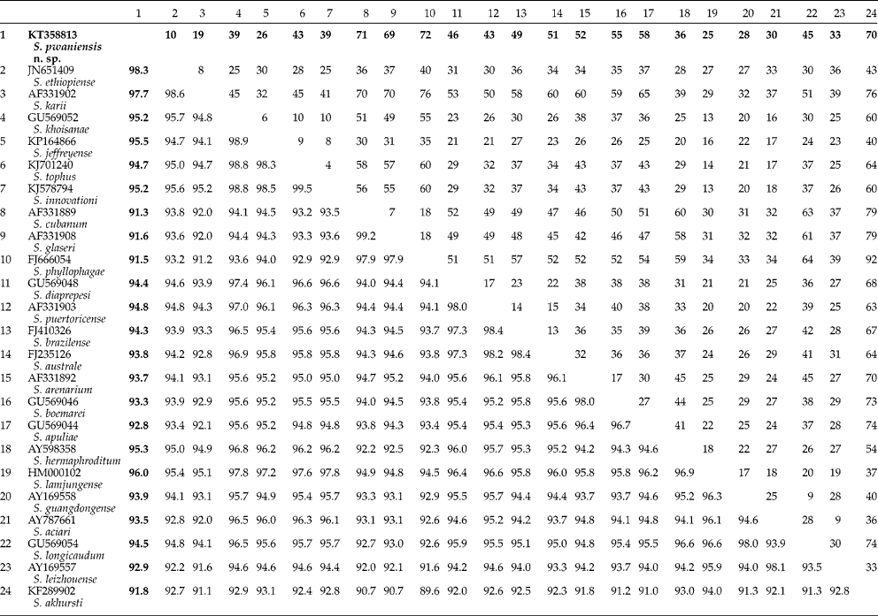
The region containing D2 and D3 expansion segments of the 28S rRNA gene, is separated by 10–72 bp from other species in the ‘glaseri’ group (table 5).
Pairwise distances of the D2–D3 expansion segments of the 28S rDNA between species of the ‘glaseri’ group. Below diagonal: percentage similarity; above diagonal: total character differences. Data for Steinernema pwaniensis n. sp. in bold.

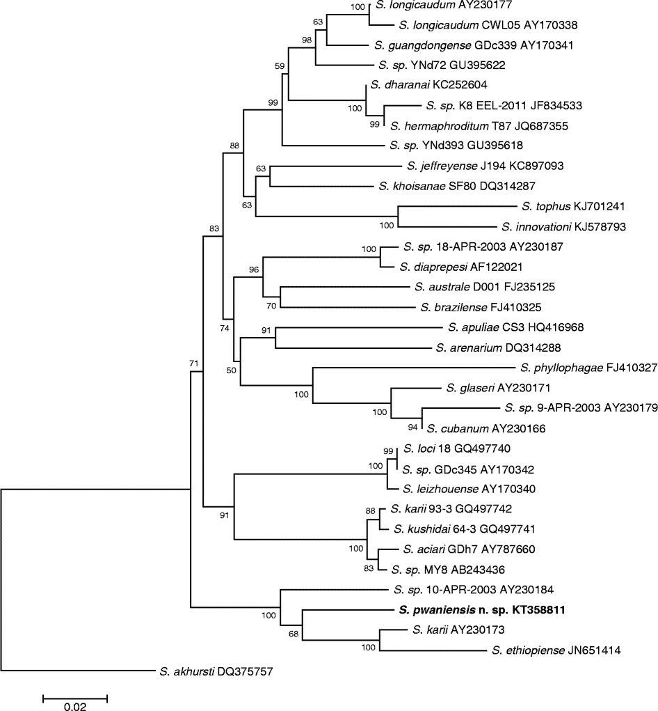
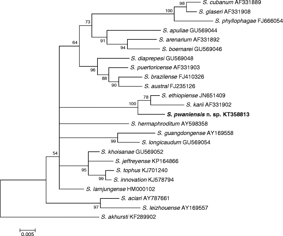
Phylogenetic analyses of the ‘glaseri’ group based on both ITS and 28S regions showed a clear separation of S. pwaniensis n. sp. from all other species (figs 4 and 5). In both analyses S. pwaniensis n. sp. formed a strongly supported group with two other East African species, S. ethiopiense and S. karii. In the ITS tree, the group of S. pwaniensis, S. ethiopiense and S. karii was at the basal position within the ‘glaseri’ group. Other African species appeared to form another group where S. tophus and S. innovationi were sister to the pair S. khoisanae and S. jeffreyense. The analysis of the D2–D3 expansion segments of the 28S rDNA sequence mainly confirmed the groups of related species, but the higher-level topology was unclear.
Phylogenetic relationships in the ‘glaseri’ group of Steinernema based on analysis of ITS rDNA regions. Steinernema akhursti was used as the outgroup taxon. The percentages of replicate trees in which the associated taxa clustered together in the bootstrap test (10,000 replicates) are shown next to the branches. Branch lengths indicate evolutionary distances and are expressed in units of number of base differences per site.

Phylogenetic relationships in the ‘glaseri’ group of Steinernema based on analysis of D2–D3 expansion segments of the 28S rDNA. Steinernema akhursti was used as the outgroup taxon. The percentages of replicate trees in which the associated taxa clustered together in the bootstrap test (10,000 replicates) are shown next to the branches. Branch lengths indicate evolutionary distances and are expressed in units of number of base differences per site.

In general, molecular data confirm the status of S. pwaniensis n. sp. as a new species according to the phylogenetic and evolutionary species concept (Adams, Reference Adams1998).
Acknowledgements
We thank Christopher Steer for language correction.
Financial support
The study was supported by the Czech Ministry of Education, Youth and Sport and Amvis in the programme KONTAKT II, project LH 12105.
Conflict of interest
None.