

Phytanic acid (PA) is a branched-chain fatty acid that is generated via oxidation of an aliphatic alcohol phytol, a constituent of chlorophyll in green plants. Some microorganisms inhabiting the rumen of ruminants can release phytol from chlorophyll (Hellgren, Reference Hellgren2010). The conversion of phytol into PA is also mediated by activities of rumen microorganisms (Hansen, Reference Hansen1966). Therefore, large amounts of PA are formed in the rumen and are translocated to adipose tissue and milk. The concentrations of PA in milk and dairy products are consequently higher than in other common foods (Brown et al., Reference Brown, Mei, Gibberd, Burston, Mayne, McClinchy and Sidey1993). Because PA cannot be synthesized de novo in the human body, PA in humans is solely derived from exogenous sources, especially foods derived from ruminant animals.
Patients with Refsum disease have a mutation in the key enzyme of α-oxidation, an essential step for PA catabolism, and show an abnormally high level of plasma PA. Therefore, elevated plasma PA concentration is considered one of the clinical signs of Refsum disease, and patients are forced to avoid PA intake. For this reason, numerous studies have investigated the relationship between PA and its pathogenic role in Refsum disease. In particular, potential toxicity of PA on neuronal cells has been studied since early times because Refsum disease is a neurocutaneous syndrome (Roca-Saavedra et al., Reference Roca-Saavedra, Marino-Lorenzo, Miranda, Porto-Arias, Lamas, Vazquez, Franco and Cepeda2017).
Research trends on PA have changed due to recent evidence that peroxisome proliferator activated receptor (PPAR) is strongly activated by PA and its metabolite pristanic acid (PrA) (Heim et al., Reference Heim, Johnson, Boess, Bendik, Weber, Hunziker and Fluhmann2002). Since PPAR modulates a wide variety of cellular functions and is a molecular therapeutic target for human metabolic diseases and immunological diseases, intake of PA or PrA may be a potential treatment to ameliorate symptoms of these diseases. Indeed, PA was reported to stimulate glucose uptake in rat hepatocytes (Heim et al., Reference Heim, Johnson, Boess, Bendik, Weber, Hunziker and Fluhmann2002). Our group has demonstrated that both PA and PrA suppressed production of autoimmune-related cytokines in mouse T-cells (Nakanishi et al., Reference Nakanishi, Anraku, Suzuki, Kono, Erickson and Kawahara2016). These findings suggest that PA is a promising research target not only as a risk factor for patients with Refsum disease but also as a health-promoting fatty acid for normal subjects. Although milk and dairy products are major PA sources for human intake, their PA concentrations vary widely depending on the feed of ruminant animals, especially the plant materials (Hellgren, Reference Hellgren2010; Che et al., Reference Che, Kristensen, Nebel, Dalsgaard, Hellgren, Young and Larsen2013). In this connection, several researchers have proposed the availability of PA as a marker for authentication of organic milk (Corazzin et al., Reference Corazzin, Romanzin, Sepulcri, Pinosa, Piasentier and Bovolenta2019), because PA cannot be synthesized by dairy cows and originates from the plant-derived chlorophyll. Thus, the production of milk and dairy products with a modified PA concentration can be considered a novel strategy to cultivate consumer demand.
Among the eight stereoisomers of PA, the 3R, 7R, 11R- and the 3S, 7R, 11R-isomers are naturally occurring PAs in milk and dairy products, because they come from chlorophyll-derived phytol (2E, 7R, 11R-isomer). However, most previous studies have used commercially available PA which includes non-natural isomers. In addition, in vitro cell-based experiments have been used in almost all studies to investigate the therapeutic or toxic effects of PA, and there is limited information about the in vivo actions (Hellgren, Reference Hellgren2010). Consequently, basic data such as the effects of dietary PA on the tissue lipid profiles have not yet been published. Recently, we succeeded in synthesizing a diastereomeric mixture of 3R, 7R, 11R- and 3S, 7R, 11R-PAs (Nakanishi et al., Reference Nakanishi, Motoba, Anraku, Suzuki, Yamaguchi, Erickson, Eto, Sugamoto, Matsushita and Kawahara2018). In this study, the accumulation of PA in adipose tissue, liver and brain was investigated using mice fed a diet containing the above diastereomeric mixture of PAs. Dietary PA-induced changes in compositions of other fatty acids in the same tissues were also investigated to provide a basis for understanding the in vivo physiological roles of PA.
Materials and methods
Preparation of PA
The chemical synthesis of a diastereomeric mixture of 3R, 7R, 11R- and 3S, 7R, 11R-PAs was carried out in our laboratory according to a previous report (Nakanishi et al., Reference Nakanishi, Motoba, Anraku, Suzuki, Yamaguchi, Erickson, Eto, Sugamoto, Matsushita and Kawahara2018). More details are given in the online Supplementary File.
Animals and treatments
Female C57BL/6J mice were purchased from Japan SLC, Inc. (Shizuoka, Japan). Mice were kept at 25°C on a 12-h dark-and-light cycle, and had free access to a commercial diet (Labo MR stock, Nosan Corporation, Yokohama, Japan). Mice aged 6 weeks at the beginning of the experiment were randomly divided into two groups of four mice each. The control group and the PA group had free access to an AIN-93M-based diet and a diet of which 0.05% soybean oil was replaced by PA, respectively (online Supplementary Table S1). After 4 weeks feeding, mice were sacrificed under anesthesia with isoflurane, and parametrial adipose tissue, liver and brain were harvested. Animals were used in accordance with the Guidelines for the Care and Use of Laboratory Animals at the University of Miyazaki as well as Law No. 105 and Notification No. 6 of the Japanese government. All experimental protocols were approved by the University of Miyazaki (approval number: 2018-019).
Lipid extraction, preparation of fatty acid methyl esters and gas chromatographic analysis
The crude lipid was extracted and fatty acid methyl esters were prepared according to a modified method described previously (Kawahara et al., Reference Kawahara, Takenoyama, Takuma, Muguruma and Yamauchi2009). The fatty acid methyl esters were analyzed on a capillary column (BPX90, 100 m × 0.25 mm × 0.25 μm, SGE Analytical Science, Austin, Texas, USA) in a GC-2010 gas chromatograph equipped with a flame-ionization detector and an automated injector (Shimadzu Corporation, Kyoto, Japan). More details are given in the online Supplementary File.
Statistical analysis
Differences between the control group and the PA group were analyzed using a non-paired Student's t test. The Mann–Whitney U test was used to compare two groups when values for one or more samples were below the detection limits and the data were nonparametric. All statistical analyses were performed using GraphPad Prism (version 6.0, GraphPad Software, San Diego, CA, USA).
Results and discussion
In this study, we investigated the effects of dietary PA on tissue accumulation of PA and on the tissue fatty acid profiles, using mice fed a diet containing a mixture of 3R, 7R, 11R- and 3S, 7R, 11R-PAs, the two isomers present in foods and the human body. Milk and dairy products are typical PA sources for human and contain PA with a few dozens of milligrams per 100 g of food (Brown et al., Reference Brown, Mei, Gibberd, Burston, Mayne, McClinchy and Sidey1993). Therefore, 0.05% PA was added to the experimental diet to approximate a situation where PA-containing milk or dairy products are consumed on a routine basis.
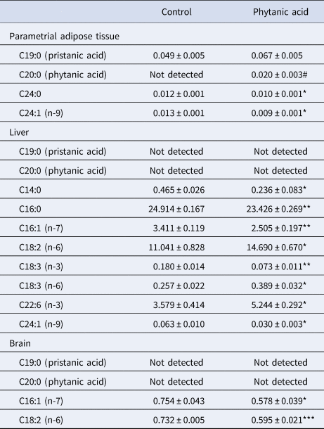
Dietary PA-induced changes in fatty acid compositions in each tissue are summarized in Table 1. Data on PA are expressed as the sum of compositions of 3R, 7R, 11R- and 3S, 7R, 11R-PAs because our gas chromatographic analysis could not discriminate between the two isomers. Our results on PA-fed mice clearly showed that dietary PA was absorbed into the mouse body and significantly accumulated in the parametrial adipose tissue. PA-fed mice also showed high composition of PrA in the adipose tissue compared with control mice, albeit without statistical significance (P = 0.06). Gas chromatographic analysis of the adipose tissue detected a total of 19 fatty acids in addition to PA and PrA. Two of the fatty acids exhibited different compositions between control and PA-fed mice, where the compositions of lignoceric acid (C24 : 0) and nervonic acid (C24 : 1, n-9) were decreased by PA intake. Since these two fatty acids can be de novo synthesized via a series of elongation and desaturation processes of palmitic acid which is the primary product of fatty acid biosynthesis, our results suggest that de novo fatty acid biosynthesis or elongation of synthesized fatty acids is inhibited by PA intake.
Dietary phytanic acid-induced changes in tissue fatty acid compositions (%)

Data are presented as means ± sem.
#P < 0.05 compared with control in Mann–Whitney U test. *P < 0.05, **P < 0.01 and ***P < 0.001 compared with control in a non-paired Student's t test.
The liver should be the organ most responsible for PA catabolism and PrA formation because the α-oxidation-mediated conversion of PA into PrA is a hepatic microsomal-peroxisomal process (Van Veldhoven et al., Reference Van Veldhoven, Mannaerts, Casteels and Croes1999). However, our results demonstrated that the hepatic concentrations of both PA and PrA in PA-fed mice were below the detection limit of gas chromatographic analysis, which may imply that a part of dietary PA was converted into PrA in the liver and the remaining PA and the resultant PrA were translocated to the adipose tissue. Our gas chromatographic analysis detected 18 fatty acids in the liver, among which 8 fatty acids showed different compositions between groups. Regardless of the undetectable levels of PA in the liver, dietary PA modified the liver fatty acid profile, with a higher impact than in adipose tissue. Our results demonstrated that the compositions of myristic acid (C14:0), palmitic acid (C16:0), palmitoleic acid (C16:1, n-7) and nervonic acid were significantly decreased by PA intake, which emphasizes the notion, that de novo fatty acid biosynthesis is inhibited by PA intake. On the other hand, dietary PA significantly increased the levels of linoleic acid (C18:2, n-6), γ-linolenic acid (C18:3, n-6) and docosahexaenoic acid (C22:6, n-3). As a consequence, the hepatic ratio of polyunsaturated fatty acids to saturated and monounsaturated fatty acids was significantly increased by PA intake (control: 16.06 ± 1.28%, PA: 21.48 ± 1.01%, P < 0.05). Because PPARα and PPARγ, which are preferentially activated by PA (Heim et al., Reference Heim, Johnson, Boess, Bendik, Weber, Hunziker and Fluhmann2002), play a critical role in lipid metabolism, these activations could be involved in PA-induced changes in the tissue fatty acid profiles, although the precise mechanism remains unclear.
Our results indicated that neither PA nor PrA accumulated in mouse brain. Among 19 fatty acids which were detected in the brain, only palmitoleic acid and linoleic acid showed significantly different compositions between groups, suggesting that dietary PA may have a relatively low impact on the fatty acid profile of the brain, at least compared with the liver.
Previous in vitro studies have suggested that improvement of glucose and lipid metabolism is a typical example of the health-promoting effects of PA. Our study supports the notion that the in vitro actions of PA can be also elicited in in vivo physiological situations by showing that PA as well as PrA accumulated in the adipose tissue and that PA modified the hepatic fatty acid profiles. Furthermore, our results showed evidence regarding the beneficial effects of PA, indicated by the significant decline of the hepatic palmitoleic acid/palmitic acid ratio in PA-fed mice (control: 0.14 ± 0.00, PA: 0.11 ± 0.01, P < 0.05). Because the palmitoleic acid/palmitic acid ratio positively correlates with symptoms of liver diseases such as non-alcoholic steatohepatitis (Yamada et al., Reference Yamada, Mizukoshi, Seike, Horii, Terashima, Iida, Kitahara, Sunagozaka, Arai, Yamashita, Honda, Takamura, Harada and Kaneko2019), our results imply that PA has beneficial properties for the treatment of these diseases. On the other hand, excess PA intake is considered a risk factor for health problems such as Refsum disease. Since neurological symptoms are commonly observed in patients with Refsum disease, many studies have evaluated the potential toxicity of PA on neuronal cells (Roca-Saavedra et al., Reference Roca-Saavedra, Marino-Lorenzo, Miranda, Porto-Arias, Lamas, Vazquez, Franco and Cepeda2017). However, PA could not be detected in the brain of PA-fed mice in our study, which indicates that the PA-induced neurotoxicity is negligible, at least in a case where PA can be normally catabolized by α-oxidation. Since this study cannot completely reject the possibility of the involvement of dietary PA in brain functions, further studies are needed to determine the overall physiological action of PA and to discuss comprehensively its therapeutic and toxic effects for human health.
In conclusion, this study demonstrated that dietary PA accumulated in the adipose tissue and modified the hepatic fatty acid profile, although the brain was relatively less affected by dietary PA. These findings provide basic information to cultivate a better understanding of the in vivo physiological role of PA, and to produce milk and dairy products with a modified PA concentration.
Supplementary material
The supplementary material for this article can be found at https://doi.org/10.1017/S0022029920001089.
Acknowledgements
This work was partially supported by a Grant-in-Aid for Scientific Research from the Japan Society for the Promotion of Science (No. 17K15364 and No. 19K06356).