Introduction
Collembola constitute one of the most important parts of soil mesofauna in density (104 to 105 specimens per meter square, Petersen and Luxton, Reference Petersen and Luxton1982) as well as in species number (20% to 60% of arthropods, Stork, Reference Stork1988; Stork and Blackburn, Reference Stork and Blackburn1993). Collembola play a vital role in the organic matter decomposition process and can have a significant impact on soil structure and composition (e.g. Seastedt, Reference Seastedt1984). Most Collembola feed on soil microflora (such as bacteria, fungi and algae) and thus have an important influence on the dynamics of their populations and the food chain (Arpin et al., Reference Arpin, Kilbertus, Ponge, Vannier and Pesson1980; Moore et al., Reference Moore, Walter and Hunt1988; Parkinson, Reference Parkinson1988). Especially in stimulating microbial activity, Collembola contribute to decomposition several times more than their metabolism (e.g. Macfayden, Reference MacFadyen, Doeksen and Drift1963). They have significant impact on N mineralization, soil respiration, leaching of dissolved organic carbon and plant growth (Filser, Reference Filser2002). As such, in that context, they have an important influence on the equilibrium of terrestrial ecosystems as a whole (Copley, Reference Copley2000).
Representatives of the genus Megalothorax (Neelidae) are commonly encountered in soil and litter. Those are small, eyeless species. Little is known regarding their biology. They are apparently feeding on microorganisms such as bacteria and microfungi, ingested with a large part of mineral elements (Arpin et al., Reference Arpin, Kilbertus, Ponge, Vannier and Pesson1980; Ponge, Reference Ponge1991). The intestinal content transits in the form of compact, spherical pellets, which is a specificity of the Neelidae. With their peculiar morphology—globular body standing above long legs, well-developed furca (jumping organ)—Megalothorax species stand out from the general morphological trends in edaphic Collembola (elongated body, short legs, reduced or missing furca). Their peculiarly small size (averaging around 500 µm) allows them to move with ease in the interstitial space of the soil. They exhibit a set of strange organs whose function is yet to be understood. These include the wax rod secretory elements (Vannier and Massoud, Reference Vannier and Massoud1967), the integumetary channels (Rusek, Reference Rusek and Striganova1987) and the so-called sensory fields.
The taxonomy of the genus has undergone recent improvements with descriptions providing more and more precise discriminatory characters (Deharveng, Reference Deharveng1978; Deharveng and Beruete, Reference Deharveng and Beruete1993; Christian, Reference Christian1998; Papáč and Kováč, Reference Papáč and Kováč2013; Schneider and D'Haese, Reference Schneider and D'Haese2013; Schneider et al., Reference Schneider, Porco and Deharveng2016) and integrating molecular analysis (Schneider et al., Reference Schneider, Cruaud and D'Haese2011; Schneider and D'Haese, Reference Schneider and D'Haese2013; Schneider et al., Reference Schneider, Porco and Deharveng2016). However, Megalothorax systematics remains crippled, due to the lack of precise descriptions for some species. Lack of accurate descriptions prevents any further gain of knowledge on those species, such as ecology, biology and biogeography, and on their contribution to soil communities and processes. Little effort has been devoted to revising those species, except for the generotype Megalothorax minimus Willem, 1900 redescribed by Schneider and D'Haese (Reference Schneider and D'Haese2013).
Denis (Reference Denis1948) described Megalothorax laevis Denis, Reference Denis1948 from Nha Thrang (Vietnam, Khánh Hòa province). Like M. minimus (generotype), M. laevis is characterized by a mucro with smooth posterior lamellae. Denis provided only one character to separate the two species, namely the supposed absence of the anterior apico-median dental spine (A1) in M. minimus. Schneider and D'Haese (Reference Schneider and D'Haese2013) redescribed M. minimus with A1 present, thus invalidating the diagnosis difference between the two species.
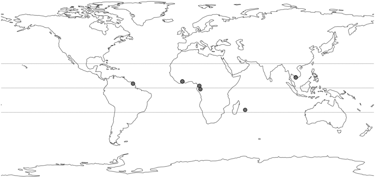
Field collections in Vietnam (including type locality of M. laevis), subequatorial Africa (Ivory Coast, Gabon), Réunion island and French Guiana yielded populations of Megalothorax displaying a blend of the character sets of the minimus-group and the incertus-group sensu Schneider and D'Haese (Reference Schneider and D'Haese2013). The re-examination of the holotype of M. laevis suggests that all these populations belong to this species, evidently widespread in the intertropical zone (Fig. 1). Megalothorax laevis is also strikingly similar to Megalothorax interruptus Hüther, Reference Hüther1967, only differing from the number of teeth on the tenaculum, the morphology of the labral chaetae and presence/absence of a pair of globular chaetae near the basis of antennae (Hüther, Reference Hüther1967).
Megalothorax laevis, known repartition.

In the present work, M. laevis is redescribed using optical and scanning electron microscopy (SEM). A pattern of pseudopore-like elements is for the first time reported on the trunk and legs of Megalothorax species. A set of DNA barcodes of the species and a key to the Megalothorax species with smooth mucro lamellae are also provided. The phylogenetic position of M. laevis within the genus is tested using molecular data: 28S rDNA d1 and d2 regions, 16S rDNA and cytochrome c oxydase I—DNA Barcode (COI).
Materials and methods
Sampling
Vietnam
The specimens were collected in two different ecosystems (suburban zone of Nha Thrang with disturbed vegetation and in the karstic caves of Hòn Chông), extracted from soil and litter using simple Berlese funnel, and collected and stored in 96% ethanol.
Ivory coast
The specimens were collected from the main types of local land-use (secondary semi-deciduous forest, savannah, fallow ground, mixed cultures, yam culture and cocoa culture) near Lamto reserve located 180 km north of Abidjan (Ivory Coast) using pitfall traps (consisting of small plastic boxes filled with one-quarter of capacity with 60% alcohol) and Berlese–Tullgren apparatus (for leaf litter and soil samples). Animals were stored in 96% ethanol.
Gabon
The specimens were collected and handed to the first author by J.-M. Betsch, they were then stored in 96% ethanol at 4 °C.
French Guiana
The specimens were collected from soil under Bromeliaceae or mosses, extracted from sieved samples using field Berlese–Tullgren apparatus, collected and preserved in 96% ethanol, and kept at 4 °C for long-term storage.
Réunion
The specimens were collected from fern roots and leaf litter in an open vegetation patch on a lava flow, extracted from samples using field Berlese–Tullgren apparatus, preserved in 96% ethanol and kept at 4 °C for long-term storage.
Morphological analysis
The specimens were cleared in lactic acid and mounted on microscope slides in Marc André II medium. They were examined using a Leica DMLB compound microscope with differential phase contrast optics at magnifications ranging from 250 to 1000. Drawings were made with a drawing tube and vectorized with Inkscape. For SEM observations, specimens were dehydrated in 100% ethanol, before critical point drying (Emitech K850) and gold coating (Jeol JFC-1200) and performing observations with a S.E.M. Jeol 840A.
Molecular analysis
The standard DNA barcode (658bp of the COI—mitochondrial c cytochrome oxidase subunit I gene) was newly obtained for five specimens of M. laevis (three from French Guiana, one from Réunion and one from Vietnam). Additionally, partial 16S rDNA and 28S rDNA (d1 and d2 regions) were obtained for a subset of those specimens. The specimens were processed in the BoEM molecular laboratory (MNHN). DNA was extracted from entire specimens with the Qiagen (Hilden, Germany) DNeasy tissue extraction kit. Tissues were digested in lysis buffer and proteinase K at 56 °C for 3 h, then extraction was carried out as advised by the manufacturer. Total DNA was finally resuspended in 120 μL of the elution buffer. Polymerase chain reaction was carried out in 25 mL volume reaction with Taq-&LOAD Mastermix 5xC reagent (MP Biomedicals, Santa Ana, CA, USA). 28S rDNA d1 and d2 regions were amplified and sequenced in one shot. Primers and temperature programs used for each marker were as follows:
• COI: LCO1490col (5’–WYT CDA CWA AYC RYA ARG AYA TYG G–3’)/HCO2198col (5’–TAN ACY TCN GGR TGN CCR AAR AAT CA–3’), specific for Collembola; 94 °C for 2 min; 5 × (94 °C for 40 s, 45 °C for 40 s, 72 °C for 1 min), 30 × (94 °C for 40 s, 51 °C for 40 s, 72 °C for 1 min), 72 °C for 5 min.
• 16S rDNA: 16SAcoll (5’–MGMMTGTTTAWCAAAAACAT–3’)/16SBcoll (5’–CGCCGGTTTGAACTCAAATCA–3’); 94 °C for 2 min; 30 × (94 °C for 40 s, 52 °C for 40 s, 72 °C for 1 min), 72 °C for 5 min.
• 28S rDNA: 28SC1p (5’–ACCCGCTGAATTTAAGCAT–3’)/28SD2 (5’–TCCGTGTTTCAAGACGGG–3’); 94 °C for 2 min; 35 × (94 °C for 40 s, 54 °C for 40 s, 72 °C for 1 min), 72 °C for 5 min.
See Greenslade et al. (Reference Greenslade, Stevens, Torricelli and D'Haese2011), Schneider and D'Haese (Reference Schneider and D'Haese2013) and Stevens and D'Haese (Reference Stevens and D'Haese2014) for more details.
Polymerase chain reaction products were sequenced by EUROFINS (France). Resulting chromatograms were interpreted using the program Sequencher 4.5 or CodonCode Aligner (CodonCode Corporation, Centerville, MA, USA). Sequences were checked manually for misreadings of the chromatograms. Unfortunately, DNA sequencing was unsuccessful for specimens from Ivory Coast and Gabon (due to ill-preservation).
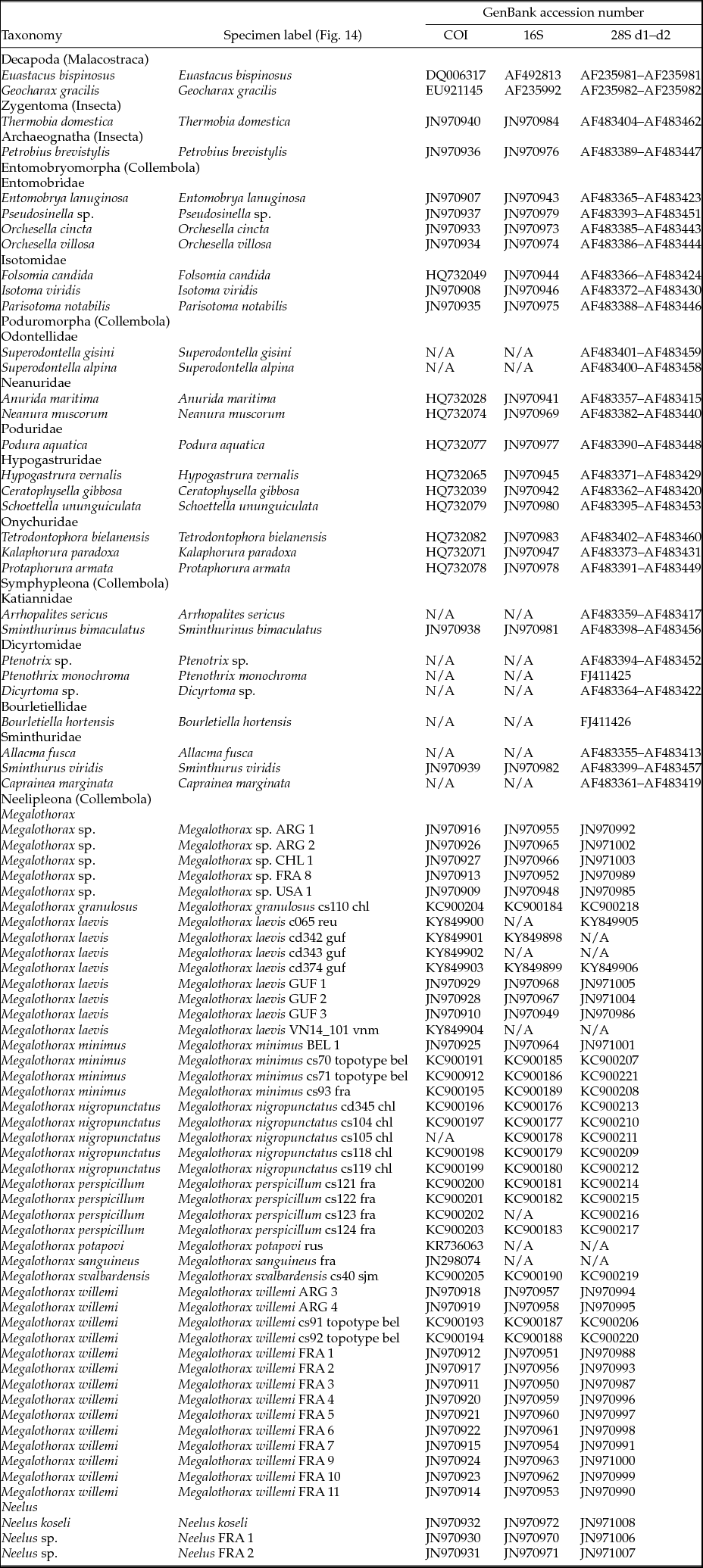
We tested the validity of the species from the available molecular data under a phylogenetic criterion. A phylogenetic analysis was performed using Schneider and D'Haese (Reference Schneider and D'Haese2013) dataset updated with the addition of Megalothorax potapovi Schneider et al., Reference Schneider, Porco and Deharveng2016 and Megalothorax sanguineus Schneider et al., Reference Schneider, Porco and Deharveng2016 (Schneider et al., Reference Schneider, Porco and Deharveng2016) and the newly obtained sequence of COI, 16S rDNA and 28S rDNA. The total dataset includes two Decapoda, two Insecta, seven species of Entomobryomorpha, eleven species of Poduromorpha, nine species of Symphypleona and forty-seven representatives of Neelipleona (three Neelus, forty-four Megalothorax). Also, three specimens from Schneider and D'Haese (Reference Schneider and D'Haese2013) dataset were identified as M. laevis from French Guiana (Megalothorax cf. interruptus). Overall, eight specimens represent M. laevis. The detailed dataset with GenBank accession numbers is provided in Table 1. The phylogeny was inferred with the parsimony criterion using POY version 5.1.1 (Wheeler et al., Reference Wheeler, Lucaroni, Hong, Crowley and Varón2015). We performed the analyses independently for COI, 16S rDNA and 28S d1 + d2 and then for the combined loci. 16S rDNA and 28S rDNA were analysed using direct optimization (dynamic homology framework; Wheeler, Reference Wheeler1996). After a manual check of the nucleic acid and translated sequences, nucleic acid sequences of COI were considered to be prealigned and analysed as such (static homology framework). The influence of gap, transition and transversion costs was explored through sensitivity analysis (Wheeler, Reference Wheeler1995). Three indel:transversion cost ratios (1, 2 and 4) and three transversion:transition cost ratios (1, 2 and 4) were explored, resulting in nine individual analyses for four datasets (16S rDNA, COI, 28S d1 + d2 and all loci combined). The analysis procedure followed a three-step strategy:
• Step 1: A starting pool of 100 Wagner trees was generated through random addition sequence. Each replicate was explored by a combination of TBR and SPR branch swapping and then submitted to ratcheting (Nixon, Reference Nixon1999). The resulting topologies were then explored by tree fusing (Goloboff, Reference Goloboff1999). A final and more thorough branch swapping was performed by retaining trees up to 10% longer than the optimal one, and the optimal trees were retained as final results of this step.
• Step 2: For each of the four combinations analyses, trees for all the nine parameter set analyses were concatenated for a supplementary tree-fusing step.
• Step 3: An additional round of tree fusing analyses using iterative pass (Wheeler, Reference Wheeler2003) for final refinement based on the trees obtained for the previous sets of analyses (steps 1 and 2). The three steps added up to 108 individual analyses for which precise command lines, scripts and results can be found in the supplementary material (available online).
Molecular dataset

To avoid an arbitrary choice of parameters, maximum character congruence among the loci was used as an optimality criterion. Congruence was measured by the incongruence length difference (ILD) metrics (Mickevich and Farris, Reference Mickevich and Farris1981). This value is calculated by dividing the difference between the overall tree length and the sum of its data components: ILD = (lengthcombined − Σ lengthindividual sets)/lengthcombined. The tree from the analysis that minimizes character conflict among all the data is taken as the best overall explanation of character variation, and thus the best estimate of the phylogeny. Node sensitivity was automatically calculated using YBYRÁ (Machado, Reference Machado2015). Bremer decay index was estimated using the swap algorithm to build the pool of suboptimal topologies; the resulting trees of the previous analyses—all loci combined—being used as the starting pool. The swap algorithm was set up with a 3 days timeout. Working directory with data, script and results are provided in Supplementary Material S1.
Terminology
The terminology follows Schneider and D'Haese (Reference Schneider and D'Haese2013) and Schneider et al. (Reference Schneider, Porco and Deharveng2016). The presence or absence of specific chaetae is described in reference to the chaetotaxy of M. minimus sensu (Schneider and D'Haese, Reference Schneider and D'Haese2013). The interpretation of the limit between the basomedian field of labium and the basolateral field labium is slightly changed in Megalothorax. The interpretation given in Schneider and D'Haese (Reference Schneider and D'Haese2013), based on Fjellberg (Reference Fjellberg1998/99) was inconsistent with what is observed in the genera Neelus and Neelides (e.g. Fjellberg, Reference Fjellberg1998/99; Kováč and Papáč, Reference Kováč and Papáč2010; Papáč et al., Reference Papáč, Lukić and Kováč2016). In practice, this concerns only a chaeta present in the incertus-group sensu (Schneider and D'Haese, Reference Schneider and D'Haese2013), attributed to the basomedian field in Schneider and D'Haese (Reference Schneider and D'Haese2013), but evidently homologous of a chaeta on the basolateral field of Neelides and Neelus. This interpretation is also more consistent with what is observed in Symphypleona (Fjellberg Reference Fjellberg2007).
Abbreviations and symbols
Crests on the anterior process of the labrum: al2, 3—anterior longitudinal, ml1–3—posterior longitudinal, amt0–2—antero-median transversal, mt2—posterior transversal. Labium: blf—basolateral field, bmf—basomedian field. Antenna: Ant. I–IV—antennomere I to IV, S1–S4—S-chaetae of Ant. III; Or—Ant. IV organite; S, Sx, Sy—S-chaetae of Ant. IV. Trunk: Th. I–III—thoracic segment I to III; Abd. I–VI—abdominal segment I to VI; av—chaetae of anal valves; s1, s2, s3—s-chaetae; τ—τ-chaetae; wrc1–wrc8—free wax rod secretory element 1 to 8. Claw: la, lp, Ba, Bp—auxiliary lamellae and crests of unguis; Ca, Cp—anterior and posterior crests of unguiculal lamella. Misc: dp—dens proximal, dd—dens distal, ps—pseudopore-like element, sf1–6—sensory field 1 to 6.
Taxonomy
Family NEELIDAE
Genus Megalothorax Willem, 1900
Revised diagnosis.
Updated interpretation of the basal fields of labium: basolateral fields of labium with 1 + 1 chaetae (being one of the characteristics of the minimus-group, also observed in Megalothorax granulosus) or 2 + 2 chaetae (being one of the characteristics of the incertus-group); basomedian fields of labium with 1 + 1 or 3 + 3 chaetae.
Megalothorax laevis (Denis, Reference Denis1948): Figs. 2–13.
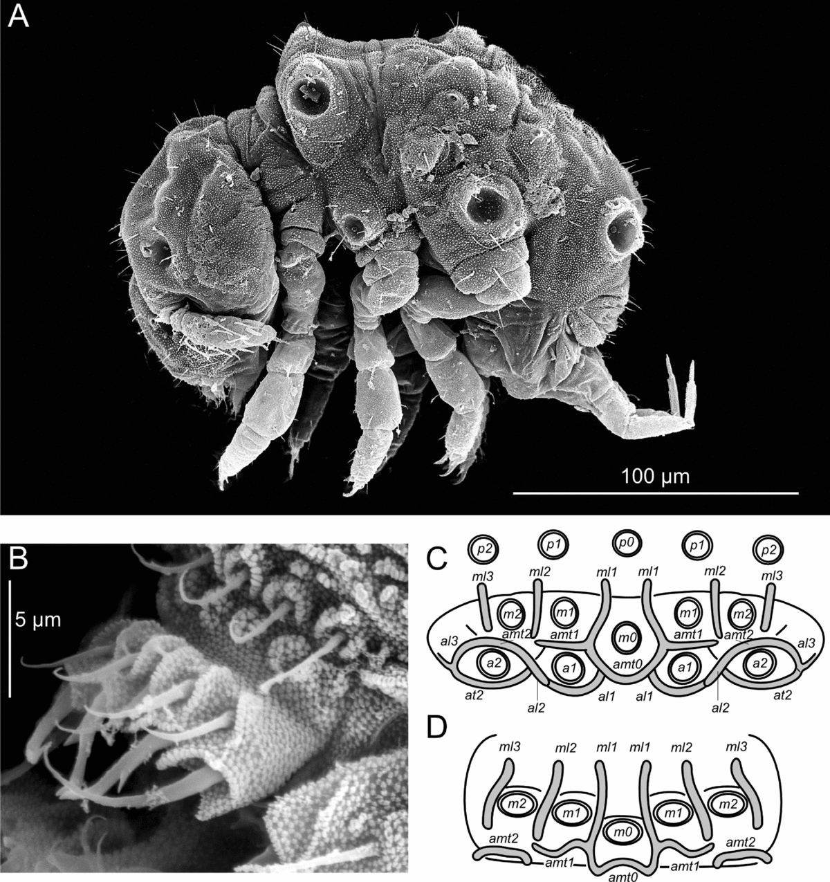
Megalothorax laevis. (A) Whole body, lateral view. (B) Labrum, dorso-lateral view. (C) Labrum, frontal diagram. (D) Labrum, dorsal diagram. Origin: (A, B)—Ivory Coast.

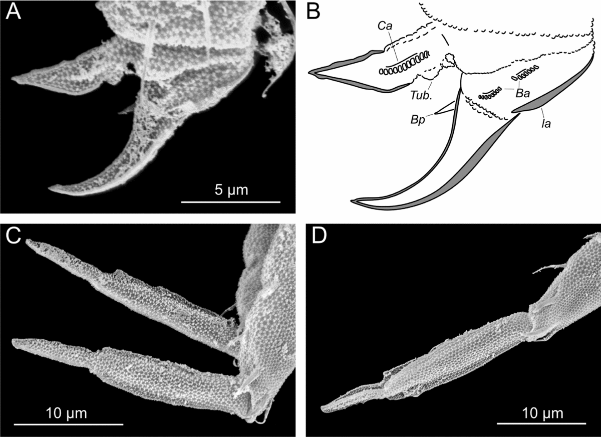
Megalothorax laevis. (A, B) Claw III, anterior view. (C, D) Mucro, lateral view. Origin: (A–D)—Ivory Coast.

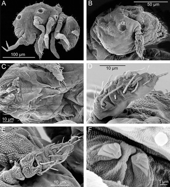
Megalothorax laevis. (A) Whole body, lateral view. (B) Head, lateral view. (C) Head, ventral view. (D) Antenna, posterior view. (E) Antenna, dorsal view. (F) Tenaculum. Origin: (A–F)—French Guiana.

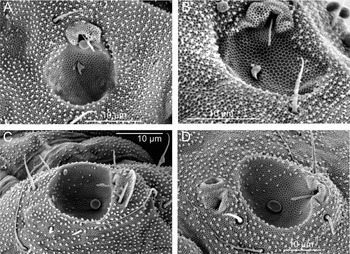
Megalothorax laevis, sensory fields. (A, B) sf2 on head. (C) sf3 on Th. II tergum. (D) sf5 on Th. III tergum (only one of the two inner chaetae visible). Origin: (A–D)—French Guiana.

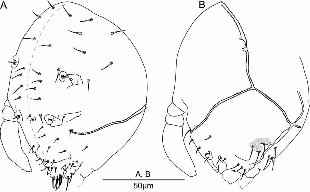
Megalothorax laevis, chaetotaxy of the head. (A) Dorso-lateral side. (B) Ventro-lateral side. Origin: (A, B)—Ivory Coast.

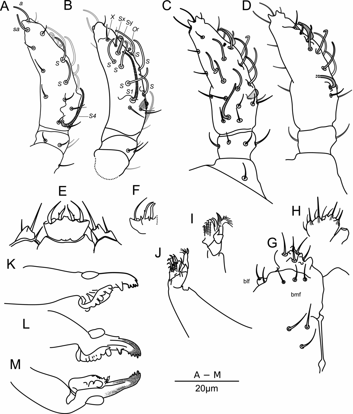
Megalothorax laevis. (A, B) Chaetotaxy of the antenna, 8 S-chaetae S phenotype. (A) Posterior side. (B) Anterior side. (C, D) Chaetotaxy of the antenna, 10 S-chaetae S phenotype. (C) Posterior side. (D) Anterior side. (E) Labrum and maxilla outer palp, ventral side. (F) Labrum a-row lateral side. (G, H) Labium and post-labial chaetae. (G) Focus on basal fields. (H) Focus on proximal papillae and hairs. (I, J) Maxilla, different views. (K, L) Right mandibula. (M) Left mandibula. Origin: (A, B, E, G, H, J, K)—Ivory Coast; (C, D, I, L, M)—French Guiana; (F)—Vietnam, holotype.

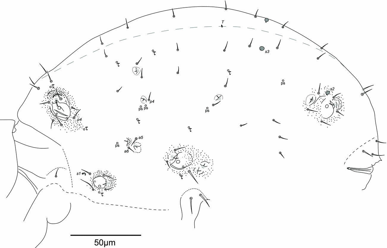
Megalothorax laevis. Chaetotaxy of the trunk, lateral side. Origin: Ivory Coast.

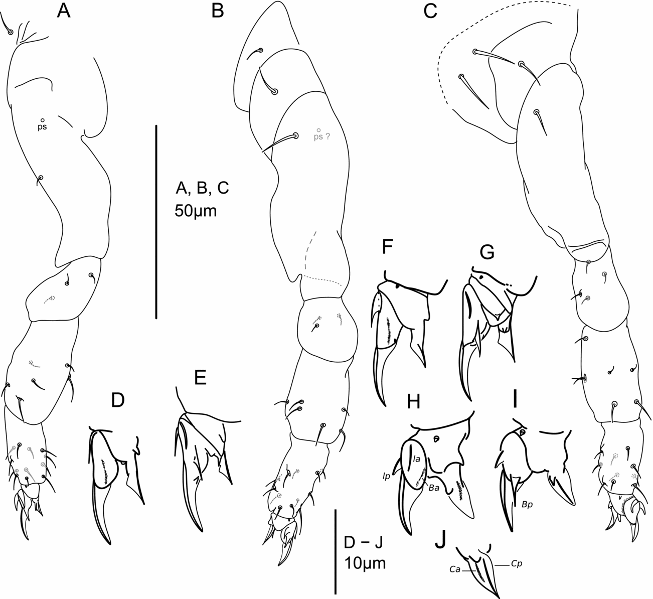
Megalothorax laevis. Legs. (A) Leg I. (B) Leg II. (C) Leg III. (D) Claw I, anterior side. (E) Claw I, posterior side. (F) Claw II, anterior side. (G) Claw II, posterior side. (H) Claw III, anterior side. (I) Claw III, posterior side. (J) Unguiculus III, internal side. Origin: (A–C)—Ivory Coast; (D, E, H, I)—Vietnam; (F, G, J)—Vietnam, holotype.

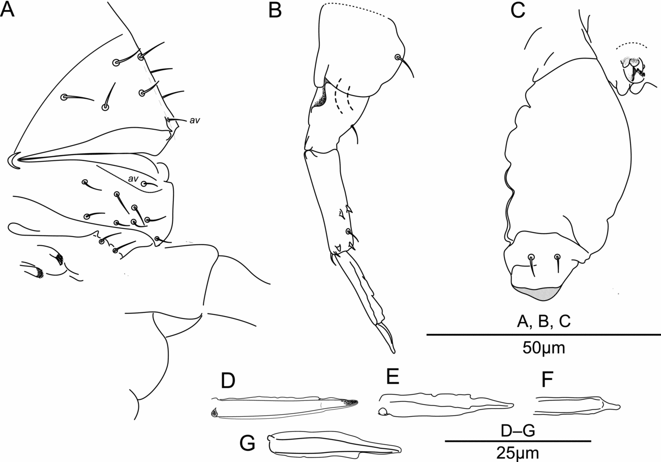
Megalothorax laevis. (A) Chaetotaxy of Abd. VI and sternum of Abd. IV. (B) Furca. (C) Ventral tube and tenaculum. (D–G) Mucro, various preparations. Origin: (A–D)—Ivory Coast; (E, F)—French Guiana; (G)—Vietnam, holotype.

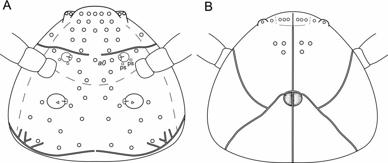
Megalothorax laevis, diagrams of the chaetotaxy and integumentary channels network of the head. (A) Dorsal side. (B) Ventral side.

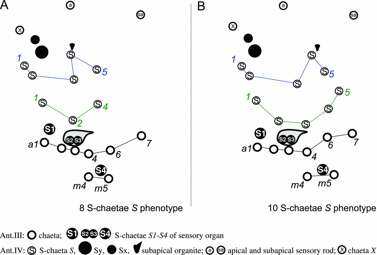
Megalothorax laevis. Diagram of the antenna chaetotaxy, 8 S-chaetae S phenotype and 10 S-chaetae S phenotype.

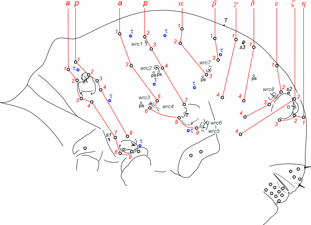
Megalothorax laevis. Diagram of the chaetotaxy of the trunk.

Megalothorax laevis (Denis, Reference Denis1948): 185, 287–289, Fig. 35.
Megalothorax minimus (Deharveng et al., Reference Deharveng, Bedos, Le C. K., Truong, Le, Truong and Ly2009): 36, Table 4.
Megalothorax cf. interruptus (Schneider and D'Haese, Reference Schneider and D'Haese2013): 328, 352–353, Figs. 1A, 2D, Tables 2 and 3.
Summary of Megalothorax laevis antennal chaetotaxy

Summary of Megalothorax laevis legs chaetotaxy

Material examined
Holotype
f on slide, Vietnam: Khánh Hòa: Nha Trang: Cầu Đá: in the bush, leg. M. C. Dawidoff (Denis, Reference Denis1948), original label: ‘Megalothorax laevis n. sp. Cauda VIII-I Daw. L’.
Topotypes
2ff on slides (MNHN-EA040269, LD-CS-VN-01), Khánh Hòa: Nha Trang: Cầu Đá: south slope oriented to the bay, X = 109.207342°, Y = 12.200963°, alt = 45 m, 16.xii.2013, Berlese extraction, soil, L. Deharveng and A. Bedos leg.; 2ff on slides (MNHN-EA040271, LD-CS-VN-02), same location and date, Berlese extraction, litter, L. Deharveng and A. Bedos leg.
Other material
Vietnam: 3ff on slides (MNHN-EA040273–275), Kien Giang: Kien Luong: Hòn Chông: Nui Bai Voi: Nui Bai Voi SW: south-west side, unnamed cave, X = 104.618153°, Y = 10.214321°; alt = 1 m, 10.x.2014, Berlese extraction, soil and roots, L. Deharveng, M. Lukic and A. Bedos leg.; 2ff on slide (MNHN-EA040276), Kien Giang: Kien Luong: Hòn Chông: Nui Khoe La: Nui Khoe La N: unnamed cave, X = 104.586645, Y = 10.207685, alt = 20 m, 13.x.2014, Berlese extraction, roots, L. Deharveng and M. Lukic leg.; 1f on slide (MNHN-EA040277), Kien Giang: Kien Luong: Hòn Chông: Nui Bai Voi: hospital-cave of Mo So, X = 104.620168°, Y = 10.218543°, alt = 4 m, 29.i.2003, Berlese extraction, hanging roots, L. Deharveng and L. C. Man leg.; 1f on slide (MNHN-EA040278), Kien Giang: Kien Luong: Hòn Chông: Nui Bai Voi: hospital-cave of Mo So, X = 104.620168°, Y = 10.218543°, alt = 4 m, 04.vi.2008, Berlese extraction, wood, L. Deharveng and A. Bedos leg.; 1f on slide (MNHN-EA040279), Kien Giang: Kien Luong: Hòn Chông: Nui Hòn Chông: Hang Kim Cuong: unnamed cave, X = 104.643702°, Y = 10.134400°, 28.xi.2006, Berlese extraction, wood and soil, L. Deharveng and A. Bedos leg.
Ivory Coast
6ff on slides (MNHN-EA040251–254, IV-VJ12L3a, IV-VJ12L3b), 3 spm on mount for S.E.M. (MNHN-EA041013), Taabo: Zougoussi terroir near Lamto reserve, X = −5.0267°, Y = 6.2572°, 27.x.2012, Berlese extraction, litter in mixed cultures, S. D. Zon leg.; 2ff on slide (MNHN-EA040260, IV-VIG11S6), Taabo: Zougoussi terroir near Lamto reserve, X = −5.0265°, Y = 6.2572°, 12.xi.2012, Berlese extraction, soil in yam cultures, S. D. Zon leg.; 1f on slide (IV-VS12L2), Taabo: Zougoussi terroir near Lamto reserve, X = −5.0309°, Y = 6.2638°, 10.xi.2012, Berlese extraction, litter in savannah, S. D. Zon leg.; 2ff on slides (MNHN-EA040258, IV-VS10P2), Taabo: Zougoussi terroir near Lamto reserve, X = −5.0302, Y = 6.2629, 29.x.2012, pitfall trap, in savannah, S. D. Zon leg.; 1f on slide (IV-VJ33L3), Taabo: Zougoussi terroir near Lamto reserve, X = −5.0267, Y = 6.2581, 12.xi.2012, Berlese extraction, litter in fallow, S. D. Zon leg.; 1f on slide (IV-VC01P7), Taabo: Zougoussi terroir near Lamto reserve, X = −5.0249°, Y = 6.2621°, 13.xi.2012, pitfall trap, in cocoa culture, S. D. Zon leg.; 1f on slide (IV-VF12L5), Taabo: Zougoussi terroir near Lamto reserve, X = −5.0265°, Y = 6.2572°, 11.xi.2012, Berlese extraction, litter in forest, S. D. Zon leg.
Gabon
3ff on slides (MNHN-EA040261–263), Bitam: Mbang-Elé: east, forest, 13.vi.2007, Berlese extraction, litter, J-M. Betsch leg.; 5ff on slides (MNHN-EA040264–268), Koulamoutou: Makoula: forest, precise GPS coordinates not available, 6.iv.2007, Berlese extraction, litter, J-M. Betsch leg.
Réunion
1f on slide (MNHN-EA040280), Sample RE22-COUL1943-2, Le Grand Brûlé national forest, open vegetation patch on the lava flow from 1943, X = 55.7920°, Y = −21.2751°, alt = 186 m, 05.x.2015, Berlese extraction, fern roots and leaf litter, C. D'Haese and S. Salmon leg.
French Guiana
1f on slide (MNHN-EA040118), Sample GUY033, Les Nouragues nature reserve: the inselberg, X = −52.6776°, Y = 4.0929°, alt = 179 m, 30.iv.2009, fauna concentrated through sample sieving followed by Berlese extraction, soil under Bromeliaceae, C. D'Haese leg.; 16ff on slides (MNHN-EA040119–124, 241–250), 8 spm on mount for S.E.M. (MNHN-EA041004), sample GUY036, Les Nouragues nature reserve: the inselberg, X = −52.6774°, Y = 4.0923°, alt = 151 m, 30.iv.2009, fauna concentrated through sample sieving followed by Berlese extraction, mosses in the limit of the ‘plateau’ and the forest; C. D'Haese leg.
Two topotypes LD-CS-VN-01, LD-CS-VN-02 will be deposited in Saigon University, Ho Chi Minh City. Slides IV-VJ12L3a, IV-VJ12L3b, IV-VIG11S6, IV-VS10P2, IV-VS12L2, IV-VJ33L3, IV-VC01P7 and IV-VF12L5 are deposited in the University Félix Houphouët-Boigny, Abidjan. All the other slides are deposited in the Apterygota Collection of the National Museum of Natural History, Paris.
Diagnosis
Whitish in alcohol
Absence of median integumentary protuberance on forehead. Ant. IV with 8–10 long S-chaetae S, chaeta X present. Labium: basomedian fields with 3 + 3 chaetae, basolateral fields with 2 + 2 chaetae. Integumentary channels as a pair of trees on posterior part of the head and a pair of simple channels on anterior part, connection of channels with linea ventralis crossed. Chaetae on head and trunk with ordinary shape. Inner chaeta of sf2 as a granule with three tips, all inner chaetae of sf3–6 globular shaped. Dorsal guard τ-chaeta of sf3 positioned anteriorly between a1 and p1. Dorsal abdominal s-chaetae s2 globular, dorsal abdominal s-chaetae s3 present. Abdominal rows α–η with 18 + 18 ordinary chaetae. Each claw of ordinary morphology, subequal, basal tubercle of unguiculus with posterior tooth feebly protruding. Tenaculum with 4 + 4 teeth. Abd. IV sternum with 2 + 2 chaetae. Mucro posterior lamellae smooth with 1–2 notches.
Description
General aspect
Habitus and segmentation typical of the genus (Figs. 2A and 4A). Length from labrum to anus: up to 390 μm. Specimens whitish in alcohol. Body chaetotaxy sparse including chaetae, s-chaetae, τ-chaetae as trichobothria, neosminthuroid chaetae, wax rod secretory elements and special swollen chaetae within sf2–6. Length of chaetae ranging from microchaetae [2.5–6 μm] to mesochaetae [6–10 μm] and macrochaetae [10–14.5 μm]. Chaetae ordinary, without any remarkable development.
Integument
Secondary granulation made of the usual dorsal rough granules (e.g. Figs. 2A and 5A–D) and of smooth and flat irregular discoid granules near the ventral, post-labial part of head. Integumentary channels extending laterally and dorsally in anterior and posterior parts of head. Posterior channels as a pair of trees with five terminal branches, the posterior branch nearly joining the mirror channel dorsally on the head, the others short and in lateral-posterior position (Fig. 11A, B); anterior channel simple (Figs. 6A, B and 11A, B). Channels connection with linea ventralis crossed (Figs. 6B and 11B). Dorsal and ventral channels on head with granular edges (Figs. 4C, E and 6A, B). Thoracic channels simple, with smooth edges, restricted to ventral part.
Sensory fields and wax rods
A total of 14 + 14 wax rod secretory crypts (2 + 2 on head, 12 + 12 on body), including the ones inserted in each 6 + 6 sensory fields (Figs. 2A, 6A, 8, 11A and 13). sf1 without inner chaeta (Figs. 4B and 6A). sf2 with one inner chaeta, like a granule with three tips (due to the small size and depending on orientation, some or all of the tips will sometimes not be visible and the chaeta will seem to be globular or as a short spine; Figs. 4B, 5A, B and 6A). Each inner chaeta of sf3–6 globular (Figs. 5C, D and 8). sf3 with three inner chaetae (Figs. 5C and 8). sf4 and 5 each with two inner chaetae (Fig. 8). sf6 with one inner chaeta (Fig. 8). wrc5 separated from sf5 border by 2–3 granules (Figs. 2A, 5D and 8).
Labrum
Chaetae (Figs. 2B and 7E, F): a1, a2 thicker than chaetae m0–2; a1 shorter than a2; m0–2 smooth, a2 externally subtly denticulate (not perceptible in light microscopy) with inward acuminate tip; a1 with an external tooth and with flattened tip pointed anteriorly spousing the mirror chaeta; m0 moved anteriorly, between rest of m-row and a-row. Integumentary crests (Fig. 2B–D): transversal crests of the anterior process of the labrum (amt0-2) separate: amt0 and amt1 fused together with ml1; amt2 fused with al2, 3 and at2, altogether forming a papilla accommodating chaeta a2. amt0 apically concave. ml2 not projecting anteriorly to amt1. Anterior side of the anterior process with 3 + 3 to 4 + 4 integumentary bulges and an axial, long bulge. Anterior process of the labrum not further studied. Ridge of the labrum with three pikes (Fig. 7E).
Other mouthparts
Oral fold with 2 + 2 mesochaetae (Fig. 6A, B). Maxilla outer lobe: palp with basal mesochaeta and apical papillate macrochaeta (11 μm), apical papilla edge with weak lobes (Fig. 7E); sublobal plate with a hair (6 μm) (Fig. 7E). Basomedian fields of labium with 3 + 3 mesochaetae, basolateral fields with 2 + 2 mesochaetae (Figs. 4C, 6B and 7G). Labial palp chaetal equipment typical of the genus, guard hairs shorter than papillate chaetae (Fig. 7G, H). Maxilla as in Fig. 7I, J. Each mandibula with five apical teeth (Fig. 7K– M), right mandibula with a strong tooth between apex and molar plate (Fig. 7K, L).
Head chaetotaxy
Posterior chaetae longer and stronger than anterior chaetae (Fig. 6A). Dorsal anterior area with 11 pairs of mesochaetae and 2 axial microchaetae (Figs. 6A and 11A); 2 + 2 pseudopore-like elements between sf1 and insertion of antenna. Lateral anterior area with 1 + 1 mesochaetae (Figs. 4C, 6A, B and 11A). Dorsal posterior area with 11 pairs of mesochaetae (Figs. 6A and 11A). Ventral side with 3 + 3 post-labial macrochaetae—the anterior axial pair 13.5 μm, the others 11.5 μm (Figs. 4C, 6B and 11B).
Antennal chaetotaxy
Figs. 4D, E and 7A–D, pattern diagram in Fig. 12. Ant. I with a microchaeta. Ant. II with 3 mesochaetae and a microchaeta (in third position from anterior side), the anterior mesochaeta subequal to the other two. Ant. III with eight mesochaetae, two long S-chaetae (S1, S4) and two short S-chaetae (S2, S3) in a deep cupule. S2 and S3 short, not or only feebly protruding from the cupule. S1–S4 ornamentation strong, perceptible in light microscopy. Tip of S1 rising up to the basis of Sy on Ant. IV, tip of S4 rising up to Ant. IV basal whorl of S-chaetae. Ant. IV with 10 to 12 S-chaetae (8 to 10 S, Sy and Sx), 5 chaetae (basal chaeta and chaeta X as mesochaetae, the others as microchaetae), a small organite (Or) with rounded tip, 2 apical and subapical rods (a, sa). S-chaetae S with blunt apex. Summary provided in Table 2.
Thoracic terga chaetotaxy
Th. II with 12 + 12 chaetae, 1 + 1 s-chaetae s1 tubular and curved and 3 + 3 τ-chaetae (Figs. 8 and 13). Chaetae including one + 1 macrochaetae (a8–14 μm) and 11 + 11 mesochaetae (as p3–6 μm<a2, p2, p4<a3, a7, a9<a1, p1, p8<a4–9 μm). Chaeta p4 postero-lateral to sf3 (Figs. 5C, 8 and 13). 2 τ-chaetae in the periphery of sf3, 1 in axial position, between a1 and p1, the other in lateral position and ~5 granules far from p4 (Figs. 5C, 8 and 13). Th. III with 10 + 10 chaetae, 6 + 6 free wax-rod generating crypts (wrc1–6) and 5 + 5 τ-chaetae (Figs. 8 and 13). Chaetae including 1 + 1 macrochaetae (a9–14.5 μm) and 9 + 9 mesochaetae (as a1, a3–6 μm<a6<a8, p2, p3, p4<a5<p7–9.5 μm). Chaeta p4 close to wrc2 (Figs. 8 and 13). Chaeta a5 stronger than a6 (Fig. 8). wrc5 separate from the edge of sf5. 3 + 3 pseudopore-like elements including 2 + 2 in lateral position to wrc2 and 1 + 1 anterior to wrc3 (Figs. 8 and 13).
Legs chaetotaxy
Legs with ordinary chaetae of variable size as in Fig. 9A–C. Subcoxa 1 I with a mesochaeta, coxa I with a microchaeta and a pseudopore-like element (Fig. 9A). Subcoxa 1 II with a microchaeta, subcoxa 2 II with a mesochaeta, coxa II with a mesochaeta (Fig. 9B). Subcoxa 1 III, 2 III and coxa III, respectively, with two macrochaetae, one mesochaeta and one mesochaeta (Fig. 9C). Anterior and posterior microchaetae present on each pretarsus. Summary provided in Table 3.
Claws
Ratio unguis length: pretarsus width on claw I–III, respectively, as 2.35, 2.06, 1.86, claw III bulkier than claw I and II (Fig. 9D–I). Claws subequal in unguis length (with a subtle trend as unguis I > unguis II > unguis III), ratio unguiculus: unguis on claw I–III, respectively, as 0.45, 0.45, 0.5 (Fig. 9D–I). Unguis basal and posterior auxiliary lamellae (la, lp and Bp) well developed, anterior crest (Ba) clear on each claw (Figs. 3A, B and 9D–I). Unguiculus III with a well-developed posterior crest (Cp), (Cp) not observed on unguiculus I and II, anterior crest (Ca) weak, short and in basal position on unguiculus I and II, stronger and not joining the internal border of the unguiculal lamella on unguiculus III, basal tubercle posterior lobe weak (Figs. 3A, B and 9D–J). Ratio unguis length: tibiotarsus length on leg I–III, respectively, as 0.52, 0.58 and 0.58.
Abd. I–V terga chaetotaxy
With a total of 18 + 18 mesochaetae (from 7 to 10 μm), 1 + 1 trichobothria, 2 + 2 free wax-rod generating crypts (wrc7, 8), 2 + 2 globular swollen chaetae s2, s3 (Figs. 8 and 13A). s3 larger than s2. Chaeta α3 close to wrc7, only slightly anterior to β3 (Figs. 8 and 13). Chaetae β2 and β4 missing. 2 + 2 pseudopore-like elements including 1 + 1 anterior to α3 and 1 + 1 anterior to wrc8 (Figs. 8 and 13).
Abd. VI and genital chaetotaxy
Abd. VI: with nine dorsal mesochaetae (Fig. 10A); each anal valve with a microchaeta av and several granular crests (Fig. 10A); mature females with 6 + 6 to 8 + 8 ventral mesochaetae (Fig. 10A). Genital plate: female with 2 + 2 microchaetae. Male unknown.
Abd. IV sternum and furca
Abd. IV sternum with 2 + 2 neosminthuroid chaetae (Fig. 10A), 2 + 2 mesochaetae and 1 + 1 integumentary bump. Manubrium with 1 + 1 posterior mesochaetae and 1 + 1 pegs with convex tip articulated with a corresponding concavity of the dens (Fig. 10B). Proximal subsegment of dens with a posterior mesochaeta (Fig. 10B); distal subsegment posteriorly with two basal spines, a median mesochaeta and two apical spines, anteriorly with three apical spines, spines without elongated apex (Fig. 10B). Mucro with a sharp narrowing in the apical 2/5 where the posterior lamellae present a steep slope, lamellae edges smooth, thin and slightly wavy (Figs. 3C, D and 10B, D–G). One or two notches visible depending on the orientation or the foldings of the posterior lamellae. Ratio dp/dd/mucro = 0.75/1/0.80.
Intra-specific variability
Every adult specimen examined from Vietnam and Ivory Coast has eight S-chaetae S on Ant. IV, while every adult specimen examined from French Guiana has ten S-chaetae S. However, we observed either eight or ten S-chaetae S on Ant. IV in adult specimens from Gabon. Furthermore, we could not report any other significant morphological differences between all the examined populations. These observations suggest that this antennal variability is intra-specific.
Affinities
M. laevis has the following characteristics shared with the minimus-group (Papáč and Kováč, Reference Papáč and Kováč2013; Schneider and D'Haese, Reference Schneider and D'Haese2013): smooth mucro lamellae and 2 + 2 chaetae on Abd. IV sterna, and the following characteristics shared with the incertus-group (Papáč and Kováč, Reference Papáč and Kováč2013; Schneider and D'Haese, Reference Schneider and D'Haese2013): connection of channels with linea ventralis on head crossed, 2 + 2 chaetae in basolateral fields of labium, inner chaetae of sensory fields globular (except sf2), tenaculum with 4 + 4 teeth, a5 stronger than a6 on Th. II, chaeta p4 close to wrc2 on Th. II and presence of s-chaeta s3. This combination of characters distinguishes the species from any others.
Distribution, ecology and biology
M. laevis is widespread in intertropical zone (South America, Africa and Southeast Asia). It is evidently a generalist edaphic species found in cultivated fields, anthropized habitats, perturbed vegetation, as well as in natural habitats. It is found in a wide range of environments (savannah, forest and cave) and habitats (mosses, litter and soil). Our samples yielded only females, thus the known populations can be supposed parthogenetic.
Results
Phylogenetic analysis
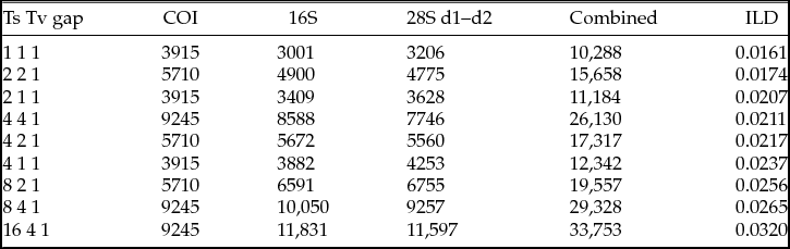
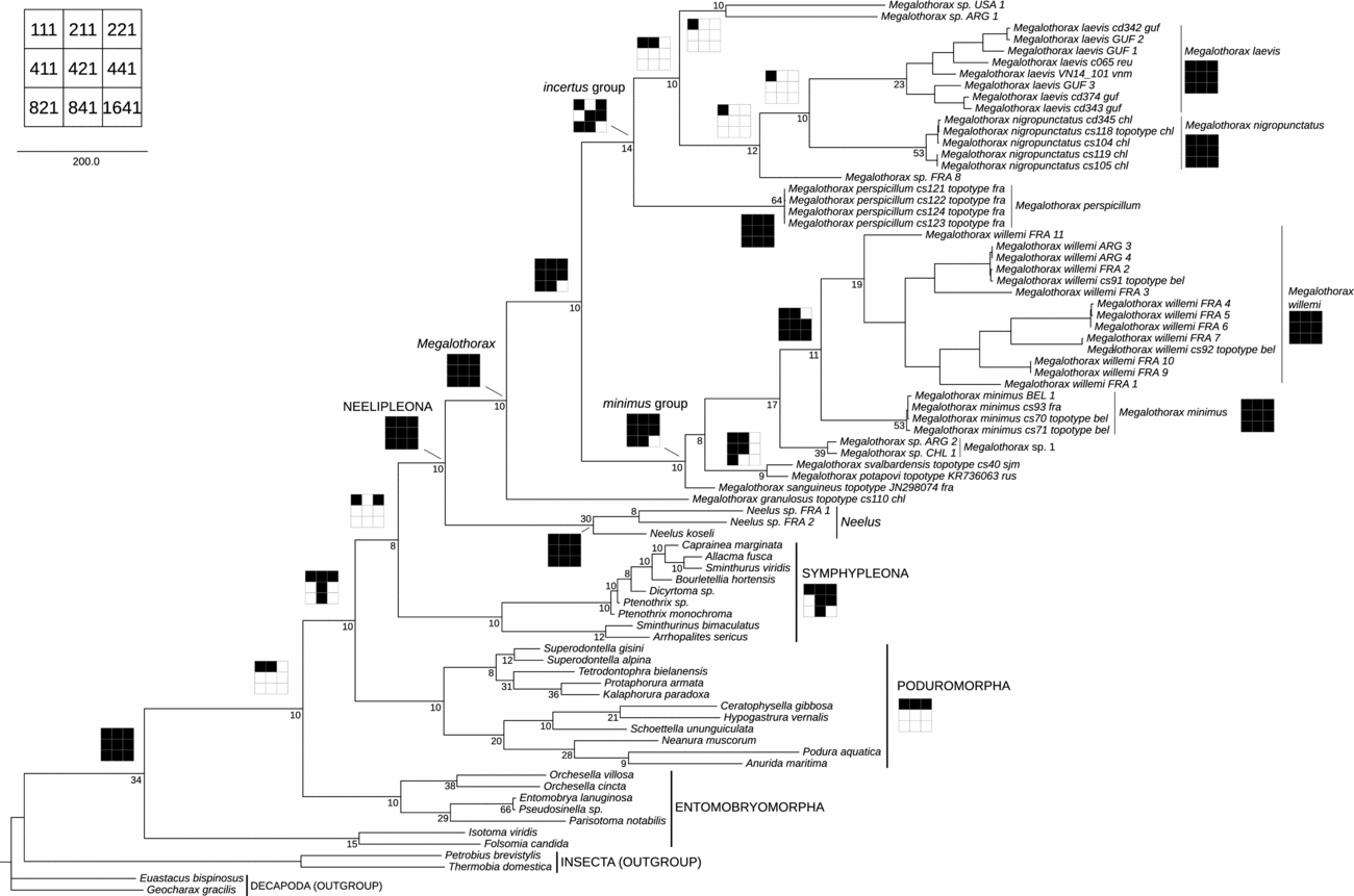
Maximum character congruence is found under the equal weighting scheme (Ts = Tv = Gap). Detailed ILD scores are provided in Table 4. The length of the most parsimonious tree equals 10,288 transformations, CI = 37.5, RI = 71.2 (Fig. 14). Poduromorpha, Symphypleona and Neelipleona are each monophyletic. The root of Collembola falls on the Isotomidae, making the Entomobryomorpha paraphyletic. The rest of Entomobryomorpha is the sister-group of the three other orders, related as (Poduromorpha, (Symphypleona, Neelipleona)). Inside Neelipleona, Neelus and Megalothorax are sister-groups. Each morphospecies of Megalothorax represented by more than one specimen is found monophyletic. Megalothorax granulosus is the sister-group of the rest of the Megalothorax (incertus and minimus groups sensu Schneider and D'Haese, Reference Schneider and D'Haese2013). The incertus and minimus groups are each monophyletic. Inside the minimus group, the relationships are: (M. sanguineus, ((M. potapovi, M. svalbardensis), (Megalothorax sp. 1, (M. minimus, M. willemi)))). Inside the incertus group, the relationships are: (M. perspicillum, ((Megalothorax sp. USA 1, Megalothorax sp. ARG 1), (Megalothorax sp. FRA 8, (M. nigropunctatus, M. laevis)))).
ILD scores

Molecular phylogeny of Collembola inferred from 16S rDNA and 28S d1 and d2 rDNA (direct optimization) and COI (prealigned)—most parsimonious phylogram under equal weighting scheme (111); 10,288 transformations, CI = 37.5, RI = 71.2. Sensitivity of the nodes of interest represented by two-dimensional diagrams (map in the left upper corner, white square = clade missing, black square = clade present). Bremer decay index reported next to the nodes, down to species level. Scale = 200 transformations.

Discussion
Phylogenetic analyses
With the present dataset, we cannot resolve the position of the root of Collembola. Thus, we will not further discuss the relationships between the orders of Collembola here. (See Schneider et al., Reference Schneider, Cruaud and D'Haese2011; Schneider and D'Haese, Reference Schneider and D'Haese2013 for a discussion.) The minimus group remains monophyletic with the addition of M. sanguineus and M. potapovi.
Megalothorax laevis is the sister species of M. nigropunctatus; however, we could not find any morphological characteristics supporting this relationship. Those two species are more similar to M. perspicillum on several aspects. Megalothorax laevis and M. perspicillum share the shape of the inner chaetae of sf2 and wrc5 rather close to sf5; M. nigropunctatus and M. perspicillum share 2 + 2 chaetae on the manubrium, a serrated mucro and 3 + 3 chaetae on sterna of Abd. IV. The relationships inside the incertus group is only weakly supported by the molecular data, as indicated by a high sensitivity to weighting parameters (Fig. 14). Suboptimal hypotheses are (M. laevis, M. perspicillum, M. nigropunctatus) for model 221 and (M. laevis, M. nigropunctatus), (M. perspicillum, minimus group) for model 211 (in supplementary material). According to the molecular data, 2 + 2 chaetae on sternum of Abd. IV and the smooth mucro are two convergences between M. laevis and the minimus group.
The phylogenetic analysis newly confirms the close relationship between M. potapovi and M. svalbardensis. This is consistent with the close similarity of the respective COI barcodes and of the antennal and abdominal chaetotaxy (Schneider et al., Reference Schneider, Porco and Deharveng2016).
Morphology
Pseudopore-like elements
Pseudopore-like structures were reported on the head of Megalothorax species in Schneider et al. (Reference Schneider, Porco and Deharveng2016). In the present work, we report additional locations of these elements on the thorax, the abdomen and the coxa I, each observed with SEM. The observation of those elements in light microscopy requires peculiarly good condition of clearness, hardly achievable in most preparations and in small or pigmented species. The whole pattern of pseudopore-like elements on head and trunk was also observed in M. aquaticus Stach, 1951, M. minimus, M. perspicillum Schneider and D'Haese, Reference Schneider and D'Haese2013, and M. potapovi and seems to be stable throughout the genus (similarly to τ-chaetae pattern). Our observations on the legs are sparse: We observed one element on coxa I of M. laevis and another seems to exist on coxa II of M. laevis (observation doubtful), and we could observe clearly the pseudopore on coxa II of M. nigropunctatus Schneider and D'Haese, Reference Schneider and D'Haese2013.
Antennal chaetotaxy variability
The interpopulational variability reported regarding the antennal chaetotaxy is unusual in Megalothorax. We initially thought to attribute the population of French Guiana to a new species until those variations could finally be observed inside the population of Gabon. We have no other characters supporting a separation. Megalothorax laevis seems widely distributed in the intertropical zone.
Sensory field 5
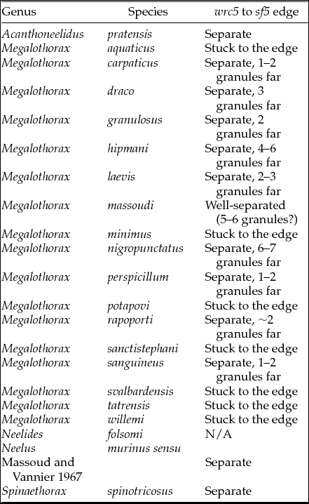
We introduced a new descriptor, namely the relative position of sf5 and wrc5 to each other. In several species of Megalothorax (e.g. M. minimus), wrc5 is right on the edge of sf5; in other species, wrc5 can be more or less separate from the edge of sf5. While this character is easily assessed with SEM., the edge of the sensory field can be difficult to observe in light microscopy. The degree of separation of wrc5 from the edge can be roughly quantified with the number of secondary integumentary granules present between the two elements. It can be noticed that in the genera Neelus, Spinaethorax and Acanthoneelidus, wrc5 is clearly separated from sf5, but the character is not applicable to Neelides. We report the state of this character in various species of Neelipleona (Table 5).
Position of wrc5 relative to sf5 among Neelipleona

Species for which the data are missing are not included, Neelides, Neelus and Spinaethorax genera are limited to their respective generotype. N/A = non-applicable. Data for Spinaethorax spinotricosus is from Papáč and Palacios-Vargas (Reference Papáč and Palacios-Vargas2016), other data are our observations.
Affinities with M. interruptus
Megalothorax interruptus was collected in 1961 in Sudan (Hüther, Reference Hüther1967). Its original description (Hüther, Reference Hüther1967) is highly consistent with M. laevis. Among the common characters between the two species are labial chaetotaxy, chaetotaxy of Ant. III, dorsal pattern of ordinary chaetae on head and trunk, chaetotaxy of manubrium, presence of s-chaetae s3 on abdomen, globular shape of sensory field inner chaetae, claw III morphology and smooth and notched posterior lamellae of mucro. Chaetotaxy of Ant. IV is similar, with 8 S-chaetae S; however, three ordinary chaetae, S-chaeta Sx and Or are missing on Ant. IV of M. interruptus (both in text and description; Hüther, Reference Hüther1967). At least Sx and Or were likely overlooked. Megalothorax interruptus would differ from M. laevis with 3 + 3 tooth on tenaculum (instead of 4 + 4); labral chaetae a2 with two clear teeth (instead of faint teeth) and presence of a pair of globular chaetae on the dorsal anterior part of the head, near antennae basis.
Adding the similarity between the two species to the evidently large repartition of M. laevis and its generalist ecology, one could suspect that M. interruptus and M. laevis are the same species. But while the fourth pair of teeth on the tenaculum is a character likely to be overlooked, the pair of globular chaetae cannot be dismissed without the reexamination of the type material (or in the lack of it, examination of topotypes of M. interruptus). Unfortunately, we could not locate the holotype of M. interruptus.
Intertropical megalothorax
Since its original description and until the present work, there was no new report of M. laevis in the literature. Other Megalothorax species known from intertropical zone are M. gabonensis Massoud and Vannier, 1965 (Gabon), M. interruptus (Sudan) and M. tristani Denis, 1933 (Costa-Rica). Megalothorax minimus was also reported from intertropical locality, e.g. Malaysia (Salmon, Reference Salmon1951), Ivory Coast (Delamare Deboutteville, Reference Delamare Deboutteville1950), Vietnam (Deharveng et al., Reference Deharveng, Bedos, Le C. K., Truong, Le, Truong and Ly2009), Cuba (Bonet, Reference Bonet1944), Brazil (Abrantes et al., Reference Abrantes, Bellini, Bernardo, Fernandes, Mendonça, Oliveira, Queiroz, Sautter, Silveira and Zeppelini2010), and India (Prabhoo, Reference Prabhoo1971). While M. gabonensis and M. tristani could hardly be confused with M. laevis, due to their serrate lamellae of the mucro, M. laevis and M. minimus were difficult to differentiate using the original descriptions. Thus, a legitimate doubt exists on the identity of the intertropical Megalothorax species with smooth mucro. For example, Bonet (Reference Bonet1947) description of M. minimus is evidently merging at least two species, one bearing s-chaetae s3 on abdomen (similarly to M. laevis), the other without s3 (similarly to M. minimus).
Megalothorax minimus redescription (Schneider and D'Haese, Reference Schneider and D'Haese2013) and the present work provide the needed disambiguation. To our knowledge, the two generalist, parthogenetic and widespread species with smooth mucro, M. minimus and M. willemi, are absent from Vietnam, Ivory Coast, Gabon, French Guiana and Réunion. While M. minimus and M. willemi are common in Europe and their presence was evidenced south of the Tropic of Capricorn (Chile, Argentina; Schneider and D'Haese, Reference Schneider and D'Haese2013), M. laevis seems to replace them as the commonest edaphic Megalothorax species in the intertropical zone. This fact is now to be verified more extensively. V. Papáč (pers. commun.) indicated to us the presence of M. willemi around the Tropic of Cancer (Mexico) and confirmed the wide distribution of M. laevis in intertropical regions of America (southern parts of Mexico, Panama, Brazil and Fernando de Noronha Island).
Identification key
The identification key is updated from Papáč and Kováč (Reference Papáč and Kováč2013) and Schneider and D'Haese (Reference Schneider and D'Haese2013) and restricted to the species with a smooth mucro. Megalothorax rubidus Salmon, 1946 was re-examined from a specimen provided by Salmon to Delamare Deboutteville and Massoud—evidently the one examined in Delamare Deboutteville and Massoud (Reference Delamare Deboutteville, Massoud, Deboutteville and Rapoport1963). Thus, a more accurate set of characters to distinguish M. granulosus Schneider and D'Haese, Reference Schneider and D'Haese2013, M. aquaticus and M. rubidus is used in the present key. Megalothorax piloli (Christiansen and Bellinger, Reference Christiansen and Bellinger1992) is described with mucro lamellae sometimes with very thin teeth but usually smooth (Christiansen and Bellinger, Reference Christiansen and Bellinger1992), and is therefore included in the key.
Key to the species of Megalothorax with smooth mucro lamellae
1. Posterior lamellae of the mucro smooth or almost smooth with possibly a notch or some irregularities . . . 2.
— Posterior lamellae of the mucro clearly serrate of crenelate . . . Not included in this key, see Papáč and Kováč (Reference Papáč and Kováč2013) and Schneider and D'Haese (Reference Schneider and D'Haese2013).
2. Mucro elliptical with wide and wavy lamellae . . . 3.
— Mucro not elliptical, with thin lamellae (no part of the mucro being clearly wider than the basis) . . . 5.
3. Head and trunk with coarse granules and a complex and dense network of integumentary channels; basomedian fields of labium with 1 + 1 chaetae; tenaculum with 4 + 4 teeth . . . M. granulosus (Schneider and D'Haese, Reference Schneider and D'Haese2013).
— Integument with thin granules; tenaculum with 3 + 3 teeth . . . 4.
4. Basomedian fields of labium with 1 + 1 chaetae; ventral, post-labial region of head with 3 + 3 chaetae, the 1 + 1 external chaetae being clearly longer and thicker than the 2 + 2 median, short chaetae; sternum of Abd. IV with 2 + 2 chaetae clearly stronger than the chaetae on sternum of Abd. VI . . . M. rubidus Salmon, 1946.
— Basomedian fields of labium with 3 + 3 chaetae; ventral, post-labial region of head with 3 + 3 thin and long chaetae, subequal; sternum of Abd. IV with 2 + 2 chaetae only slightly longer than the chaetae on sternum of Abd. VI and subequal in length . . . M. aquaticus Stach, 1951.
5. S-chaetae S1 and S4 missing on Ant. III . . . M. poki (Christiansen and Bellinger, Reference Christiansen and Bellinger1992).
— S-chaetae S1 and S4 present on Ant. III . . . 6.
6. Inner chaetae of sf2–6 with globular shape . . .7.
— Inner chaetae of sf2–6 either flam-shaped or T-shaped . . .9.
7. Labrum with forked chaetae a1; posterior lamellae of the mucro usually smooth, potentially with irregularities or very thin teeth . . . M. piloli (Christiansen and Bellinger, Reference Christiansen and Bellinger1992).
— Chaetae a1 of the labrum not forked; posterior lamellae of the mucro smooth with a distal notch . . . 8.
8. Presence of globular organ between antennae; labral chaetae a2 with 2 clear teeth; tenaculum with 3 + 3 teeth . . . M. interruptus (Hüther, Reference Hüther1967).
— Absence of globular organ between antennae; labral chaetae a2 with very faint teeth (almost smooth aspect); tenaculum with 4 + 4 teeth . . . M. laevis (Denis, Reference Denis1948).
9. Ant. IV with chaeta X missing; on head anterior integumentary channels covering the clypeal area with multiple forks . . . 10.
— Ant. IV with chaeta X present; on head anterior integumentary channels simple and not reaching the clypeal area . . . 11.
10. Without integumentary process in the expected position of chaeta X on Ant. IV, Ant. III with six ordinary chaetae; dorsal, anterior chaeta a0 missing, replaced with an integumentary, oval protuberance; unguis of ordinary morphology . . . M. sanctistephani (Christian, Reference Christian1998).
— Chaetae X missing, replaced with an integumentary process on Ant. IV, Ant. III with eight ordinary chaetae; dorsal, anterior chaeta a0 present, oval protuberance missing; unguis long and thin . . . M. tuberculatus (Deharveng and Beruete, Reference Deharveng and Beruete1993).
11. On head, dorsal, posterior chaetae thin or ordinary, subequal to anterior chaetae . . . 12.
— On head, dorsal, posterior chaetae thickened, stronger than anterior chaetae . . .14.
12. On head, dorsal, posterior chaetae thin and short; presence of a peculiar pattern of the granulation on the abdomen; unguis of ordinary shape . . . M. svalbardensis (Schneider and D'Haese, Reference Schneider and D'Haese2013).
— On head, dorsal posterior chaetae ordinary; no specific pattern of the granulation on the abdomen . . . 13.
13. Whitish with black and brownish pigments; channels on head without cycles; unguis elongated . . . M. tatrensis (Papáč and Kováč, Reference Papáč and Kováč2013).
— Adult with a deep red pigmentation; channels on head as a network; unguis ordinary . . . M. sanguineus (Schneider et al., Reference Schneider, Porco and Deharveng2016).
14. sf3–6 each with at least one T-shaped inner chaeta, the other inner chaetae flam-shaped; terga of Abd. I–V with 20 + 20 chaetae (total of α–η rows) . . . M. minimus Willem, 1900 sensu Schneider and D'Haese (Reference Schneider and D'Haese2013).
— Every inner chaetae of sf2–6 flam-shaped . . . 15.
15. Dorsal, posterior region of head with lanceolate macrochaetae, thorax with enlarged macrochaetae; clypeal area with an integumentary protuberance chaetae in front of chaeta a0 . . . M. potapovi (Schneider et al., Reference Schneider, Porco and Deharveng2016).
— Dorsal, posterior region of head and terga of thorax with ordinary or thickened chaetae; clypeal area without integumentary protuberance chaetae in front of chaeta a0 . . . 16.
16. Inner chaetae of sf6 long; maxilla outer lobe with a sublobal hair; unguis ordinary . . . M. willemi (Schneider and D'Haese, Reference Schneider and D'Haese2013).
— Inner chaetae of sf6 short; maxilla outer lobe with two sublobal hairs; unguis elongated . . . M. carpaticus (Papáč and Kováč, Reference Papáč and Kováč2013).
Acknowledgements
The laboratory work was done in D'Haese's BoEM laboratory and the cluster of the Muséum National d'Histoire Naturelle (CNRS UMS 2700, http://ums2700.mnhn.fr/pcia/acces) provided the computing resources. Microphotographs were taken at the Microscopies and Imagery Platform (MNHN). Field-collection in French Guiana (2009) was supported by CaFoTrop (www.cafotrop.com). Many thanks to Sandrine Salmon who we collected the material with in La Réunion Island and to Pierre-Michel Forget (UMR7179 CNRS-MNHN) and Dominique Strasberg (UMR pvbmt) and to the Parc national de la Réunion that made that project possible, and also to Action Transversale du Muséum DS-E and MECADEV UMR7179 CNRS-MNHN for the financial support. The specimens from Gabon were provided by Jean-Marie Betsch. The 2013 Vietnamese specimens (including topotypes) were collected during the project ‘Biodiversité de la cordillère annamitique: endémisme et gradients altitudinaux’ funded by the LABEX BCDIV of the Muséum national d'Histoire naturelle (Paris). We are grateful to Truong Quang Tam (Institute of Tropical Biology, Ho Chi Minh City) and to Louis Deharveng (MNHN) who organized the field trip and obtained the research permits, and to Nguyen Hanh, director of the Hon Ba Nature Reserve and the staff of the Reserve who provided guiding and facilities for the field work in Hon Ba. The 2014 Vietnamese specimens were collected during a biodiversity assessment trip in Hon Chong area supported by Holcim Co and IUCN. The other Vietnamese specimens were collected during field trips organized by Louis Deharveng (MNHN), Anne Bedos (MNHN) in collaboration with Pr. Le Cong Kiet (Vietnam National University, Ho Chi Minh City), Le Cong Man (Vietnam National University, Ho Chi Minh City) and Truong Quang Tam (Institute of Tropical Biology, Ho Chi Minh City). We thank Vladimír Papáč and anonymous reviewer for the helpful comments and corrections that improved the manuscript.
Supplementary material
To view supplementary material for this article, please visit https://doi.org/10.1017/S1742758418000024