Introduction
Transsphenoidal pituitary surgery has continued to evolve since being described in the early twentieth century. Initially, it was performed with a headlight and speculum,Reference Patel, Husain, Eloy, Couldwell and Liu1 then using a microscope, and is now often performed entirely endoscopically. In 1992, Jankowski et al. described an entirely endoscopic transnasal approach.Reference Jankowski, Auque, Simon, Marchal, Hepner and Wayoff2 This is currently the most common technique for pituitary surgery, and it involves a direct transostial approach with a posterior septectomy.Reference Jankowski, Auque, Simon, Marchal, Hepner and Wayoff2 However, a technique via an endoscopic transseptal route has also been described that avoids a posterior septectomy, but it has its own disadvantages.Reference Papay, Stein, Rhoten, Luciano, Zins and Hahn3,Reference Favier, Boetto, Cartier, Segnarbieux and Crampette4
We describe a modification of the endoscopic transseptal technique. Hull ENT Ethics Committee ethical approval was not required for this technical description.
Materials and methods
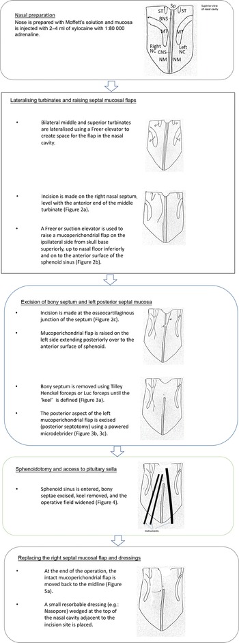
A schematic diagram of the steps involved in the modified transseptal approach for endoscopic transsphenoidal pituitary surgery is shown in Figure 1.
Schematic diagram of steps involved in the modified transseptal approach in endoscopic transsphenoidal pituitary surgery. Sp = sphenoid; ST = superior turbinate; BNS = bony nasal septum; MT = middle turbinate; NC = nasal cavity; CNS = cartilaginous nasal septum; NM = nasal mucosa

Nasal preparation
The nose is prepared with Moffett's solution, and the mucosa is injected with 2–4 ml of xylocaine with 1:80 000 adrenaline.
Lateralising turbinates and raising septal mucosal flaps
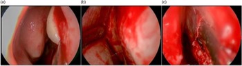
Surgery commences with a 0-degree endoscope. Bilateral middle and superior turbinates are lateralised using a Freer elevator to create space for the flap in the nasal cavity (we do not routinely resect the middle turbinates as some do). The incision is made on the nasal septum, using a number 15 scalpel blade, level with the anterior end of the middle turbinate (Figure 2a). It is our preference to perform this on the right side, and further descriptions below assume the initial incision is made on the right. However, if required, the principle can be reversed with the initial incision on the left.
Intra-operative endoscopic view showing the mucosal incision level with the middle turbinate (a), raising of right mucosal flap (b), and incising of the osseocartilaginous junction prior to excision of the bony septum (c).

The incision starts approximately 1 cm below the skull base superiorly (to avoid transecting the olfactory region)Reference Gomez, Yanez-Siller, Carrau, Montaser, Lima and Servian5 and continues down to the nasal septal floor inferiorly. The latter is important because the septal flap will then be anchored at the floor rather than the inferior part of the septum itself, resulting in: (1) a wider operative field because the septal flap can be pushed more laterally into the nasal cavity; and (2) a reduced risk of inadvertently ‘dividing’ the flap's inferior attachment whilst raising the mucoperichondrial flap.
A Freer or suction elevator is used to raise a mucoperichondrial flap on the ipsilateral side from the skull base superiorly, up to nasal floor inferiorly and on to the anterior surface of the sphenoid sinus (Figure 2b). The sphenoid sinus opening comes into view at the end of this step. Further surgery is performed with instruments placed medial to the mucoperichondrial flap.
Excision of bony septum and left posterior septal mucosa
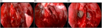
An incision is made at the osseocartilaginous junction of the septum, from a superior to inferior direction (Figure 2c). A mucoperichondrial flap is raised on the contralateral side, extending posteriorly over to the anterior surface of the sphenoid on the contralateral side. Bony septum is removed using Tilley Henckel forceps or Luc forceps until the ‘keel’ is defined (Figure 3a). At this point, we excise the posterior aspect of the contralateral mucoperichondrial flap (posterior septotomy) using a powered Medtronic microdebrider (Figure 3b, 3c). The size and position of this window can be adapted according to tumour features and surgeon preference, allowing a binasal approach to the skull base, as is standard practice for an endoscopic transnasal approach.
Intra-operative endoscopic view showing the ‘keel’ following removal of bony septum (a), microdebridement of left septal mucosa (b) and both sphenoid ostia following these steps (c).

Sphenoidotomy and access to pituitary sella
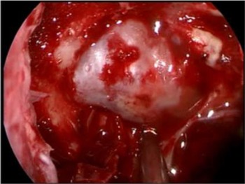
The sphenoid sinus is entered, bony septae excised, keel removed, and the operative field widened to obtain optimum access to anterior and middle skull base structures (Figure 4).
Intra-operative endoscopic view of pituitary fossa dura following sphenoidotomy and removal of the ‘keel’, creating a wide access to the pituitary fossa.

Replacing right septal mucosal flap and dressings
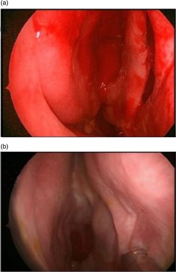
At the end of the operation, the lateralised intact mucoperichondrial flap is moved back to the midline (Figure 5a). We do not suture the incision line. We insert a small resorbable dressing (e.g. NasoPore®), wedged at the top of the nasal cavity adjacent to the incision site, to ensure the flap does not lateralise, and to allow end-to-end contact at the incision. Alternatively, tissue glue can be used to ‘stick’ the incision back together.
Intra-operative endoscopic view showing replacement of right septal mucosa at the end of the procedure (a) and its appearance six months later (b).

Discussion
An endoscopic transseptal transsphenoidal approach to the pituitary fossa has been previously described by Papay et al. (in 1997)Reference Papay, Stein, Rhoten, Luciano, Zins and Hahn3 and Favier et al. (in 2018).Reference Favier, Boetto, Cartier, Segnarbieux and Crampette4 Our modified transseptal approach differs from the erstwhile described methods and has several advantages, as described below.
Classical transseptal approach incision is a hemitransfixion incision at the caudal margin of the cartilaginous nasal septum. The incision we describe is level with the anterior end of the middle turbinate. However, the incision can be made further posteriorly or anteriorly, or extended to include mucosa of the floor and lateral wall if one anticipates a larger anterior skull base defect, or is indeed unexpectedly faced with one after tumour resection, providing more versatility and choice.
Making the incision level with the anterior middle turbinate leaves 2 cm (or more) of septal cartilage caudal strut with intact mucoperichondrium bilaterally, which is reliable to maintain vascularity and minimise the cartilage resorption risk.
A hemitransfixion incision often requires the release of the nasal septum from its attachment to the maxillary crest and spine. Although the septum can be reattached, it will have become unstable, and might heal in a variable position to the right or left, with resultant columellar deviation or indrawing.
If there is significant anterior septal deviation, the mucosal incision can be made more anteriorly, to allow septoplasty for access.
A classical transseptal approach attempts to maintain the septum, which creates a significantly narrower operative field. The modified approach uses the space in both nasal cavities. Furthermore, lateralisation of bilateral middle and superior turbinates at the beginning of the procedure allows the intact right mucoperichondrial flap to be lateralised to the lateral nasal wall. (The initial mucosal incision superiorly to the nasal floor allows the flap to be lateralised to the lateral wall.) These steps, together with the posterior bony septectomy and resection of the contralateral flap, provide sufficient access for instruments via both nostrils.
The septum heals with no perforation in our experience (Figure 5b). It ensures nasal airflow in two separate nasal cavities that is physiological which may result in less crust formation, reduced risk of whistling due to septal perforation and improved nasociliary transport of mucous.
In the event of a previously unplanned need for a nasoseptal flap to repair the skull base, the flap can be released from its superior and inferior attachments, and used without compromise to the dimensions or vascularity. This holds true in case of a return to the operating theatre for a recalcitrant cerebrospinal fluid leak in the post-operative period. If revision surgery is required in the future, it is still possible to raise a nasoseptal flap.
At least 20 cases have been carried out by the authors using this technique. Although this technique aims to avoid septal perforation, this can still occur, especially if there is a tear in the flap.
Competing interests
None declared