Introduction
Early “ptychoparioid” trilobites are notoriously difficult to identify to genus and supergeneric groups primarily due to their relatively simple and similar morphologies and the lack of detailed information. They are typically thought to be the ancestral stock for the more derived trilobite clades (Fortey, Reference Fortey and Kaesler1997, Reference Fortey2001), and thus occupy a pivotal place in trilobite evolution. Unfortunately, there are several barriers preventing accurate placement of these early trilobites into a phylogenetic scheme, particularly the typical absence of information regarding non-cranidial sclerites and the limited number of specimens used to name taxa.
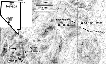
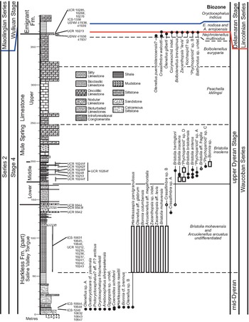
Described herein are “ptychoparioid” trilobites found in the middle to upper Dyeran Stage of Laurentia (Cambrian Series 2, Stage 4) from the upper Harkless Formation, Mule Spring Limestone, and lowermost Emigrant Formation at Clayton Ridge, Nevada (Figs. 1, 2). This paper is complimentary to a report on the corynexochines and biostratigraphy of the same interval (Sundberg and Webster, Reference Sundberg and Webster2021). Some of the new collections contain abundant specimens, including articulated exoskeletons and/or associated sclerites. These specimens alone do not solve the taxonomic mess of early “ptychoparioids,” but they do provide data that represent a significant step towards potential resolution of early libristomate trilobite phylogeny.
Location map for the trilobites from the Harkless Formation and Mule Spring Limestone in Clayton Ridge, Nevada (part of the Split Mountain 7.5′ topographic quadrangle map, U.S. Geological Survey, 1991, 1:24,000 scale).

Composite stratigraphic section of the Saline Valley Tongue, Harkless Formation; Mule Spring Limestone; and lowermost Emigrant Formation, Clayton Ridge, Nevada. Section modified from Sundberg and McCollum (Reference Sundberg and McCollum2003b), Webster (Reference Webster, Hollingsworth, Sundberg and Foster2011b, Reference Websterc), Sundberg (Reference Sundberg2018), and Mike and Linda McCollum (personal communication, 2020). Trilobite occurrences are compiled from the various localities around Split Mountain (Fig. 1). Open boxes indicate specific stratigraphic position is unknown; open circles indicate questionable species identification; “f” after collection number indicates material was float, vertical bars for these collections indicate most likely interval of stratigraphic occurrence of the material. Information for some taxa from the Mule Spring Limestone and the lowermost Emigrant Formation is derived from Sundberg and McCollum (Reference Sundberg and McCollum2003b) and Webster (Reference Webster, Hollingsworth, Sundberg and Foster2011b, Reference Websterc). Olenelloid and corynexochine trilobite stratigraphic ranges are from Sundberg and Webster (Reference Sundberg and Webster2021). Blue line represents the base of the global Miaolingian Series; red line represents the base of the Laurentian Lincolnian Series.

Geologic background
Clayton Ridge is a relatively small, northeast-southwest trending fault block west of the Montezuma Range, in Esmeralda County, Nevada (Fig. 1). In the Split Mountain area of Clayton Ridge, Cambrian strata range from the pre-trilobitic upper Deep Springs Formation to the uppermost Cambrian Emigrant Formation. This paper focuses on “ptychoparioids” from the mid to upper Dyeran succession, comprising the uppermost Harkless Formation, Mule Spring Limestone, and lowermost Emigrant Formation in the Split Mountain area. Much of the geologic background presented here is more fully documented in Sundberg and Webster (Reference Sundberg and Webster2021).
The Harkless Formation (Nelson, Reference Nelson1962) is a thick succession of siltstone, sandstone/quartzite, and minor carbonates. Ahn et al. (Reference Ahn, Babcock and Hollingsworth2011) subdivided the Harkless Formation into two members: the lower Weepah Member and upper Alkali Member. The upper portion of the Alkali Member contains ~100 m of pale brownish to gray crystalline limestones and thin sandstone beds, which are referred to as the Saline Valley Tongue (Ahn et al., Reference Ahn, Babcock and Hollingsworth2011; = upper Harkless Formation of Webster, Reference Webster2011c). Trilobites from the section in the 145 m thick Saline Valley Tongue at Clayton Ridge represent the mid-Dyeran (no trilobite zonation) and the overlying Arcuolenellus arcuatus and Bristolia mohavensis biozones (Webster, Reference Webster, Hollingsworth, Sundberg and Foster2011a, Reference Websterc).
The overlying Mule Spring Limestone (Nelson, Reference Nelson1962) is a carbonate unit that includes fenestral, micropeloidal, and oncolitic beds (Webster, Reference Webster2011c; Cordie et al., Reference Cordie, Dornbos and Marenco2020). The transition between the Harkless Formation and the Mule Spring Limestone is gradational; the latter formation has a total thickness of ~224 m (Webster, Reference Webster2011c). The trilobite faunas in this formation represent (in ascending order) the Bristolia insolens, Peachella iddingsi, and probably the Bolbolenellus euryparia biozones (Webster, Reference Webster, Hollingsworth, Sundberg and Foster2011b).
Only the first two meters of shales and limestones of the Emigrant Formation contain faunas of the uppermost Dyeran Nephrolenellus multinodus Biozone (Sundberg and McCollum, Reference Sundberg and McCollum2003b; Webster, Reference Webster, Hollingsworth, Sundberg and Foster2011b; Sundberg, Reference Sundberg2018).
The ptychoparioid problem
As pointed out by Fortey (Reference Fortey and Kaesler1997) and Adrain (Reference Adrain2011), the order Ptychopariida Swinnerton, Reference Swinnerton1915, as presently recognized, is probably paraphyletic. Even the relationships among confamilial genera within “Ptychopariida” are unclear, primarily due to the lack of knowledge of the complete exoskeletons, ontogeny, and phylogenetic analyses, especially for lower and middle Cambrian trilobites (e.g., Sundberg, Reference Sundberg2004). As a result, we agree with Adrain (Reference Adrain2011) that the ordinal assignment of the taxa discussed below is unknown, and we use “ptychoparioid” in a phylogenetically informal sense to refer to any of the early libristomates previously encompassed within the abandoned order Ptychopariida. The familial assignment of the “ptychoparioid” taxa discussed below is equally problematic, as was stated by Rasetti (Reference Rasetti1963, p. 575) nearly 60 years ago: “In the writer's opinion, many of the proposed suprageneric taxa are based at best on superficial similarities, with little consideration for time and space distribution, and are not indicative of relationship. Hence the decision of publishing the descriptions at the present time is not due to the existence of a satisfactory classification, but rather to belief that no substantial improvement will be forthcoming in the foreseeable future. Considering the pretense of ordering the genera into ‘families’ in this group rather misleading than useful the genera are arranged in alphabetical order.”
Determining the phylogenetic relationships between early “ptychoparioids,” or even assigning them to a genus, is difficult and, at present, largely subjective. Dealing with these taxa is complicated by several factors: (1) these trilobites have a simple morphology, with a few exceptions; (2) many of the genera are poorly diagnosed and type species are illustrated by drawings or retouched photos; (3) some of the type species are poorly preserved; (4) the complete exoskeleton of many species is unknown, and most are named from cranidia only; and (5) when multiple species are observed, recognized genera appear to grade from one to another. These factors are briefly discussed below.
Simple morphology
Most early “ptychoparioids” possess a semi-circular cephalon, a posteriorly tapering multisegmented thorax, and are micropygous. Generally, the glabella is anteriorly tapered with shallow lateral glabellar furrows, the frontal area has a convex anterior border, and the palpebral lobes are relatively horizontal. Species and genera have been discriminated by relatively minor morphological variation (often in continuously gradational traits) within this conservative morphology (Lochman, Reference Lochman1947; Rasetti, Reference Rasetti1951, Reference Rasetti1955; Shaw, Reference Shaw1962; Palmer and Halley, Reference Palmer and Halley1979; Blaker and Peel, Reference Blaker and Peel1997; Fortey Reference Fortey and Kaesler1997, Reference Fortey2001; Cotton, Reference Cotton2001; Webster, Reference Webster2011d).
Poor diagnoses and illustrations
The epitome of poor diagnoses and the lack of illustrations is represented by Resser's “Contribution to nomenclature on Cambrian trilobites” (Resser, Reference Resser1935, Reference Resser1936, Reference Resser1937, Reference Resser1938, Reference Resser1942; see Sundberg, Reference Sundberg2007, for discussion of Resser's work). This series of publications provided abbreviated diagnoses and comparison of genera, and relied upon previous illustrations. The result is that taxonomists have either assumed that the genera are valid or, in most recent works, needed to obtain type material and/or recollect type localities to determine the morphology of the type species. For example, the diagnosis of Syspacephalus Resser, Reference Resser1936, gave only the general characteristics of the glabella, palpebral lobes, and sutures, and made no comparison to similar taxa. The referenced illustration of the type species, S. charops (Walcott, Reference Walcott1917, pl. 13, fig. 2) is a single retouched photo and a line drawing of the side profile.
As a result of Resser's poor choice of a type species and poor description, the genus Syspacephalus has had a rather long and confusing history. Lochman (Reference Lochman1947, p. 64) revised Syspacephalus, but based only on the cranidium of the type specimen of S. charops and the associated eight other cranidia found in the same rock or an associated rock. She assigned only the type species to the genus. Rasetti (Reference Rasetti1951, pl. 8, figs. 11–13) illustrated another cranidium of the species from the lower Cambrian of British Columbia, but added six more species to the genus that included articulated specimens and/or additional sclerites other than cranidia. Five of the new species are from the middle Cambrian Wenkchemnia-Stephenaspis Zone of the Mount Whyte Formation and one is possibly from the Albertella zone of the Cathedral Formation. Palmer and Halley (Reference Palmer and Halley1979) recognized Syspacephalus and Elrathina as valid genera and named addition middle Cambrian species. In contrast, Babcock (Reference Babcock1994, p. 110) synonymized Elrathina with Syspacephalus, based on Robison's (Reference Robison, Robison and Rowell1976, p. 102) comment that the two genera are potentially the same. Furthermore, Blaker and Peel (Reference Blaker and Peel1997, p. 124) synonymized Syspacephalus, Elrathina, and Eoptychparia Rasetti, Reference Rasetti1955, with the genus Ptychoparella Poulsen, Reference Poulsen1927. The type species, P. brevicauda Poulsen, Reference Poulsen1927, is at least known from three specimens (a cranidium, partial librigena, and pygidium), all isolated sclerites from one or more boulders on the beach at Cape Frederick VII (Poulsen, Reference Poulsen1927, p. 341). Blaker and Peel (Reference Blaker and Peel1997) placed this material in the Cape Wood Formation of North-West Greenland. This synonymy was rejected by Sundberg and McCollum (Reference Sundberg and McCollum2003a, Reference Sundberg and McCollumb) and Geyer and Peel (Reference Geyer and Peel2017). Sundberg (Reference Sundberg2018) further discussed the differences between Syspacephalus and Elrathina and discussed the taxa that had been assigned to the former.
It is interesting to note that the type specimen of S. charops has never been illustrated since Walcott's (Reference Walcott1917) retouched photograph, despite the extensive work on the genus that has been undertaken. Furthermore, the non-cranidial sclerites are still unknown for the type species. Thus, assignment of species to Syspacephalus cannot be made with confidence because of the very limited morphological information available for the type species.
This previous example is not isolated. Lochman (Reference Lochman1947) revised 10 other “lower” Cambrian genera based on cranidial features, including: Antagmus Resser, Reference Resser1936; Litocodia Resser, Reference Resser1938, Onchocephalus Resser, Reference Resser1937; Periomma Resser, Reference Resser1937; Periommella Resser, 1938; Plagiura Resser, Reference Resser1935; Plagiurella Resser, Reference Resser1937; Proliostracus Poulsen, Reference Poulsen1932; and Ptychoparella Poulsen, Reference Poulsen1927, as well as Syspacephalus. She attempted to provide more detailed diagnoses based on observations of the type material and additional material from the same collections. In addition, she proposed generic assignments for previously named, non-type species. Unfortunately, she only illustrated the type specimens of her two new genera, Piaziella and Crassifimbra.
Rasetti (Reference Rasetti1955) subsequently revised some lower Cambrian “ptychoparioid” genera using well-preserved cranidia found in boulders in the Ville Guay Conglomerate (= Sillery Formation) in Quebec. These taxa included Antagmus; Onchocephalus; Crassifimbra; Austinvillia Resser, 1938; Periomma; Periommella; and Rimouskia Resser, 1938. In addition, Rasetti (Reference Rasetti1955) recognized the new genera Eoptychoparia, Luxella, and Bicella. Unfortunately, virtually all of Rasetti's specimens were cranidia, with only two species also being illustrated with librigenae. Thus, like for Lochman (Reference Lochman1947), taxa were diagnosed primarily on cranidial features.
Poor preservation
Some genera are based on species that are poorly preserved and are inadequate for reliable diagnosis. For example, Litocodia was based on a small, laterally distorted cranidium preserved in shale (Lochman, Reference Lochman1947), and Antagmus is based on a limited number of specimens preserved as internal molds in a limonite-cemented, friable, medium-grained sandstone (Sundberg, Reference Sundberg2007, fig. 9).
Taxonomic problems can extend beyond the generic level. Many early “ptychoparioids” have been placed into the Family Antagmidae, which is based on the genus Antagmus. Unfortunately, the type specimens of the type species, Antagmus teucer (Walcott, Reference Walcott1886), are poorly preserved and consist only of cranidia. As a result, the species is a nomen dubium (see Sundberg, Reference Sundberg2007), which in turn places the genus Antagmus, the subfamily Antagminae, and 24 previously named genera and 104 previously named species (Adrain, Reference Adrain2011) in open taxonomic assignment.
Poor knowledge of morphology
The vast majority of early “ptychoparioids” are known from only a very few specimens, and primarily from cranidia. This paucity of anatomical information has led to significant problems in the taxonomic assignment of these forms to generic and higher taxonomic groups (see comments in Rasetti, Reference Rasetti1951, Reference Rasetti1955; Palmer, Reference Palmer1968; Blaker and Peel, Reference Blaker and Peel1997; Fortey, Reference Fortey and Kaesler1997; Geyer and Malinky, Reference Geyer and Malinky1997; Cotton, Reference Cotton2001; Sundberg, Reference Sundberg2004; Webster, Reference Webster2011d).
A prime example of the importance of knowing more than just cranidial morphology is the Family “Ehmaniellidae” proposed by Sundberg (Reference Sundberg1994). Taxa assigned to this family previously had been included in Alokistocaridae Resser, Reference Resser1939 (Resser, Reference Resser1939) (Howell in Harrington et al., Reference Harrington, Henningsmoen, Howell, Jaanusson, Lochman-Balk and Moore1959), but were reassigned to the new family based on their overall similarity and the paucity of morphological data on the type species (Alokistocare subcoronatum [Hall and Whitfield, Reference Hall and Whitfield1877]) of the genotype (Alokistocare Lorenz, Reference Lorenz1906) of Alokistocaridae (Sundberg, Reference Sundberg1994). Subsequent collecting of the type area of Alokistocare subcoronatum produced additional specimens, including librigenae, hypostomes, rostral plates, thoracic segments, and pygidia. With more complete knowledge of the trilobite morphology, especially the pygidium, Sundberg (Reference Sundberg1999) recognized Ehmaniellidae to be a junior synonym of Alokistocaridae.
Gradational morphology
Both Lochman (Reference Lochman1947) and Rasetti (Reference Rasetti1955) noted the existence of morphologically intermediate species that blurred distinctions between many purported taxa. Rasetti (Reference Rasetti1955) commented on the similarities and gradational boundaries among cranidia of the genera Antagmus, Austinvillia, Crassifimbra, Eoptychoparia, Luxella, Onchocephalus, and Piaziella. These gradations were evident in features examined in dorsal view and in the convexity of the intraocular fixigena and glabella.
To highlight just one further example, Elrathiella? intermedia Sundberg, Reference Sundberg1994, is a morphological (and stratigraphic) intermediate between older Elrathiella species and younger Ehmaniella species. This species is most similar to Elrathiella in having a moderately rounded frontal lobe, narrower preglabellar field, and long pygidial axis having several rings. However, Elrathiella? intermedia does have longer palpebral lobes and a less-tapered glabella, as seen in Ehmaniella. This combination of features places this species morphologically between the two established genera.
Recommendations
Establishment of new species and genera of early “ptychoparioid” trilobites should be based on examination of numerous, well-preserved specimens covering a wide range of sizes; ideally, multiple sclerites (the cranidium, librigena, pygidium, and, hopefully, the thorax) should be represented. Fortunately for early “ptychoparioid” systematists, interspecific differences among the three species studied by Webster (Reference Webster2011d) were already evident at cranidial length smaller than 1.75 mm, which is good news. It suggests that “ptychoparioid” species can be distinguished even at very small cranidial size. The fact that these species exhibit divergent ontogenetic trajectories of shape change confirms that diagnosis of early “ptychoparioid” species is easier when comparing large cranidia. Poorly diagnosed genera should be restricted to the type species.
Material and methods
Material
The trilobites from the lowest limestones of the Saline Valley Tongue of the Harkless Formation reported here are preserved as finely silicified specimens in laminated clayey micrites. The methods used to prepare these silicified specimens are presented in Sundberg and Webster (Reference Sundberg and Webster2021). Other non-silicified specimens from the Harkless Formation, Mule Spring Limestone, and Emigrant Formation were mechanically prepared.
Illustrated specimens were coated with colloidal graphite and then ammonium chloride sublimate. Specimen orientation for photography and measurements is primarily with the cranidial anterior border and/or palpebral lobes, librigenal border, or pygidial border in a horizontal plane.
Morphometric data and analyses
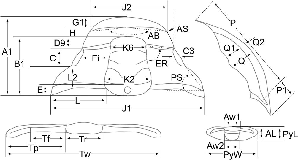
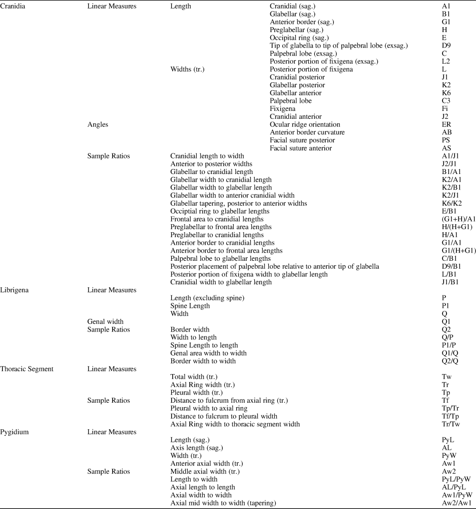
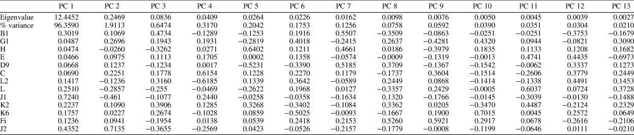
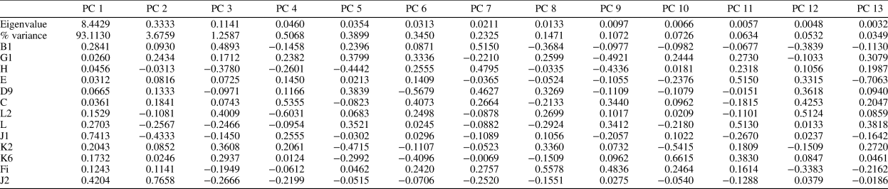
Linear and angular measurements are illustrated in Figure 3 (also see Table 1) and were extracted from digital images using Photoshop. Tectonically distorted specimens were not measured. Morphometric analyses were undertaken using Excel and principal component analysis (PCA) of linear cranidial measurements using a variance-covariance matrix in PAST (Hammer et al., Reference Hammer, Harper and Ryan2001). Two linear measurements not included in the PCA were the cranidial length (A1), due to its duplication of the other sagittal measurements (E + B1 + H + G1 = A1), and the palpebral lobe width (C3), which is missing for several specimens due to breakage. Three PCAs were undertaken: (1) all taxa including smaller specimens; (2) all taxa including specimens >3.0 mm to compare the morphology of larger specimens; and (3) the meraspides, Anebocephalus silverpeakensis n. gen. n. sp. and Coenoides scholteni n. gen. n. sp., from locality ICS-10647 to determine changes in morphology due to ontogeny.
Measures and angles used to describe morphologic characteristics of “ptychoparioid” trilobites. Abbreviations are listed in Table 1. Modified from Sundberg and McCollum (Reference Sundberg and McCollum2003b).

Morphological features, angles, and ratios used in this study and their abbreviations (see Fig. 3).

Diagnosis of new “ptychoparioid” genera from the Harkless Formation necessitates comparison to other taxa. However, most early “ptychoparioids” are known only from cranidia. This limits complete comparisons and thus diagnoses. The new taxa Coenoides, Anebocephalus, and Harklessaspis n. gen. are compared not only to each other, but also to the Dyeran taxon Crassifimbra (previously assigned to Antagmidae, Jell in Jell and Adrain, Reference Jell and Adrain2002) and the Delamaran taxa Eokochaspis and Elrathina (presently assigned to Ptychopariidae, Jell in Jell and Adrain, Reference Jell and Adrain2002), which share similar morphologies and represent potential outgroups. Taxa considered herein are: Coenoides = Co. scholteni (type species) and Co. freucheni (Geyer and Peel, Reference Geyer and Peel2011); Anebocephalus = A. silverpeakensis (type species) and A. pearylandica (Geyer and Peel, Reference Geyer and Peel2011); Harklessaspis = H. rasettii n. gen. n. sp. (type species) and H. parvigranulosus n. gen. n. sp.; Elrathina = El. cordillerae (Rominger, Reference Rominger1887; type species), all preserved flattened in shale (data from Rasetti, Reference Rasetti1951, and Geyer and Peel, Reference Geyer and Peel2017, not figs. 6.6, 6.9, 6.10, which probably = Elrathia permulta [Walcott, Reference Walcott1918]), El. aphrodite Geyer and Peel, Reference Geyer and Peel2017, El. athena Geyer and Peel, Reference Geyer and Peel2017, and El. hera Geyer and Peel, Reference Geyer and Peel2017; Crassifimbra = Cr. walcotti (Resser, Reference Resser1937; type species) and Cr. metalaspis (Sundberg and McCollum, Reference Sundberg and McCollum2000), all data from Webster (Reference Webster2011d); Eokochaspis = Eo. nodosa Sundberg and McCollum, Reference Sundberg and McCollum2000 (type species), all data from Webster (Reference Webster2011d), and due to the paraphyletic nature of the genus (Sundberg, Reference Sundberg2004), only the type species was included.
Repositories and institutional abbreviations
Specimens discussed within this paper are housed in the Field Museum, Chicago (FMNH), the United States Natural History Museum (USNM), and the Geology Museum at the University of California, Riverside (UCR). Figured specimens from collections initially housed in the ICS (Institute for Cambrian Studies, University of Chicago) have been reaccessioned into the FMNH. Specimens from UCR collections are numbered sequentially using the collection number as a prefix (e.g., UCR 10273-1).
Results
All three principal component analyses of cranidial data for the studied taxa illustrate that principal component (PC) 1 represents 93–98% of sample variance (Tables 3–5) and is strongly correlated to sagittal cranidial length (R2 = 0.93–0.98, p << 0.0001). PC1 can be interpreted as essentially representing specimen size. In the analysis that includes specimens of all sizes and species (Table 2), PC2 represents 1.9% of sample variance and has relatively high positive loadings (>0.2) for the anterior border length (G1), palpebral lobe length (C), and anterior border width (J2), and strongly negative loadings (< −0.2) for posterior portion of the fixigena width (L) and cranidial width (J1). PC3 represents 0.6% of sample variation and has relatively high positive loadings for glabellar length (B1), posterior portion of the fixigenal length (L2), and glabellar widths (K2, K6), and strongly negative loadings for preglabellar field length (H), posterior portion of the fixigena width (L), and anterior border width (J2).


In the analysis that includes specimens >3 mm in length (Table 3), PC2 and PC3 represent 3.7% and 1.3% of sample variation, respectively, and have similar morphological loadings as the analysis of all the specimens.
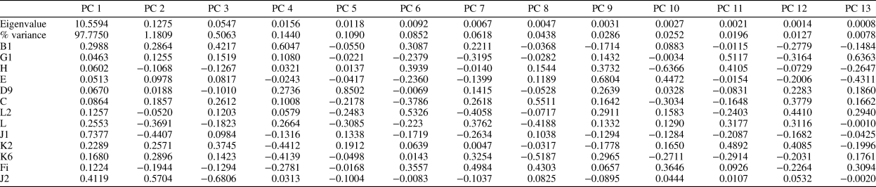
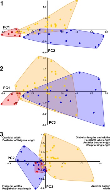
In the PCA that includes only the meraspides, Anebocephalus silverpeakensis and Coenoides scholteni from locality ICS-10647 (Table 4), PC2 represents 1.2% of sample variance and has relatively high positive loadings (>0.2) for glabellar length (B1), glabellar widths (K2, K6), and anterior cranidial width (J2), and strongly negative loadings (< −0.2) for cranidial widths (L, J1). PC3 represents 0.5% of sample variation and has relatively high positive loadings for glabellar length (B1), palpebral lobe length (C), and posterior glabellar width (K2), and negative loadings for the anterior border width (J2).
Eigenvectors showing loadings of variables on each principal component for “ptychoparioid” meraspides, Coenoides scholteni n. gen. n. sp. and Anebocephalus silverpeakensis n. gen. n. sp. Variables are shown in Figure 3; projections of morphospace defined by first three PC axes are shown in Figure 7.

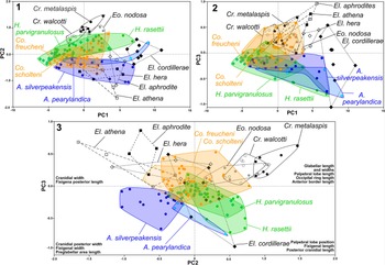
The gradational boundary between genera is evident in the representative taxa studied here using the standard measurements of the cranidia (see below; Fig. 4). Although some genera are clearly separated from each other along some PC axes (e.g., Elrathina vs. Crassifimbra along PC 2; Fig. 4.1, 4.3), the addition of representatives of other genera (Anebocephalus, Coenoides Harklessaspis, and Eokochaspis) bridge those differences with a considerable degree of overlap. Part of this overlap is the result of similarity among small specimens in different taxa, as seen in the increased overlap at negative PC1 values and near the origin of the PC2 vs. PC3 plot (Fig. 4, Table 2) A reanalysis of the data with the exclusion of small specimens (<3.0 mm; Table 3) illustrates a very similar pattern (Fig. 5.1) to the PC2 vs. PC3 plot that includes the small specimens (Fig. 4). Further addition of other genera only obscures the morphological fields of genera (results not presented). It appears that, for these taxa, cranidial morphology alone is insufficient for unambiguous genus-level diagnosis.
Scatter plots of the first three principal component axes obtained from PCA of cranidial measurements of the “ptychoparioids” Coenoides n. gen. (orange), Anebocephalus n. gen. (blue), Harklessaspis n. gen. (green), Elrathina (black dashed line), Crassifimbra (black solid line), and Eokochaspis (gray line). Note the gradation of morphologies between the different taxa. Representatives of the new taxa are in color; previously named genera are in black/gray. (1) Bivariate plot of PC1 (size) and PC2; (2) bivariate plot of PC1 (size) and PC3; (3) bivariate plot of PC2 and PC3; variables that load strongly in each quadrant are listed (see also Table 2).

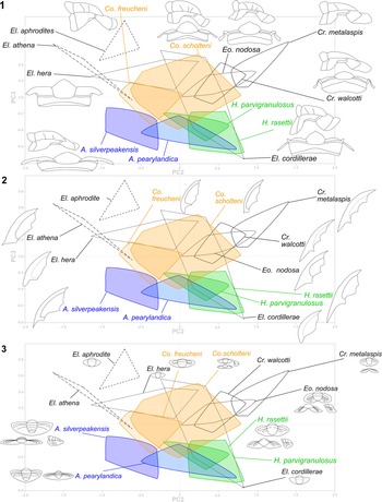
Distribution of different morphological features (drawings) relative to the PC2 and PC3 from an analysis of cranidial specimens 3.0 mm or larger (Table 3). The polygons depict the species occupation (in a projection of PC2 versus PC3) of a morphospace defined by PCA of cranidial linear measurements. Outlines of sclerites are shown for each species to indicate how other aspects of morphology (not used in generation of the morphospace) correspond to species occupation of that morphospace. Drawings are derived either from the specimens reported herein or from Webster (Reference Webster2011d) and Geyer and Peel (Reference Geyer and Peel2011, Reference Geyer and Peel2017). A lack of drawings for some taxa is the result of either the sclerites having not been reported or profiles that have not been illustrated in previous publications. (1) Illustrates glabellar lateral profile and convexity, fixigenal intraocular curvature and slope, and overall convexity of the cranidium; (2) illustrates librigenal shape and spine length; (3) illustrates pygidial shapes, convexity, and development of the postaxial notch.

Analysis of potential generic character states
Cranidia are generally considered to be the most important feature of lower and middle Cambrian “ptychoparioids” (Lochman, Reference Lochman1947; Rasetti, Reference Rasetti1951, Reference Rasetti1955). However, considerable difficulty has resulted from the identification of genera based solely on cranidia (see Rasetti, Reference Rasetti1951, Reference Rasetti1955; Sundberg, Reference Sundberg2004). Based on the taxa studied here, there are some commonalities in cranidial, librigenal, and pygidial features (Fig. 5) that have potential for use in determining generic assignments. Further testing with new information is needed to verify that these character states are valid to use for generic diagnoses.
In general, the features of the glabella appear to be indistinguishable among the species studied, with the exception of Crassifimbra walcotti, Cr. metalaspis, and Eokochaspis nodosa (Figs. 4, 5). The two Crassifimbra species appear to have a longer occipital ring relative to the glabella at 23–25% and the species of both Crassifimbra and Eokochaspis have a more tapered glabella with the anterior width 70–79% of the posterior width; Eokochaspis nodosa also has well-developed S1–S3 lateral glabellar furrows. In addition, unlike the other taxa, Coenoides scholteni and Co. freucheni have a glabella without the slight constriction at the S2 position. Convexity and lateral profile of the glabella (Fig. 5.1) has also played an important role in past classification of genera (e.g., Lochman, Reference Lochman1947; Rasetti, Reference Rasetti1955). Whereas the species of Crassifimbra and Anebocephalus both have lower convexities (in lateral view), the other represented taxa have only minor differences in convexity.
The position and shape of the palpebral lobe and ocular ridges have been used in the past to separate genera (Lochman, Reference Lochman1947; Rasetti, Reference Rasetti1951, Reference Rasetti1955). Generally, the representatives here have faint to moderately defined ocular ridges, which is variable among species with overall similar cranidia. The shape of the palpebral lobe is generally lenticular, with little curvature; however, Harklessaspis rasettii, H. parvigranulosus, Cr. walcotti, Cr. metalaspis, and especially Eo. nodosa have more strongly curved lobes. The relative length of palpebral lobes does differ between some taxa, with Elrathina species having the proportionally shortest lobes. The palpebral lobe furrows are well defined in all taxa, but generally shallower anteriorly. This is different in Eo. nodosa, where the furrows are more uniform in depth. The position of the palpebral lobe on the edge of the cranidium is related to the orientation of the ocular ridge and the width of the fixigena. The angle of divergence of the ocular ridge relative to the sagittal axis is generally ~80°, but taxa with a wider fixigena have the anterior end of the palpebral lobe set more posteriorly (e.g., species H. rasettii and H. parvigranulosus).
The nature of the anterior border and border furrow is more commonly assigned to specific differences (see Lochman, Reference Lochman1947). The swelling in the middle part of the border can be slight or absent (e.g., A. silverpeakensis, El. aphrodite, and El. athena) or much more pronounced forming a “V” shaped curve back towards the frontal lobe (e.g., Cr. metalaspis and Cr. walcotti); however, overall it appears not to be a distinctive character for generic identification (e.g., variation seen in Co. scholteni, H. rasettii, and H. parvigranulosus).
The course of the facial sutures is also considered important in identifying taxa (Lochman, Reference Lochman1947; Rasetti, Reference Rasetti1951, Reference Rasetti1955); however, this varies within the species studied herein. In general, all representatives have anteriorly convergent anterior branches of the facial suture; however, some specimens of some species show slightly divergent sutures (e.g., Eo. nodosa). Some variation in the course of the anterior branch of the facial suture may be non-biological (i.e., some samples are taphonomically compacted, including most specimens of A. silverpeakensis, El. aphrodite, and El. athena) even though they are in limestone and others have a very short distance between the palpebral lobe and the anterior border furrow, which makes it difficult to accurately determine the angle of convergence (e.g., Cr. walcotti, Cr. metalaspis, and Eo. nodosa). The form of the posterior branch of the facial suture is influenced not only by the position of the palpebral lobe, but also by the transverse width of the posterior area of the fixigena. In general, the angle measured from the base of the palpebral lobe to the posterior border furrow is ~50° to a transverse line.
The transverse width of the fixigena has also been used to characterize genera (Lochman, Reference Lochman1947; Rasetti, Reference Rasetti1951, Reference Rasetti1955; Geyer and Peel, Reference Geyer and Peel2017). This feature was measured from the middle part of the palpebral lobe furrow to the axial furrow and is related to the position and length of the palpebral lobe. In general, this width is 50% of the basal glabellar width. The convexities of the intraocular region and its slope towards the palpebral lobe also have been used as a key characteristic. Among the taxa studied herein, only Cr. walcotti and Cr. metalaspis have an outwardly downsloping intraocular regions (Fig. 5.1), with the other taxa having a more-or-less horizontal intraocular region. The lateral termination of the posterior area of the fixigena is also related to the path of the posterior branch of the facial suture; generally termination generates a wide cranidial width (e.g., A. silverpeakensis, A. pearylandica, El. aphrodite, El. athena, El. cordillerae, and El. hera), but in Cr. walcotti and Cr. metalaspis, the rearmost portion of the suture flexes into an almost directly posterior orientation generating a transversely narrower posterior area of the fixigena and giving the cranidium a subquadrate shape. The anterolateral inflection in the distal portion of the posterior cranidial margin in El. aphrodite, El. athena, El. cordillerae, and El. hera is a distinctive character that is not seen in the other taxa.
The librigenae appears to be useful in characterizing genera (Fig. 5.2). One key characteristic is the presence of a moderately long genal spine that is not laterally deflected (e.g., H. rasettii, A. silverpeakensis, Cr. walcotti, and Eo. nodosa). The other group of taxa have either no spine or a very short genal spine deflected laterally (i.e., not forming a smooth lateral margin of the librigenae; e.g., Co. scholteni and El. hera). Another feature that appears to be useful is the width of the genal area compared to the width of the librigenae. In Cr. walcotti and Cr. metalaspis, this width is narrower than in the other taxa.
The thorax also displays some characteristics that can be useful (e.g., Webster, Reference Webster2011d). In contrast to the other species studied for which the thorax is known, A. silverpeakensis has a proportionally wider thorax (pleura and axial ring), which matches its wider posterior area of the fixigena and cranidium.
Pygidia of almost all taxa are sublenticular (Fig. 5.3), but differ in the axial ring number; axis width and length; development of the pleural furrows and pygidial border; and the anterior lateral corner position relative to the axis. Axial rings (not including the terminal piece) range from 1–4, with 1–2 rings in El. aphrodite, El. hera, and El. cordillerae, and 2–4 rings in the other species studied. Axis length is 100% of sagittal pygidial length in the three species of Elrathina and 90–95% in the other taxa; maximum axial width compared to maximum pygidial width is also greater in the species of Elrathina. Pleural furrows are generally absent in the Elrathina species and present in the other taxa. A pygidial border is generally poorly developed, but can be defined by a shallow border furrow and the termination of the pleural lobes (e.g., H. rasettii, A. silverpeakensis, Cr. metalaspis, and Eo. nodosa). The posterior medial notch development, as viewed from the posterior, may be tied to the overall convexity of the axial lobe (e.g., A. silverpeakensis and A. pearylandica have the lowest convexity and have the weakest medial notch, if present). The position of the anterolateral corners of the pygidium relative to the axis is also useful in identifying taxa. In A. silverpeakensis and A. pearylandica, this position is transversely opposite the anterior half of the axis; in H. rasettii, H. parvigranulosus, Co. scholteni, Co. freucheni, Cr. metalaspis, El. hera, El. cordillerae, and El. aphrodite, the corners are transversely opposite the middle portion of the axis; and in Eo. nodosa the corners are transversely opposite a point behind the axis.
Systematic paleontology
Order Unknown Family Unknown
Remarks
The “ptychoparioids” described herein typically would be assigned to the Ptychopariidae Matthew, Reference Matthew1887, in the broad sense of Fortey (Reference Fortey and Kaesler1997, p. O302). We agree with Adrain (Reference Adrain2011) that the “order Ptychopariida” is at best paraphyletic and probably polyphyletic, and should therefore be abandoned. We also agree with Geyer and Peel (Reference Geyer and Peel2017) that family-level assignments of “ptychoparioids” are impossible at present (e.g., Ptychopariidae versus Antagmidae).
The generic diagnoses are extensive due to the diversity of taxa used for comparison. As future phylogenetic analyses reveal distinct clades, we expect these diagnoses to be shortened to their key synapomorphies. Discussions first present the generic diagnostic features compared to the other genera described herein. Then the other different features between the genera are presented. Percentages and angles have been presented as ranges based on μ ± σ.
“Ptychoparioid” Meraspides Figure 6
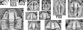
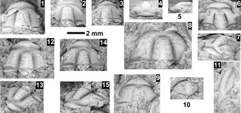
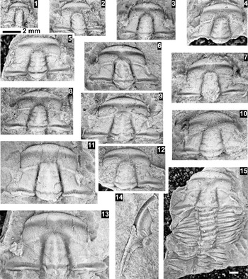
“Ptychoparioid” meraspides unidentifiable at the species level, from the mid-Dyeran, Harkless Formation, Clayton Ridge, Nevada. All specimens are silicified and from ICS-10647; in dorsal view unless specified; 1.0 mm scale bar applies to all. (1) Cranidium FMNH PE58745; (2) 1st degree meraspid FMNH PE58741, ventral view; (3) ?1st degree meraspid FMNH PE58740; (4) 3rd degree meraspid FMNH PE58742, ventral view; (5) 3rd degree meraspid FMNH PE58744; (6) 6th degree meraspid FMNH PE58743; (7) ?8th degree meraspid FMNH PE58751; (8) cranidium and partial thorax FMNH PE58739; (9) cranidium and thoracic segment FMNH PE58749; (10) partial cranidium FMNH PE58748; (11) cranidium FMNH PE58747; (12) cranidium FMNH PE58750; (13) cranidium FMNH PE58746.

Occurrence
Harkless Formation (mid-Dyeran), Clayton Ridge, Nevada.
Remarks
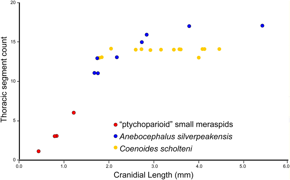
Two taxa of “ptychoparioids” are recognized within collection ICS-10647: Coenoides scholteni and Anebocephalus silverpeakensis Associated with these taxa are several small meraspides (<1.5 mm in sagittal cranidial length) that cannot be unambiguously assigned to one species or the other. A principal component analysis (PCA) of cranidial features illustrates that the smallest meraspides that can be unambiguously assigned to either of the two species (>1.5 mm in sagittal cranidial length) have similar morphologies to these smaller, unidentifiable meraspides (Fig. 7, Table 4). At which meraspid stage this cranidial changes to a more adult morphology is unknown, but is presumed to occur after M6 stage. This presumption is based on the number of thoracic segments observed in the largest of these early meraspides, which has a sagittal cranidial length of 1.17 mm (Fig. 8). The upper bracket for when the cranidial transformation occurs is less constrained. The lowest thoracic segment count observed among complete specimens unambiguously representing either Co. scholteni is 12 (M12; a specimen with sagittal cranidial length of 1.64 mm) or A. silverpeakensis is 11 (M11; a specimen with sagittal cranidial length of 1.55 mm).
Scatter plots of the first three PC axes following PCA of cranidial data of the “ptychoparioid” meraspides, Coenoides scholteni n. gen. n. sp. and Anebocephalus silverpeakensis n. gen. n. sp., from collection ICS-10647. (1) PC1 (size) versus PC2; (2) PC1 (size) versus PC3; (3) PC2 versus PC3; variables that load strongly in each quadrant are listed (see also Table 4).

Bivariate plot of the number of thoracic segments vs. sagittal cranidial length in the unidentifiable “ptychoparioid” meraspides, Coenoides scholteni n. gen. n. sp., and Anebocephalus silverpeakensis n. gen. n. sp. All specimens are from collection ICS-10647.

Genus Anebocephalus new genus
Type species
Anebocephalus silverpeakensis n. gen. n. sp. from the Harkless Formation (mid-Dyeran), Clayton Ridge, Nevada.
Other species
Eoptychoparia pearylandica Geyer and Peel, Reference Geyer and Peel2011.
Diagnosis
Cranidium trapezoidal, length 54–61% of width, anterior width 48–58% of cranidial width. Glabella slightly tapered with anterior width 74–88% of posterior width, constricted at S2; lateral profile evenly curved from S1–S4 and a slightly flattened frontal lobe, low convexity; occipital ring short at 16–21% of glabellar length; incision of lateral glabellar furrows moderate (S1) to shallow (S2–S4). Palpebral lobes slightly curved; moderately long at 26–36% of glabellar length; defined by shallow furrow that deepens posteriorly; anterior end located 16–24% of glabellar distance from the front of the glabella. Ocular ridges weakly to moderately defined, directed posterolaterally at 74–82° to exsagittal line. Anterior border with no or very slight medial swelling. Fixigena horizontal to palpebral lobes, posterior lateral termination sharp, distally directed slightly posterolaterally.
Librigenae with moderately long genal spine, ~15% of librigenal length, spine not deflected laterally, genal field moderately wide at 45–64% of librigenal width.
Third thoracic segment with wide pleural lobe width 137–174% of axial ring, axial ring 22–27% of entire width of segment, fulcrum weakly developed at 64–68% of the distance from axial furrow.
Pygidium sublenticular in outline, anterolateral corners located transversely opposite anterior portion of axis; posterior margin nearly uniformly curved; postaxial notch slight to absent in posterior view; axis 28–32% pygidial width, and 75–88% pygidial length, four axial rings plus terminal piece; pleural furrows and bands moderately defined; border weak to moderately developed.
Occurrence
Waucoban Series, Dyeran Stage, United States (Nevada) and Greenland.
Etymology
Anebocephalus is derived from Greek anebos, meaning young, not yet mature, for the similarity in glabella outline (with a strong constriction) to those of the “ptychoparioid” meraspides reported herein.
Remarks
Anebocephalus differs from the other genera studied herein in having a sharp posterior lateral termination of the fixigena, relatively wide thoracic pleurae, narrower axial ring width, a weaker fulcrum, pygidia with four axial rings, and anterolateral corners located transversely opposite the axis anterior margin. Anebocephalus is most similar to Harklessaspis, with the latter differing in having a proportionally narrower cranidium (length at 63–71% width), a proportionally wider cranidial anterior margin (60–70% of cranidial width), more posteriorly located palpebral lobes (20–30% glabellar distance from frontal lobe), more posterolaterally directed ocular ridges (68–78° to exsagittal line), and a more rounded and less posterolaterally directed posterior lateral termination of the fixigena. The pygidium of Harklessaspis also bears a more strongly defined postaxial notch and has an axis that is proportionally wider (34–38% pygidial width) and longer (88–94% pygidial length), with only three axial rings.
Anebocephalus differs from Crassifimbra with the latter having a subquadrate cranidium (length 75–85% width and anterior width at 71–78% of posterior width); an anterior border with a more prominent medial swelling; a glabella with more tapered sides (glabellar anterior width 71–79% of posterior width), lateral profile evenly curved from S1 to frontal lobe, a shorter occipital ring (16–21% glabellar length); longer palpebral lobes (36–41% of glabellar length); fixigena that downslope outwards to the palpebral lobes; a more rounded posterolateral termination of the fixigena (triangular in some species), with a terminus nearly perpendicular to the posterior margin, and distally directed slightly posterolaterally. The librigenae of Crassifimbra have a narrower genal field (28–40% of librigenal width), and the pygidium has a strong postaxial notch.
Anebocephalus differs from Eokochaspis with the latter having a more rectangular shape (length at 62–71% width and anterior width 62–72% of posterior width); glabella with more distinctly tapered sides (glabellar anterior width 70–77% of posterior width), lateral profile evenly curved from S1 to frontal lobe, and deeper lateral glabellar furrows; moderately curved and longer palpebral lobes (35–41% of glabellar length), and defined adaxially by a shallow and uniform-depth palpebral furrow; and the posterior lateral termination of the fixigena moderately rounded, triangular, and distally directed posterolaterally. The librigenae of Eokochaspis have a narrower genal field (30–32% of librigenal width), and the pygidium has a dumbbell-like outline with a straight to anteriorly bent posterior margin, strong postaxial notch, and broader axis (35–40% pygidial width).
Anebocephalus differs from Coenoides in the latter having a proportionally greater cranidial length (59–70% of width); an anterior border with a more pronounced medial swelling; a glabella that is not constricted at S2, with a lateral profile that is evenly curved from S1 to the frontal lobe, and with deeper lateral glabellar furrows; and with a posterolateral termination of the fixigena that is bluntly rounded and directed laterally. The librigenae of Coenoides bear a shorter genal spine (~0–7% of librigenal length) that is deflected laterally. The pygidial axis of Coenoides is proportionally wider (33–38% pygidial width) and longer (88–98% pygidial length).
Anebocephalus is also dissimilar to Elrathina in the latter having a glabellar lateral profile that is evenly curved from S1 to the frontal lobe and shallower lateral glabellar furrows; smaller palpebral lobes (20–26% of glabellar length); and a posterior lateral termination of the fixigena that is moderately sharp (triangular in some species) with a posterior margin that bends anteriorly to form a sharper angle, and distally directed posterolaterally. The librigenae of Elrathina have a rounded posterolateral corner or a short genal spine (0–11% of librigenal length) that is deflected laterally. The pygidium of Elrathina has an axis that is proportionally broader (39–51% pygidial width) and longer (100% pygidial length), and pleurae with very shallow or absent furrows and that lacks a clear border and border furrow.
Anebocephalus silverpeakensis new species Figures 9, 10, 11.1
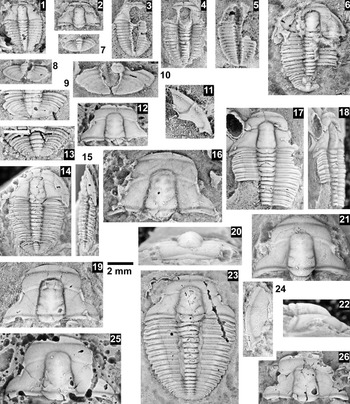
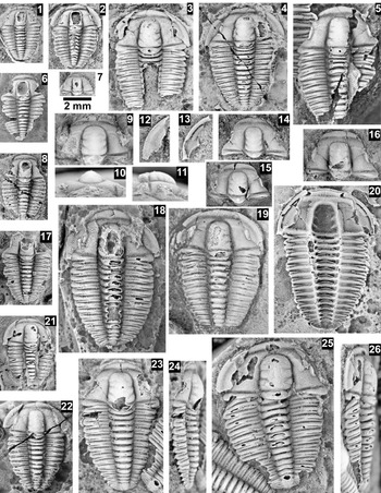
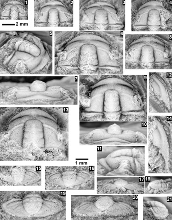
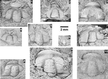
Anebocephalus silverpeakensis n. gen. n. sp., from the mid-Dyeran, Harkless Formation, Clayton Ridge, Nevada. All specimens are from ICS-10647, silicified, and in dorsal view unless noted otherwise; 2 mm scale bar applies to all except 13. (1) Meraspid paratype exoskeleton (M11) FMNH PE58576; (2) small paratype cranidium FMNH PE58573; (3) partial meraspid paratype exoskeleton (M13) FMNH PE58578; (4) partial meraspid paratype exoskeleton (M13) FMNH PE58582; (5) partial meraspid paratype exoskeleton (M11) FMNH PE58577, ventral view; (6) partial paratype exoskeleton FMNH PE58580; (7) small paratype pygidium FMNH PE58590; (8) paratype pygidium FMNH PE58581; (9) paratype pygidium of FMNH PE58571; (10) paratype pygidium FMNH PE58589; (11) paratype librigena FMNH PE58575; (12) partial paratype cranidium FMNH PE58587; (13–15) partial exoskeleton FMNH PE58587, close up of paratype pygidium, white scale bar 1 mm; dorsal and lateral views; (16) paratype cranidium FMNH PE58593; (17, 18) partial paratype exoskeleton FMNH PE58579, dorsal and lateral views; (19) paratype cranidium FMNH PE58574; (20–22) paratype cranidium FMNH PE58597 preserved as a limestone specimen from ICS-10623, anterior, dorsal, and lateral views; (23) holotype exoskeleton FMNH PE58569, lacking librigena; (24) paratype librigena FMNH PE58599 preserved as a limestone specimen from ICS-10623; (25) paratype cranidium FMNH PE58583; (26) paratype cranidium FMNH PE58586.

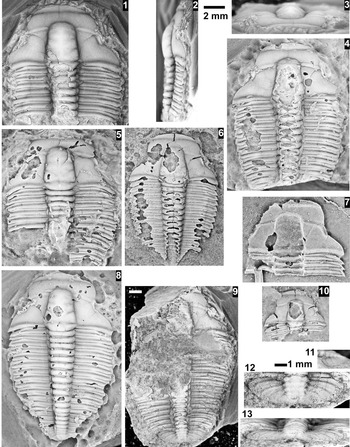
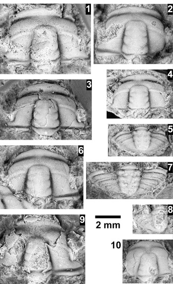
Anebocephalus silverpeakensis n. gen. n. sp., from the mid-Dyeran, Harkless Formation, Clayton Ridge, Nevada. Specimens use the 2.0 mm black scale bar, from ICS-10647, silicified, and in dorsal view unless otherwise mentioned. (1–3) Hypotype partial exoskeleton FMNH PE58594 from ICS-10643, limestone specimen, dorsal, lateral, and anterior views; (4) large partial paratype partial exoskeleton FMNH PE58598 from ICS-10623; (5) paratype partial exoskeleton FMNH PE58588; (6) hypotype partial exoskeleton FMNH PE58595 from ICS-10643; (7) large paratype cranidium FMNH PE58569, ventral view; (8) paratype exoskeleton FMNH PE58600 from ICS-10623; (9, 11–13) large partial paratype exoskeleton FMNH PE58572, white scale bar = 2 mm, close up of pygidium using the adjacent black 1 mm scale bar, dorsal, anterior, and lateral views; (10) hypotype cranidium FMNH PE58596 from ICS-10643.

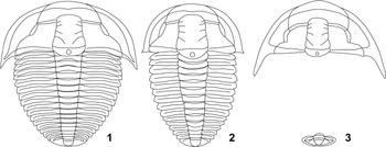
Reconstruction of the type species of new genera of “ptychoparioid” trilobites from the Harkless Formation. (1) Anebocephalus silverpeakensis n. gen. n. sp. (2) Coenoides scholteni n. gen. n. sp., (3) Harklessaspis rasettii n. gen. n. sp.

Holotype
Cranidium FMNH PE 58569 from the upper Harkless Formation, Clayton Ridge, Nevada (ICS-10647).
Paratypes
Specimens FMNH PE 58570–58593 (ICS-10647) and 58597–58600 (ICS-10632) from the upper Harkless Formation, Clayton Ridge, Nevada.
Diagnosis
Frontal area length 23 ± 3% of cranidial length; glabella 77 ± 3% of cranidial length; preglabellar field 50 ± 8% of frontal area.
Occurrence
Harkless Formation (mid-Dyeran), Clayton Ridge, Nevada (see Appendix 1).
Description
Cranidium of small to moderate size, length ~35–40% exoskeleton length; length 3.7 ± 1.5 mm (N = 18); subtrapezoidal, moderately low convexity (sag. and trans.); anterior margin slightly and evenly curved, not arched dorsally; posterior margin (excluding occipital ring) directed slightly posterolaterally. Anterior branches of facial sutures moderately convergent by 20 ± 10° to anterior border, more convergent to anterior margin; posterior branches moderately divergent at 54 ± 6°. Glabella moderately elongate, length 77 ± 3% cranidial length; width 29 ± 2% cranidial width; slightly tapered, width at anterior end 82 ± 8% of posterior glabellar width; moderately convex (sag. And trans.); frontal lobe bluntly rounded. Axial furrows moderately deep, with constriction at the L2; preglabellar furrow moderately deep. Lateral glabellar furrows moderately shallow, S1 bifurcated, directed slightly posteriorly; S2 bifurcated directed transverse to slightly posteriorly; S3 and S4 very faint or absent, directed anteriorly. Occipital ring not elevated above rest of glabella, moderately convex; length 18 ± 2% glabellar length; posterior margin slightly convex posteriorly, less curved posteriorly; small occipital node. SO straight to very slightly curved anteriorly medially; deepest laterally, moderate depth medially. Frontal area nearly equally divided; length 23 ± 3% cranidial length. Preglabellar field slightly convex, downsloping, length 12 ± 3% cranidial length, 50 ± 8% frontal area length. Anterior border slightly convex, level, length 11 ± 1% cranidial length, tapering laterally, no or slight medial inbend or swelling. Anterior border furrow narrow and of moderate depth, shallower medially, shallower than axial furrows. Fixigena slightly convex, nearly level, anterior area downsloping anteriorly, width 63 ± 6% of posterior glabellar width. Palpebral lobes nearly straight, level, moderately narrow and moderately long, length 30 ± 3% glabellar length; anterior margin located transversely opposite of S3 or L4. Ocular ridge moderately defined, very slightly curved anteriorly, directed slightly posterolaterally from glabella at 79 ± 3° to sagittal axis. Posterior area of fixigena triangular, terminated with sharp corner; length 40 ± 3% glabellar length; width 82 ± 5% glabella length.
Librigenae small, length ~2.8–4.9 mm (N = 3); moderately wide at ~35–45% length without spine; lateral margin moderately curved. Genal field slightly convex. Border slightly convex, uniform in width, width ~20–25% librigenal width; border furrows shallow, which persists into the librigenal spine. Librigenal spine short, ~15% librigenal length based on incomplete spine; directed posterolaterally.
Rostral plate and hypostome unknown.
Thorax with variably 16–18 segments, tapering towards small pygidium. Pleurae width 153 ± 17% of axial ring width (N = 11), inner portion oriented transversely, directed slightly posterolaterally and downwards at weakly developed fulcrum. Fulcrum positioned 66 ± 3% of pleural distance away from axial ring at third pleural segment and 57 ± 9% at tenth pleural segment. Anterior and posterior pleural bands moderately convex and uniform in length. Pleural furrow moderately deep and uniform depth, narrows distally. Pleural spine very short, termination pointed.
Pygidium very small, length ~5–10% exoskeleton length, length 0.9 ± 0.5 mm (N = 11); alate, length 31 ± 3% width; margin smooth, anterior margin nearly straight and directed slightly posterolaterally; anterolateral corners sharply rounded, transversely opposite medial portion of axis; no postaxial notch; moderately convex (sag.). Axis moderately tapered, mid-width 81 ± 8% anterior width, anterior width 30 ± 1% pygidial width; length 83 ± 6% pygidial length, extends to nearly posterior margin; four or possibly five axial rings, moderately convex; terminal piece small, rounded posteriorly; axial ring furrows shallow. Pleural regions very slightly convex, downsloping; anteriormost pleural furrow narrow and shallow, extending to near margin; other pleural furrows very shallow. Border narrow and defined by shallow furrow.
Fine granules on the entire exoskeleton. Very faint genal caeca on preglabellar regions of larger specimens.
Etymology
Named after the nearby town of Silver Peak, Nevada.
Remarks
The single cranidium and pygidium of Syspacephalus? sp. illustrated by Palmer (1964, pl. 3, figs. 15, 20) from the Saline Valley tongue of the Alkali Member, Harkless Formation (Sundberg, Reference Sundberg2014) are similar to the species described here, with the exception of the former having a wider anterior border and more parallel sided glabella. These two specimens probably represent a separate species of Anebocephalus, but additional specimens are needed.
Changes occur during the ontogeny of later meraspides (sagittal cranidial length >1.5 mm) to holaspides in A. silverpeakensis (Fig. 12). In general, the outline of the cranidium becomes less trapezoidal (anterior width to posterior width changes from 50% to 55%), the glabella becomes proportionally slightly shorter (80% to 75% cranidial length) and wider (63% to 73%), and the degree of glabellar tapering increases (anterior width relative to posterior width 90% to 75%). The anterior border width relative to frontal area width decreases (60% to 40%), and the angle of the ocular ridges relative to the sagittal axis slightly decreases (82° to 75°). Eighteen thoracic segments is the maximum number of observed (Fig. 8). This number is present in a specimen with a poorly preserved cranidium. Specimens with 16 or 17 segments have cranidial lengths ≥2.8 mm.
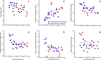
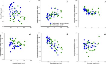
Bivariate plots of different biometrics of Anebocephalus silverpeakensis n. gen. n. sp. and A. pearylandica (Geyer and Peel, Reference Geyer and Peel2011). (1) Ocular ridge angle vs. sagittal cranidial length; (2) glabellar width/glabellar length vs. sagittal cranidial length; (3) glabellar length/cranidial length vs. sagittal cranidial length; (4) glabellar anterior width/glabellar width vs. sagittal cranidial length; (5) anterior border length/frontal area length vs. sagittal cranidial length; (6) fixigenal width/glabellar width vs. sagittal cranidial length.

The specimens from the Harkless Formation are similar to A. pearylandica from North Greenland (Geyer and Peel, Reference Geyer and Peel2011; Peel et al., Reference Peel, Streng, Geyer, Kouchinsky and Skovsted2016; Fig. 12), but differ in possessing a shorter frontal area length (22 ± 3% vs. 30 ± 2% of cranidial length) and corresponding longer glabella (78 ± 3% vs.70 ± 2% of cranidial length; Fig. 12.3) and a more equal division of the frontal area (preglabellar field 49 ± 8% vs. 30 ± 2% of frontal area; Fig. 12.5).
Genus Coenoides new genus
Type species
Coenoides scholteni n. gen. n. sp. from the Harkless Formation (mid-Dyeran), Clayton Ridge, Nevada.
Other species
Onchocephalus? freucheni Geyer and Peel, Reference Geyer and Peel2011.
Diagnosis
Cranidium trapezoidal, length 59–70% of width, anterior width 51–62% of posterior width. Glabella slightly tapered with anterior width 75–88% of posterior width, not constricted at S2; lateral profile evenly curved from S1 to frontal lobe, moderate convexity; occipital ring short at 17–22% of glabellar length; incision of lateral glabellar furrows moderate (S1, S2) to shallow (S3, S4). Palpebral lobes slightly curved; moderately long at 27–36% of glabellar length; defined by shallow furrow that deepens posteriorly; anterior end located 13–21% of glabellar distance from the front of the glabella. Ocular ridges weakly to moderately defined, directed posterolaterally at 79–86° to exsagittal line. Anterior border with slight to moderate medial swelling. Fixigena slightly downsloping to palpebral lobes; posterior lateral termination bluntly rounded, distally directed laterally.
Librigenae with a rounded posterolateral corner or a short genal spine, 0–7% of librigenal length, spine deflected laterally; genal field moderately wide at 40–55% of librigenal width.
Third thoracic segment with width of pleural lobe moderately wide at 108–140% of axial ring, axial ring 27–32% of entire width of segment, fulcrum well developed at 47–57% of the distance from axial furrow.
Pygidium sublenticular in outline, anterolateral corners located transversely opposite middle of axis; posterior margin nearly uniformly curved; slight postaxial notch in posterior view; axis 33–38% pygidial width, and 88–96% pygidial length, one or two axial rings; pleurae furrows and bands moderately defined; border weak to moderately developed.
Occurrence
Waucoban Series, Dyeran Stage, United States (Nevada) and Greenland.
Etymology
Coenoides is derived from Greek koinos, meaning common, for its typical cranidial shape among early “ptychoparioids”.
Remarks
Coenoides differs from the other genera studied herein in not having a constriction at the S2 position on the glabella, and in having moderately deep S1 and S2 lateral glabellar furrows. Coenoides is most similar to Elrathina, but differs in that the latter has an anterior border either without or with only a very slight medial swelling; smaller palpebral lobes (20–26% of glabellar length); and a sharper posterior lateral termination of the fixigena, triangular in some species, with the posterior margin bending anteriorly to form a sharper angle, distally directed posterolaterally. The pygidium of Elrathina bears a moderate postaxial notch, with an axis of sagittal pygidial length, the pleural band and furrows are very weak, and the border and furrow are not apparent.
Coenoides is also similar to Eokochaspis, but differs in that the latter has a proportionally wider anterior width of the cranidium (62–72% of cranidial width); an anterior border with no or only a very slight medial swelling; a glabella with more strongly tapered sides (glabellar anterior width 70–75% of posterior width) and deep lateral glabellar furrows; moderately curved palpebral lobes that are longer (35–41% of glabellar length) and defined adaxially by a shallow and completely developed palpebral furrow; and a posterior lateral termination of the fixigena that is moderately rounded, triangular, and distally directed posterolaterally. The librigenae of Eokochaspis bear a longer genal spine (24–42% of librigenal length) that is not deflected laterally and a narrower genal field (30–32% of librigenal width). The pygidium of Eokochaspis is dumbbell-shaped in outline, with a posterior margin that is straight to anteriorly bent, a strong postaxial notch, anterolateral corners that are transversely opposite the posterior portion of the axis, and a shorter axis (81–88% pygidial length).
Coenoides differs from Harklessaspis in that the latter has a greater cranidial anterior width (60–70% of cranidial width); an anterior border with a very slight medial swelling; a glabella with a lateral profile that is evenly curved from S1 to slightly flattened in the frontal lobe, and shallower lateral glabellar furrows; palpebral lobes that are more posteriorly set (20–30% of glabellar distance from the front of the glabella); and ocular ridges that are directed more strongly posterolaterally (68–78° from the glabella). The librigenae of Harklessaspis bear a longer genal spine (41–45% of librigenal length) that is not deflected laterally. The pygidium of Harklessaspis has a slightly to moderately large postaxial notch and three axial rings.
Coenoides differs from Crassifimbra in that the latter has a subquadrate cranidium (length 75–85% width, anterior width 71–78% of posterior width); an anterior border with a prominent medial swelling; a glabella with more tapered sides (anterior width 71–79% of posterior width), shallower lateral glabellar furrows, a longer occipital ring (23–25% glabellar length); moderately curved palpebral lobes that are longer (36–41% of glabellar length); and a posterior lateral termination of the fixigena that is more rounded, triangular, and distally directed posterolaterally. The librigenae of Crassifimbra bear a longer genal spine (21–41% of librigenal length) that is not deflected laterally, and a narrower genal field (28–40% of librigenal width). The thoracic segments of Crassifimbra are narrower (trans.; pleural width 87–99% of axial ring width). The pygidium of Crassifimbra has a distinct postaxial notch, a shorter axis (74–88% pygidial length), and three axial rings.
Differences between Coenoides and Anebocephalus are discussed under the latter genus.
Coenoides scholteni new species Figures 11.2, 13, 14
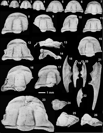
Coenoides scholteni n. gen. n. sp., from the mid-Dyeran, Harkless Formation, Clayton Ridge, Nevada. All specimens are from ICS-10643, silicified (except 9–11), and use the 2.0 mm scale bar; all in dorsal view unless specified. (1) Meraspid paratype exoskeleton (M12) FMNH PE58677; (2) meraspid paratype exoskeleton (M13) FMNH PE58623; (3) partial s paratype exoskeleton FMNH PE58655; (4) holaspid paratype shield FMNH PE58628; (5) partial paratype exoskeleton FMNH PE58634; (6) partial paratype exoskeleton FMNH PE58627; (7) small paratype cranidium FMNH PE58639; (8) partial paratype exoskeleton FMNH PE58647; (9–11) paratype cranidium FMNH PE 58637 (not silicified), dorsal, anterior, and lateral views; (12) paratype librigena FMNH PE58640; (13) paratype librigena FMNH PE58624, ventral view; (14) paratype cranidium FMNH PE58676; (15) paratype cranidium FMNH PE58629; (16) paratype cranidium FMNH PE58661; (17) partial paratype exoskeleton FMNH PE58650; (18) holaspid paratype exoskeleton FMNH PE58618; (19) holaspid paratype exoskeleton FMNH PE58646, lacking pygidium; (20) holaspid paratype exoskeleton FMNH PE58670, ventral view; (21) meraspid (M13) paratype exoskeleton FMNH PE58648; (22) holaspid paratype exoskeleton FMNH PE58619; (23, 24) holaspid holotype exoskeleton FMNH PE58617, dorsal and lateral views; (25, 26) meraspid? (M13) paratype exoskeleton FMNH PE58658, dorsal and lateral views.

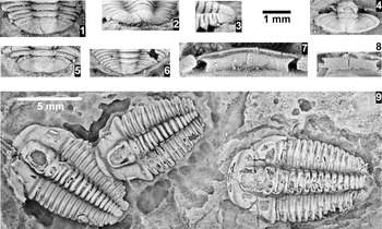
Coenoides scholteni n. gen. n. sp., from the mid-Dyeran, Harkless Formation, Clayton Ridge, Nevada. All specimens are from ICS-10643, use the 1.0 mm scale bar, and in dorsal view, unless otherwise mentioned. (1–3) Close-up of pygidium of holotype FMNH PE58617, dorsal, posterior, and lateral views; (4) paratype pygidium FMNH PE58660; (5) close-up of pygidium of paratype FMNH PE58621; (6) close-up of paratype pygidium of FMNH PE58653; (7) rostral plate of paratype exoskeleton FMNH PE58670, ventral view; (8) rostral plate of paratype exoskeleton FMNH PE58662, ventral view; (9) paratype exoskeletons (left to right) FMNH PE58673–58675, are from ICS-10623, white bar scale = 5 mm.

Holotype
Nearly complete exoskeleton FMNH PE58617 from the upper Harkless Formation, Clayton Ridge, Nevada (ICS-10647).
Paratypes
Specimens FMNH PE58618–58672, 58676, and 58677 (ICS-10647) and 58673–58675 (ICS-10632) from the upper Harkless Formation, Clayton Ridge, Nevada.
Diagnosis
Cranidium with a moderately curved anterior border, fixigena 51 ± 8% glabellar width, prominent occipital node, palpebral lobe length 34 ± 3% glabellar length; librigena width 31 ± 4% length, with a very small librigenal spine; 13 or 14 thoracic segments; and pygidium with one or two axial rings and effaced pygidial furrows.
Occurrence
Harkless Formation (mid-Dyeran), Clayton Ridge, Nevada.
Description
Cranidium of small to moderate size, length ~35–40% exoskeleton length; length 3.2 ± 0.9 mm (N = 40); subtrapezoidal, moderate convexity (sag. and trans.); anterior margin slightly and evenly curved, not arched dorsally; posterior margin, excluding occipital ring, nearly straight, directed very slightly posterolaterally. Anterior branches of facial suture moderately convergent by 23 ± 6° to anterior border, more convergent to anterior margin; posterior branches moderately divergent at 53 ± 7°. Glabella moderately elongate, length 78 ± 2% cranidial length; width 33 ± 3% cranidial width; slightly tapered, width at anterior end 82 ± 7% of posterior glabellar width; moderately convex (sag. and trans.); frontal lobe bluntly rounded. Axial furrows deep, nearly straight, and slightly convergent anteriorly; preglabellar furrow moderately deep. Lateral glabellar furrows moderately shallow; S1 bifurcated, directed posteriorly; S2 directed transverse; S3 and S4 very faint or absent. Occipital ring slightly elevated above glabella, moderately convex; length 20 ± 2% glabellar length; posterior margin convex posteriorly, evenly curved; moderately large occipital node. SO straight to very slightly curved anteriorly medially; deepest laterally, moderate depth medially. Frontal area unequally divided; length 22 ± 2% cranidial length. Preglabellar field slightly convex, downsloping, length 8 ± 2% cranidial length, 35 ± 6% frontal area length. Anterior border moderately convex, level, length 14 ± 1% cranidial length, tapering laterally, slight medial inbend and swelling. Anterior border furrow of moderate width and depth, shallower medially, shallower than axial furrows. Fixigena slightly convex, slightly downsloping laterally, anterior area downsloping anteriorly, width 51 ± 8% of posterior glabellar width. Palpebral lobes nearly straight, level, moderately narrow, and moderately long, length 34 ± 3% glabellar length; anterior margin located transversely opposite L4. Ocular ridge very faint to absent, straight to slightly curved anteriorly, directed slightly posterolaterally from glabella at 82 ± 3° to sagittal axis. Posterior area of fixigena triangular, terminated with rounded corner; length 36 ± 3% glabellar length; width 68 ± 6% glabella length.
Librigenae small, length ~3.2 ± 0.9 mm (N = 10); moderately wide at 31 ± 4% librigenal length without spine; lateral margin moderately curved. Genal field moderately convex. Border slightly convex, uniform in width, width 40 ± 9% librigenal width; border furrows shallow. Librigenal spine very short, 5 ± 2% librigenal length, and slightly deflected posterolaterally.
Rostral plate (Fig. 14.7, 14.8) subrectangular, longest (sag.) medially; wider (trans.) than the frontal lobe, approximately as long (sag.) as the anterior border.
Hypostome unknown.
Thorax with variably 13 or 14 segments, tapering towards small pygidium. Pleurae width 121 ± 14% of axial ring width (N = 23), inner portion oriented transversely, directed slightly posterolaterally and downwards at strongly developed fulcrum. Fulcrum positioned 53 ± 6% of pleural distance away from axial ring at third pleural segment and 50 ± 8% at tenth pleural segment. Anterior and posterior pleural bands moderately convex and uniform in length. Pleural furrow moderately deep and uniform depth. Pleural spine very short, termination pointed.
Pygidium very small, length ~5–7% exoskeleton length, length 0.6 ± 0.1 mm (N = 9); alate, length 28 ± 4% width; margin smooth; anterior margin nearly straight and directed slightly posterolaterally; anterolateral corners sharply rounded, located transversely opposite anterior portion of axis; no postaxial notch; moderately convex (sag.). Axis slightly tapered mid width, 90 ± 6% anterior width, anterior width 36 ± 3% pygidial width; length 91 ± 6% pygidial length, extends nearly to posterior margin; one or possibly two axial rings, moderately convex; terminal axial piece moderate size, rounded posteriorly; axial ring furrows effaced. Pleural regions slightly convex, downsloping; anteriormost pleural furrow narrow and very shallow, extending to near margin; other pleural furrows effaced. Border not defined.
Fine granules on the entire exoskeleton. Faint genal caeca on preglabellar regions of larger specimens.
Etymology
Named in honor of Peter Scholten for field assistance and support in FAS's research on Cambrian trilobites.
Remarks
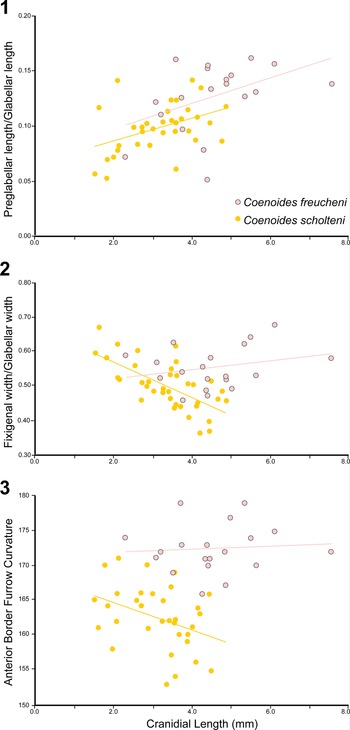
The specimens from the Harkless Formation differ from Co. freucheni (Geyer and Peel, Reference Geyer and Peel2011) from North Greenland (=Ptychoparella sp. A of Blaker and Peel, Reference Blaker and Peel1997, in part) in that the former possesses a cranidium with slightly more curved anterior border (Fig. 15.3), proportionally narrower fixigena (Fig. 15.2), a slightly larger occipital node, longer palpebral lobes; narrower librigenae with a very small librigenal spine; and a pygidium with two axial rings and less-pronounced pygidial axial furrows; and fewer thoracic segments (13 or 14 vs. 17). The preglabellar field also appears to be proportionally shorter, but this could be due to ontogenetic changes (the Harkless specimens are typically smaller than those from Greenland; Fig. 15.1).
Bivariate plots of Coenoides scholteni n. gen. n. sp. and Co. freucheni (Geyer and Peel, Reference Geyer and Peel2011). (1) The preglabellar length/glabellar length vs. sagittal cranidial length; (2) fixigenal width/glabellar width vs. sagittal cranidial length; (3) anterior border furrow curvature (degrees) vs. sagittal cranidial length.

Ontogenetic changes between later meraspides (sagittal cranidial length >1.5 mm) and holaspides in Co. scholteni include the following: the outline of the cranidium becomes less trapezoidal (anterior width to posterior width changes from 50% to 60%); the glabella becomes proportionally slightly shorter (80% to 77% cranidial length) and wider (63% to 75% of glabellar length), and more strongly tapered (anterior width relative to posterior width 90% to 75%); the anterior border width relative to frontal area width decreases (75% to 58%); fixigenal width relative to glabellar width decreases (60% to 42%); and the angle between the ocular ridges and the sagittal axis slightly increases (80° to 83°). Fourteen thoracic segments is the maximum number of observed (Fig. 8). This number is achieved in specimens with cranidial length 2.0 mm or larger. A single larger specimen (4.0 mm in cranidial length) has 13 segments and two specimens smaller than 2.0 mm also have 13 segments. The holaspid period therefore is entered at a small size, and considerably before the cranidium attains its morphological maturity.
Genus Crassifimbra Lochman, Reference Lochman1947
Type species
Onchocephalus walcotti Resser, Reference Resser1937, from the Pioche Formation (probably Combined Metals Member; Nephrolenellus multinodus Biozone), Highland Range, Nevada.
Other species
Crassifimbra metalaspis (Sundberg and McCollum, Reference Sundberg and McCollum2000).
Diagnosis
Cranidium subquadrate, length 75–85% width, anterior width 71–78% of cranidial width. Glabella moderately tapered with anterior width 71–79% of posterior width; very slightly constricted at S2; lateral profile evenly curved from S1 to frontal lobe, low convexity; occipital ring short at 22–25% of glabellar length; incision of lateral glabellar furrows shallow (S1–S4). Palpebral lobes moderately curved; moderately long at 36–41% glabellar length; defined by shallow furrow that deepens posteriorly; anterior end located 17–21% of glabellar distance from the front of the glabella. Ocular ridges weak to moderate strength, directed posterolaterally at 67–75° to exsagittal line. Anterior border with prominent medial swelling. Fixigena downsloping to palpebral lobes, posterior lateral terminations moderately rounded, triangular in some species, with terminus nearly parallel to posterior margin, distally directed slightly posterolaterally.
Librigenae with moderately long genal spine at 21–41% of librigenal length, spine not deflected laterally, genal field narrow at 28–40% of librigenal width.
Thoracic segments with narrow pleural lobe narrow at 87–99% of axial ring, axial ring 34–36% of entire width of segment, fulcrum well developed at 55–64% of the distance from axial furrow.
Pygidium sublenticular in outline, anterolateral corners located transversely opposite midlength of axis; posterior margin nearly uniformly curved; strong postaxial notch in posterior view; axis 26–40% pygidial width, length 74–88% pygidial length, three axial rings; pleurae furrows and bands moderately defined; border weakly to moderately developed.
Remarks
Only small articulated exoskeletons of species of Crassifimbra have been found in addition to isolated sclerites (see Webster, Reference Webster2011d). As a result, larger isolated thoracic segments were used for the segment characteristics instead of the third segment that were used for other taxa.
Lochman (Reference Lochman1947) assigned Antagmus arizonaensis Resser, Reference Resser, McKee and Resser1945, Ptychoparia cleon Walcott, Reference Walcott1917, and Ptychoparia cuneas Walcott, Reference Walcott1917, to Crassifimbra. All three species are known only from cranidia, with the latter two known only from a single specimen; thus their placement within Crassifimbra is at least questionable. In addition, A. arizonaensis has divergent anterior branches of the suture and longer posterior limbs, giving the cranidium an hour-glass shape, unlike that of Cr. walcotti or Cr. metalaspis. Ptychoparia cleon does appear to have similar cranidial features to Crassifimbra, but without other specimens and sclerites it is not reasonable to make a generic assignment. Ptychoparia cuneas has a relatively wide fixigena, anteriorly placed palpebral lobes, and wide (tr.) posterior area of the fixigena that are unlike Crassifimbra.
Crassifimbra differs from the other genera studied herein in having a subquadrate cranidium (length to width ratio of 75–85%; anterior width 71–78% cranidial width); moderate length occipital ring (23–25% glabellar length); anterior border with moderate medial swelling, and downsloping fixigena. Crassifimbra is most similar to Eokochaspis, with the latter differing in having deep lateral glabellar furrows; palpebral lobes that are moderately curved and defined adaxially by a shallow and uniform-depth palpebral furrow; and the posterior lateral termination of the fixigena being nearly perpendicular to the posterior margin and distally directed slightly posterolaterally. The thoracic segments of Eokochaspis have wider (trans.) pleural lobes (~116% of axial ring width) with the fulcrum well developed at 64–68% distance from axial furrow. The pygidium of Eokochaspis is dumbbell-shaped in outline, with a straight to anteriorly bent posterior margin, and two axial rings.
Differences among Crassifimbra, Anebocephalus, and Coenoides are discussed above.
Crassifimbra differs from Elrathina in that the latter has a glabella with less-tapered sides (anterior width 79–88% of posterior width); palpebral lobes that are only slightly curved and shorter (20–26% of glabellar length); ocular ridges that are directed more strongly posterolaterally (76–84° to exsagittal line); and a posterior lateral termination of the fixigena that is moderately sharp, triangular in some species, with the posterior margin bending anteriorly to form a sharper angle, and distally directed posterolaterally. The librigenae of Elrathina can bear a shorter genal spine (0–11% of librigenal length) that is deflected laterally; and a wider genal field (46–62% of librigenal width). Thoracic segments of Elrathina have wider (trans.) pleurae (115–144% of axial ring width) and a narrower axial ring (26–30% thoracic width). The pygidium of Elrathina has a moderate postaxial notch, an axis that is broader (39–51% pygidial width) and longer (100% pygidial length), one to two axial rings, pleurae without a well-developed segmentation, and lacking a border and furrow.
Crassifimbra differs from Harklessaspis in that the latter has a less-tapered glabella (anterior width 79–87% of posterior width) with a lateral profile that is evenly curved from S1–S4 and a slightly flattened frontal lobe; palpebral lobes that are more posteriorly placed (anterior end located 20–30% from the front of the glabella); and the posterior lateral termination of the fixigena is rounded and distally directed laterally. The librigenae of Harklessaspis bear a longer genal spine (41–45% of librigenal length) and a wider genal field (53–55% of librigenal width). The pygidium of Harklessaspis has a longer axis (88–94% pygidial length).
Crassifimbra walcotti (Resser, Reference Resser1937) Figure 16.1–16.5
- Reference Resser1937
Onchocephalus walcotti Resser, p. 21.
- Reference Webster2011d
Crassifimbra walcotti; Webster, p. 205, figs. 2–4, 22, 23 (also see for further synonymy).
Crassifimbra species, all specimens use the 2.0 mm scale bar; all in dorsal view unless specified. (1–5) Crassifimbra walcotti (Resser, Reference Resser1937) from UCR 10276, Nephrolenellus multinodus Biozone, Emigrant Formation, Clayton Ridge, Nevada: (1) partially testate hypotype cranidium UCR 10276-4, (2) exfoliated hypotype cranidium UCR 10276-5 illustrating pitted surface, (3–5) partial hypotype testate cranidium UCR 10276-3, dorsal, anterior, and lateral views. (6–11) Crassifimbra sp. B from the Bristolia mohavensis Biozone, Harkless Formation, Clayton Ridge, Nevada; specimens are slightly tectonically distorted: (6, 7) exfoliated hypotype cranidium UCR 10243-1, dorsal and oblique views; (8) exfoliated? hypotype cranidium UCR 10243-4; (9) exfoliated hypotype cranidium UCR 10243-5; (10) mostly exfoliated partial hypotype pygidium UCR 10243-87; (11) exfoliated hypotype librigena with punctuate texture, UCR 10243-2. (12–15) Crassifimbra? sp. A from the Bristolia mohavensis Biozone, Harkless Formation, Clayton Ridge, Nevada: (12, 13) exfoliated cranidium UCR 9942-2, dorsal and oblique view; (14, 15) testate cranidium UCR 9942-1, dorsal and oblique views.

Holotype
Cranidium USNM 61642 from the Pioche Formation, Highland Range, Nevada (USNM loc. 30).
Occurrence
Emigrant Formation (Nephrolenellus multinodus Biozone), Clayton Ridge, Nevada. Combined Metals Member, Pioche Formation (Nephrolenellus multinodus Biozone), Highland Range and Oak Spring Summit, Nevada (Webster, Reference Webster2011d).
Remarks
The cranidia from the lower Cambrian portion of the Emigrant Formation match well to the limestone type material (see Sundberg and McCollum, Reference Sundberg and McCollum2000, fig. 11) and silicified specimens studied by Webster (Reference Webster2011d).
Crassifimbra? sp. A Figure 16.12–16.15
Remarks
Specimens from collection UCR 9942 are similar to Crassifimbra walcotti and Cr. metalaspis, but the limited number of specimens (N = 4) and the absence of associated sclerites prevent confident generic identification. Differences observed include a less-curved anterior border; wider, less-defined, and straighter anterior border furrow; a glabella that is shorter (~65% vs. 71 ± 2% of cranidial length) and narrower (~34% vs. 40 ± 1% cranidial width), with broader glabellar furrows; wider fixigenae (~63% vs. 51 ± 6% cranidial width), and slightly shorter palpebral lobes (~35% vs. 38 ± 2% of glabellar length).
Crassifimbra sp. B Figure 16.6–16.11
- Reference Palmer and Halley1979
Ptychopariid sp. undet. 2, Palmer in Palmer and Halley, p. 118, pl. 5, fig. 14.
- ? Reference Sundberg and McCollum2003b
Ptychopariid sp. B, Sundberg and McCollum, p. 983, pl. 1, fig. 7.
Occurrence
Harkless Formation, and Mule Springs Limestone (Bristolia mohavensis Biozone), Clayton Ridge, Nevada. Thimble Limestone, Carrara Formation (Bristolia mohavensis or B. insolens Zone; Webster, Reference Webster, Hollingsworth, Sundberg and Foster2011a), Funeral Mountains, California (Palmer and Halley, Reference Palmer and Halley1979).
Remarks
Specimens from collection UCR 10243 are similar to Crassifimbra walcotti, but the limited number of specimens and the tectonic distortion of the specimens make it difficult to confirm the assignment. Differences observed (N = 2) include a narrower anterior cranidial width (~63% vs. 72 ± 3% of cranidial width), shorter glabella (~66% vs. 71 ± 2% of cranidial length), and narrower (~33% vs. 40 ± 1% cranidial width) and shorter palpebral lobes (~31% vs. 38 ± 2% of glabellar length). The single librigena is similar to Cr. walcotti and the pygidium has slightly better developed plural bands and furrows.
Specimens of ptychopariid sp. B reported by Sundberg and McCollum (Reference Sundberg and McCollum2003b) are poorly preserved, but match the characteristics that of Cr. sp. B reported herein.
Genus Harklessaspis new genus
Type species
Harklessaspis rasettii n. gen. n. sp. from the Harkless Formation (mid-Dyeran), Clayton Ridge, Nevada.
Other species
Harklessaspis parvigranulosus n. gen. n. sp.
Diagnosis
Cranidium subtrapezoidal, length 63–71% width, anterior width 60–70% of posterior width. Glabella slightly tapered with anterior width 79–87% of posterior width; very slightly constricted at S2; lateral profile evenly curved from S1–S4 and a slightly flattened frontal lobe, occipital ring short at 18–24% of glabellar length; incision of lateral glabellar furrows shallow (S1–S4). Palpebral lobes moderately curved; moderately long at 31–37% of glabellar length; defined by shallow furrow that deepens posteriorly; anterior end located 20–30% of glabellar distance from the front of the glabella. Ocular ridges weakly to moderately defined, directed moderately posterolaterally at 68–78° to exsagittal line. Anterior border with slight medial swelling. Fixigena horizontal to palpebral lobes, posterior lateral termination rounded, distally directed laterally.
Librigenae with a moderately long genal spine, 30–64% of librigenal length, spine not deflected laterally, genal field moderately wide at 33–55% of librigenal width.
Third thoracic segment with moderately wide pleural lobe at 113–123% of axial ring, axial ring 29–31% of entire width of segment; fulcrum well developed at 57–67% of the distance from axial furrow.
Pygidium sublenticular in outline, anterolateral corners located transversely opposite midpoint of axis; posterior margin nearly uniformly curved; slight to moderate postaxial notch in posterior view; axis 32–42% of pygidial width and 87–94% pygidial length; three axial rings; pleurae furrows and bands moderately developed; border weak to moderately developed.
Occurrence
Waucoban Series, Dyeran Stage, United States (Nevada) and Greenland.
Etymology
Named for the Harkless Formation, which contains the type species.
Remarks
The diagnosis of Harklessaspis is based on a unique combination of morphological character states; however, no individual character state distinguishes this genus from the other genera. Harklessaspis differences from Anebocephalus, Coenoides, and Crassifimbra are documented above.
Harklessaspis differs from Elrathina in that the latter has a cranidium that is proportionally shorter (53–61% cranidial width) and narrower anteriorly (anterior width 48–63% of cranidial width); a glabella lateral profile that is evenly curved from S1 to frontal lobe; palpebral lobes that are shorter (20–26% of glabellar length) and more anteriorly placed (13–23% glabellar distance from the front of the glabella); ocular ridges that are directed less posterolaterally (76–84° to exsagittal line); and a posterior lateral termination of the fixigena that is moderately sharp, triangular in some species, with a posterior margin that bends anteriorly to form a sharper angle, distally directed posterolaterally. The librigenae of Elrathina either bear a shorter genal spine (0–15% of librigenal length) that is deflected laterally or lack a genal spine. The pygidial axis of Elrathina is wider (39–51% pygidial width) and longer (100% pygidial length), bears one or two axial rings, has pleurae without well-defined furrows and border and furrow not apparent.
Harklessaspis differs from Eokochaspis in that the latter has a glabella with greater tapering (anterior width 70–77% of posterior width) and a lateral profile that is evenly curved from S1 to the frontal lobe with deep lateral glabellar furrows; palpebral lobes that are more strongly curved, positioned more anteriorly (anterior end located 17–24% glabellar distance from frontal lobe), and defined adaxially by a shallow and uniform-depth palpebral furrow; and a posterior lateral termination of the fixigena that is moderately rounded, triangular, and distally directed posterolaterally. Librigenae of Eokochaspis bear a shorter genal spine (24–42% of librigenal length) and a narrower genal field (30–32% of librigenal width). The pygidium of Eokochaspis is dumbbell-shaped in outline, with a straight to anteriorly bent posterior margin, a stronger postaxial notch; and two axial rings.
Harklessaspis rasettii new species Figures 11.3, 17
Harklessaspis rasettii n. gen. n. sp. from the mid-Dyeran, Harkless Formation, Clayton Ridge, Nevada. All specimens are from ICS-10648 and are in dorsal view unless specified; 1–14 use the 2.0 mm scale bar, 15–21 use the 1.0 mm scale bar. (1) Small, partially exfoliated paratype cranidium FMNH PE58711; (2) testate paratype cranidium FMNH PE58719; (3) fractured testate paratype cranidium FMNH PE58713; (4) testate paratype cranidium FMNH PE58712; (5–7) mostly testate paratype cranidium FMNH PE58716, oblique, dorsal, and anterior views; (8) mostly exfoliated paratype cranidium FMNH PE58715; (9–11) testate holotype cranidium FMNH PE58707, dorsal, anterior, and lateral views; (12) testate paratype librigena FMNH PE58718; (13) partial, testate paratype cranidium FMNH PE58714; (14) testate paratype librigena FMNH PE58717; (15) partial, exfoliated? paratype pygidium FMNH PE58708; (16–18) testate paratype pygidium FMNH PE58709, dorsal, posterior, and lateral views; (19–21) testate paratype pygidium FMNH PE58710, dorsal, posterior, and lateral views.

Holotype
Cranidium FMNH PE58707 from the upper Harkless Formation, Clayton Ridge, Nevada (ICS-10648).
Paratypes
Specimens FMNH PE58708–58738 from the upper Harkless Formation, Clayton Ridge, Nevada (ICS-10648).
Diagnosis
Surface of exoskeleton finely granular with scattered coarser granules.
Occurrence
Harkless Formation (mid-Dyeran), Clayton Ridge, Nevada.
Description
Cranidium of small to moderate size, length 3.9 ± 1.3 mm (N = 21); subtrapezoidal, moderate convexity (sag. and trans.); anterior margin evenly curved, slightly arched dorsally; posterior margin (excluding occipital ring) directed very slightly posterolaterally. Anterior branches of facial sutures slightly convergent by 10 ± 10° to anterior border, less convergent to anterior margin; posterior branches moderately divergent at 48 ± 7°. Glabella moderately elongate, length 74 ± 3% cranidial length; width 33 ± 2% cranidial width; slightly tapered, width at anterior end 83 ± 4% of posterior glabellar width; moderately convex (sag. and trans.); frontal lobe bluntly rounded to rounded. Axial furrows moderately deep with very slight constriction at the L2; preglabellar furrow moderately deep. Lateral glabellar furrows of moderate depth, S1 bifurcated, directed posteriorly; S2 bifurcated, directed transverse to slightly posteriorly; S3 and S4 faint, directed anteriorly. Occipital ring not elevated above glabella, moderately convex; length 19 ± 2% glabellar length; posterior margin moderately convex posteriorly; moderate size occipital node. SO straight to very slightly curved anteriorly; deepest laterally, moderate depth medially. Frontal area unequally divided; length 26 ± 3% cranidial length. Preglabellar field slightly convex, downsloping, length 9 ± 3% cranidial length, 34 ± 9% frontal area length; very slight sulcus in front of glabella. Anterior border moderately convex, level, sagittal length 17 ± 2% cranidial length, tapering laterally, very slight medial swelling. Anterior border furrow narrow and of moderate depth, shallower medially, deeper laterally than axial furrows. Fixigena moderately convex, level, anterior area downsloping anteriorly, width 61 ± 7% of posterior glabellar width. Palpebral lobes slightly curved, tilted upwards, moderately narrow and moderately long, length 34 ± 4% glabellar length; anterior margin located opposite of S3 or L4. Ocular ridge clearly defined, very slightly curved anteriorly, directed moderately posterolaterally from glabella at 70 ± 5° to sagittal axis. Posterior area of fixigena subtriangular, terminated with rounded corner; length 31 ± 3% glabellar length; width 69 ± 7% glabella length.
Librigenae moderate size, length 8.8 ± 1.7 mm (N = 6); moderately wide at 44 ± 4% length without spine; lateral margin moderately curved. Genal field slightly convex. Border slightly convex, broadening posteriorly, width ~34 ± 5% librigenal width; border furrow deeper anteriorly and shallows posteriorly, does not persist into the librigenal spine. Librigenal spine broad based, moderately long at 43 ± 2% librigenal length; directed posterolaterally.
Rostral plate, hypostome, and thorax unknown.
Pygidium very small, length 0.6–1.6 mm (N = 3); suboval, length ~35% width; margin smooth, anterior margin nearly straight adjacent to axis, then directed slightly posterolaterally; anterolateral corners sharply rounded, transversely opposite or slightly anterior of the medial portion of axis; postaxial notch present; moderately convex (sag.). Axis moderately tapered, mid-width, ~85% anterior width, anterior width ~35% pygidial width; length ~90% pygidial length, extends nearly to posterior margin; two or possibly three axial rings, moderately convex; terminal piece moderate size, rounded posteriorly; axial ring furrows shallow. Pleural regions very slightly convex, downsloping; anteriormost pleural furrow narrow and moderately deep to shallow, extending to anterior border; other pleural furrows very shallow. Border narrow and defined by shallow furrow.
Entire exoskeleton covered with fine granules; coarser granules scattered on genal areas of cranidium and librigenae.
Etymology
Named in honor of Franco Rasetti for his work to provide some clarity to previously named lower Cambrian “ptychoparioids.”
Remarks
Ontogenetic changes between cranidia of length (sag.) >1.5 mm to morphological maturity in H. rasettii (Fig. 18) include: the glabella becomes proportionally slightly shorter (changing from 80% to 70% cranidial length), wider (posterior width changing from 63% to 73% of length), and more strongly tapered (anterior width relative to posterior width changing from 90% to 77%); and the anterior border length relative to frontal area width decreases (changing from 75% to 50%).
Bivariate plots of different biometrics of Harklessaspis rasettii n. gen. n. sp. and Harklessaspis parvigranulosus n. gen. n. sp. (1) Ocular ridge angle vs. sagittal cranidial length; (2) glabellar width/glabellar length vs. sagittal cranidial length; (3) glabellar length/cranidial length vs. sagittal cranidial length; (4) glabellar anterior width/glabellar width vs. sagittal cranidial length; (5) anterior border length/frontal area length vs. sagittal cranidial length; (6) fixigenal width/glabellar width vs. sagittal cranidial length.

The difference between H. rasettii and H. parvigranulosus is the presence of larger granules, which are absent on the cranidia of the latter.
Harklessaspis parvigranulosus new species Figures 19–21
Harklessaspis parvigranulosus n. gen. n. sp. from the Arcuolenellus arcuatus and/or Bristolia mohavensis biozones, Harkless Formation, Clayton Ridge, Nevada. All specimens are silicified and in dorsal view unless specified. (1) Paratype cranidium UCR 10241-17; (2) paratype cranidium UCR 10241-20; (3) paratype cranidium UCR 10241-21; (4) paratype cranidium UCR 10241-23; (5) paratype cranidium UCR 10241-33; (6) paratype cranidium UCR 10241-30; (7) paratype cranidium UCR 10241-32; (8) paratype cranidium UCR 10241-43; (9) paratype cranidium UCR 10241-38; (10) paratype cranidium UCR 10241-39; (11) paratype cranidium UCR 10241-37; (12–14) paratype cranidium UCR 10241-44, dorsal, anterior, and lateral views; (15) paratype cranidium UCR 10241-42; (16) paratype cranidium UCR 10241-47; (17) paratype cranidium UCR 10241-46; (18) paratype librigena UCR 10241-62; (19) paratype librigena UCR 10241-59; (20) paratype librigena UCR 10241-60; (21) broken, large paratype librigena UCR 10241-64; (22) paratype librigena UCR 10241-58; (23) holotype cranidium UCR 10241-49, assembled from two separate photographs due to the specimen having been broken into two pieces; (24) paratype pygidium UCR 10241-51; (25, 26) paratype pygidium UCR 10241-53, dorsal and posterior views.

Harklessaspis parvigranulosus n. gen. n. sp. from the mid-Dyeran (ICS-10643) or Arcuolenellus arcuatus and Bristolia mohavensis undifferentiated biozones (ICS-10645), Harkless Formation, Clayton Ridge, Nevada. All figures use the 2.0 mm scale bar. (1) Partially testate paratype cranidium FMNH PE58687 from ICS-10643; (2) mostly testate paratype cranidium FMNH PE58683 from ICS-10643; (3) mostly testate paratype cranidium FMNH PE58684 from ICS-10643; (4) mostly testate paratype cranidium FMNH PE58685 from ICS-10643; (5) testate paratype pygidium FMNH PE58688 from ICS-10643; (6) mostly testate paratype cranidium FMNH PE58682 from ICS-10643; (7) testate paratype pygidium FMNH PE58681 from ICS-10643; (8) testate hypotype pygidium FMNH PE58691 from ICS-10645; (9) partially hypotype cranidium FMNH PE58689 from ICS-10645; (10) testate hypotype cranidium FMNH PE58690 from ICS-10645.

Harklessaspis parvigranulosus n. gen. n. sp. from the Arcuolenellus arcuatus and Bristolia mohavensis undifferentiated biozones, Harkless Formation, Clayton Ridge, Nevada. All specimens are from ICS-10646, preserved in shale, exfoliated with limonite residue, and slightly tectonically distorted. All figures use the 2.0 mm scale bar, unless otherwise mentioned. (1) Hypotype cranidium FMNH PE58705; (2) hypotype cranidium FMNH PE58706; (3) hypotype cranidium FMNH PE58704; (4) hypotype cranidium FMNH PE58703; (5) hypotype cranidium FMNH PE58697; (6) hypotype cranidium FMNH PE58698; (7) hypotype cranidium FMNH PE58695; (8) hypotype cranidium FMNH PE58695; (9) hypotype cranidium FMNH PE58699; (10) hypotype cranidium FMNH PE58696; (11) hypotype cranidium FMNH PE58694; (12) hypotype cranidium FMNH PE58702; (13) hypotype cranidium FMNH PE58693; (14) hypotype librigena FMNH PE58700; (15) paratype cranidium and partial thorax FMNH PE58692, white scale bar is 2 mm.

Holotype
Cranidium UCR10241-49 from the upper Harkless Formation, Clayton Ridge, Nevada (collection UCR 10241).
Paratypes
Specimens UCR 10241-18–48 (collection UCR 10241); FMNH PE58681–58688 (ICS-10645) and FMNH PE58692 (ICS-10646) from the upper Harkless Formation, Clayton Ridge, Nevada.
Diagnosis
Exoskeleton with fine granulations, lacking coarser granules.
Occurrence
Harkless Formation (Arcuolenellus arcuatus and Bristolia mohavensis undifferentiated biozones), Clayton Ridge, Nevada.
Description
Cranidium of small to medium size, length 2.5 ± 1.1 mm (N = 32; without smaller meraspides 2.9 ± 1.0, N = 28); subquadrate to subtrapezoidal, moderate convexity (sag. and trans.); anterior margin evenly curved, slightly arched dorsally; posterior margin (excluding occipital ring) directed slightly posterolaterally. Anterior branches of facial sutures convergent by 21 ± 7° to anterior border, more strongly convergent to anterior margin; posterior branches moderately divergent at 58 ± 4°. Glabella moderately elongate; length 73 ± 3% cranidial length; width 33 ± 2% cranidial width, slightly to moderately tapered, width at anterior end 82 ± 4% of posterior glabellar width; moderately convex (sag. and trans.); frontal lobe bluntly rounded. Axial furrows moderately deep, with very slight constriction at S2; preglabellar furrow moderate depth. Lateral glabellar furrows moderate depth to shallow; S1 bifurcated, directed posteriorly; S2 bifurcated, directed transversely to slightly posteriorly; S3 and S4 faint, directed anteriorly. Occipital ring slightly elevated above glabella, moderately convex; length 22 ± 2% glabellar length; posterior margin moderately convex posteriorly; small occipital node. SO straight to very slightly curved anteriorly; deepest laterally, moderate depth medially. Frontal area unequally divided; length 27 ± 2% cranidial length. Preglabellar field slightly convex, downsloping, length 9 ± 3% cranidial length, 32 ± 10% frontal area length; slight to prominent sulcus in front of frontal lobe. Anterior border moderately convex, level, length 18 ± 2% cranidial length, tapering laterally, slight to moderately strong medial swelling. Anterior border furrow narrow, of moderate depth, shallower medially, deeper laterally than axial furrows. Fixigena moderately convex, slightly downsloping, anterior area downsloping anteriorly, width 63 ± 3% of posterior glabellar width. Palpebral lobes slightly curved, tilted upwards, moderately narrow and moderately long, length 34 ± 6% glabellar length; anterior margin located transversely opposite of S3 or L4. Ocular ridge moderately to weakly defined, very slightly curved anteriorly, directed moderately posterolateral from glabella at 75 ± 5° to sagittal axis. Posterior area of fixigena subtriangular, terminated with rounded corner; length 41 ± 5% glabellar length; width 67 ± 5% glabella length.
Librigenae small size, length approximately 3.2 ± 0.3 mm (N = 6); moderately wide at 37 ± 2% length without spine; lateral margin moderately curved. Genal field slightly convex. Border slightly convex, broadening in width posteriorly, width about 33 ± 0.1% librigenal width; border furrow deepest anteriorly, does not persist into the librigenal spine. Librigenal spine broad based, moderate long at 47 ± 17% librigenal length; directed posterolaterally.
Rostral plate and hypostome unknown.
Thorax incompletely known (Fig. 21.15; compressed in shale and partially disarticulated, N = 2) with a minimum of 14 or 15 segments, tapering. Pleurae width 113–158% of axial ring width, inner portion oriented transversely, directed slightly posterolaterally at weakly developed fulcrum. Fulcrum positioned 57–67% of pleural distance away from axial ring at third pleural segment and 55–63% at tenth pleural segment. Anterior and posterior pleural bands moderately convex and uniform in length. Pleural furrow moderately deep and uniform depth, narrows distally. Pleural tip sharply rounded.
Pygidium very small, length 1.6 ± 0.5 mm (N = 6); suboval, length 41 ± 7% width; margin smooth, anterior margin nearly straight adjacent to axis, then directed slightly posterolaterally; anterolateral corners sharply rounded, transversely opposite to a point slightly anterior of the medial portion of axis; postaxial notch present; strongly convex (sag.). Axis slightly to moderately tapered mid-width 37 ± 5% anterior width, anterior width 37 ± 5% pygidial width; length 92 ± 2% pygidial length, extends to near posterior margin; two or possibly three axial rings, strongly convex; terminal axial piece moderate size, rounded posteriorly; axial ring furrows shallow. Pleural regions moderately convex, downsloping; anterior pleural furrow narrow and moderately deep, extending to lateral border; other pleural furrows moderately shallow. Border narrow and poorly defined.
Fine granules on the entire exoskeleton, few larger granules on axis of large pygidium.
Etymology
Named based on its fine granular prosopon.
Additional material
Specimens FMNH PE 58689– 58691 (ICS-10644), and FMNH PE58693–58706 (ICS-10646) from the upper Harkless Formation, Clayton Ridge, Nevada.
Remarks
Percentages and angles given above exclude the smallest cranidia (e.g., Fig. 18.1–18.4). Morphological changes occurring during the late stages of ontogeny (cranidial lengths >1.5 mm to mature cranidia) in Harklessaspis parvigranulosus (Fig. 18) include: the cranidium becomes more trapezoidal (length to width increases from 50% to 70%); the glabella becomes slightly shorter (changing from 77% to 70% cranidial length) and wider (width to length ratio changing from 65% to 75%); and the anterior border length relative to frontal area length decreases (from 80% to 50%).
Specimens of H. parvigranulosus from ICS-10646 (Fig. 21) and collections UCR 10212, UCR 10235, UCR 10236, and UCR 10242 are tectonically distorted and were not used to determine ratios discussed above in the description and comparison to similar species. The specimens preserved in shale have their exoskeletons preserved with limonite generally obscuring the fine surface ornamentation. However, the external molds show no evidence of coarse granules, only fine granules. Specimens preserved in shale (Fig. 21) illustrate the effects of compaction and mild tectonic distortion of the morphological features of the species. In general, the frontal lobe can become more rounded (Fig. 21.3, 21.6, 21.8, 21.13), glabellar furrows become indistinct (Fig. 21.3, 21.6, 21.13), the frontal area and anterior facial sutures become more elongate (Fig. 21.11, 21.13), the anterior border furrow becomes longer (sag.) and sometimes deeper (Fig. 21.6, 21.7, 21.10, 21.11, 21.13), and the swelling of the anterior border increases with an more pronounced sulcus extending to the frontal lobe (Fig. 21.6, 21.9, 21.13).
Comparison of H. parvigranulosus to H. rasettii illustrates minor differences in the overall measurements (Figs. 4, 5, 18). Most notably, H. parvigranulosus has a more tapered glabella (Fig. 18.4). Other possible differences include H. parvigranulosus having a more transversely directed ocular ridge (Fig. 18.1); a proportionally narrower anterior border in larger specimens (Fig. 18.5); and a proportionally shorter glabella (Fig. 18.3); but the variation in the data and the lack of larger specimens (of sagittal cranidial length >4.0 mm) hinders robust statistical testing of these putative differences. The consistent difference between the two species is the presence of larger granules on the cranidia of H. rasettii.
Undetermined “Ptychoparioids”
Remarks
Four taxa represented by only a few cranidia are similar to the genus Luxella Rasetti, Reference Rasetti1955, or Piaziella, Lochman, Reference Lochman1947, which, like most of the taxa discussed in his paper, lack associated sclerites to fully characterize the genus.
“Ptychoparioid” sp. A Figure 22.1–22.5
- Reference Palmer and Halley1979
Ptychopariid sp. undet. 1, Palmer in Palmer and Halley, p. 117, pl. 5, figs. 12, 13.
“Ptychoparioid” spp. from the Bristolia mohavensis Biozone, Harkless Formation, Clayton Ridge, Nevada. All specimens use the 2.0 mm scale bar. (1–5) “Ptychoparioid” sp. A; (1) exfoliated cranidium UCR 10248-4; (2) mostly exfoliated cranidium UCR 10248-3; (3) exfoliated cranidium UCR 10253-8; (4) mostly exfoliated cranidium UCR 10242-3; (5) mostly exfoliated cranidium UCR 10242-2; (6, 7) “Ptychoparioid” sp. B from ICS 1338, (6) mostly exfoliated small cranidium FMNH PE58789, (7) exfoliated cranidium FMNH PE58788; (8) “Ptychoparioid” sp. C, internal mold of cranidium UCR 10262-1; (9, 10) “Ptychoparioid” sp. D, (9) latex cast of internal surface of cranidium UCR 10244-6; (10) latex cast of poorly preserved cranidium UCR 10246-6.

Occurrence
Harkless Formation (Bristolia insolens Biozone), Clayton Ridge, Nevada. Thimble Limestone, Carrara Formation (Bristolia mohavensis or B. insolens Zone; Webster, Reference Webster, Hollingsworth, Sundberg and Foster2011a), Desert Range, Nevada. Mule Spring Limestone (Bristolia mohavensis or B. insolens Zone; Webster, Reference Webster, Hollingsworth, Sundberg and Foster2011a), Last Chance Range, California (Palmer and Halley, Reference Palmer and Halley1979).
Remarks
The five cranidia are most similar to Luxella lux (Walcott, Reference Walcott1917) from the Bonnia-Olenellus Zone (= Dyeran Stage), Peyto Limestone, Canada (Rasetti, Reference Rasetti1955), in their glabellar tapering, long frontal area, direction of ocular ridges, more posteriorly placed palpebral lobes, and wider posterior area of the fixigena. The cranidia are almost entirely exfoliated. The internal mold is finely punctuate (similar to Luxella crassimarginata Rasetti, Reference Rasetti1955), but the testate anterior border furrow (Fig. 22.1, 22.2) has a finely granular surface. Ptychopariid sp. undet. 1 illustrated by Palmer and Halley (Reference Palmer and Halley1979) has similar cranidial morphologies. Ptychopariid sp. undet. 3 also of Palmer and Halley (Reference Palmer and Halley1979) is mostly exfoliated and has the punctuate texture seen in the Harkless specimens, but has shallower anterior border and axial furrows.
“Ptychoparioid” sp. B Figure 22.6, 22.7
Occurrence
Mule Spring Limestone (Nephrolenellus multinodus Biozone), Clayton Ridge, Nevada.
Remarks
The two cranidia are most similar to Luxella crassimarginata Rasetti, Reference Rasetti1955, from limestone boulders of Dyeran age contained within the Ville Guay Conglomerates (Ordovician), Canada (Rasetti, Reference Rasetti1955). The larger specimen (Fig. 22.7) is similar in the prominent constriction in the glabellar outline, posterior placement of the palpebral lobes, and wide posterior area of the fixigena. However, it differs from L. crassimarginata in having a proportionally longer preglabellar field with a more-pronounced medial swelling and in the ocular ridge being directed more strongly posterolaterally away from the glabella. The smaller specimen from the same location (Fig. 22.6) appears to have a finely granular ornamentation. The larger specimen is exfoliated and has a fine punctate texture on the internal mold. The larger specimen is also similar in cranidial shape to kochaspid? sp. reported by Sundberg and McCollum (Reference Sundberg and McCollum2003b). This latter specimen has coarse granulation.
“Ptychoparioid” sp. C Figure 22.8
Occurrence
Mule Spring Limestone (Bristolia insolens Biozone), Clayton Ridge, Nevada.
Remarks
The partial cranidium is most similar to Piaziella? rara Fritz, Reference Fritz1972, from the Bonnia-Olenellus Zone (= Dyeran Stage), Sekwi Formation, Canada, in the slightly tapered glabella, simple glabellar furrows, shallow depression in the preglabellar field and curvature of the anterior border. However, it differs from P.? rara in having a proportionally longer (sag.) anterior border.
“Ptychoparioid” sp. D Figure 22.9, 22.10
Occurrence
Mule Spring Limestone (Bristolia insolens Biozone), Clayton Ridge, Nevada.
Remarks
Two cranidia are most similar Piaziella pia (Walcott, Reference Walcott1917) illustrated by Fritz (Reference Fritz1972) from the Bonnia-Olenellus Zone (= Dyeran Stage), Sekwi Formation, Canada, in the tapered glabella, shallow glabellar furrows, relatively wide fixigena and posteriorly placed palpebral lobes. However, they differ from P. pia in having a longer (sag.) anterior border and border furrow, stronger curvature of the border, and a more truncated frontal lobe.
Acknowledgments
Appreciation is given to L.B. and M. McCollum for their assistance in the field and permission to use a portion of their measured stratigraphic section in this paper. Field assistance was also provided to FAS by P.G. Scholten (Show Low, AZ), and to MW by M. Hopkins, C. Madison-Bell, E. Sperling, L. Webster, and the participants of the University of Chicago Spring Break Field Trip (2008). G. Geyer and B. Pratt provided valuable reviews of the manuscript, which was greatly appreciated.
Appendix: Localities
Split Mountain (general area)
Two bulk collections utilized herein include fossils pooled from more than one locality in the Split Mountain area. Although the stratigraphic provenance of each of those collections is constrained to some degree, the geographic point of collection of particular fossiliferous slabs within each collection cannot be constrained.
ICS-10631
Fossils collected from float in green shale in uppermost Harkless Formation, pooled from various areas around Split Mountain. From same stratigraphic interval as UCR 10212, UCR 10213, UCR 10214, UCR 10234–10236. Within 60–0 m below base of measured section presented by Webster (Reference Webster2011c, fig. 2), and ~55–115 m above the base of the Saline Valley Tongue (86–26 m below base of Mule Spring Limestone). Collected by MW, Melanie Hopkins, and Erik Sperling; 3/17/2005. Bristolia mohavensis (Crickmay in Hazzard, Reference Hazzard1933).
UCR 10264
Float blocks from middle unit of Mule Spring Limestone, pooled from various sites around Split Mountain. Collected by MW. Bonnia sp.
Split Mountain East hillside
The sections documented by Webster (Reference Webster2011c, p. 239–242, figs. 2, 3, 4.1–4.4, 5.1) were measured on the northern face of this hillside, spanning from the uppermost Harkless Formation to the middle unit of the Mule Spring Limestone. Extensive fossil collections have also been made from various horizons below the base of the measured section (see Webster, Reference Webster2011c; herein), but poor exposure and faulting mean that the stratigraphic positions of such collections can only be crudely estimated. Fossil collections mentioned herein are noted below.
ICS-10643
Upper part of exposure of dark gray, laminated limestone in upper part of Harkless Formation. Estimated to lie ~106.5 m below base of measured section presented by Webster (Reference Webster2011c, fig. 2), and ~3.5 m above the base of the Saline Valley Tongue (132.5 m below base of Mule Spring Limestone). Collected by FAS, 10/13/2007. Oryctocephalops frischenfeldi Lermontova, Reference Lermontova and Vologdin1940, Anebocephalus silverpeakensis, Coenoides scholteni.
ICS-10644
Uppermost part of exposure of dark-gray, fractured, bioclastic limestone in upper part of Harkless Formation. Estimated to lie ~105 m below base of measured section presented by Webster (Reference Webster2011c, fig. 2), ~5 m above the base of the Saline Valley Tongue (130 m below base of Mule Spring Limestone). Collected by FAS, 6/23/2018. Olenellus sp. B, Bonnia cf. B. brennus (Walcott, Reference Walcott1916), Harklessaspis parvigranulosus, Coenoides scholteni.
ICS-10645
Limestones interbedded with green shales on northern face of hillside. Probably from same stratigraphic interval as UCR 10212–10214 and UCR 10234–10236; estimated to lie within 60–0 m below base of measured section presented by Webster (Reference Webster2011c, fig. 2), and ~55–115 m above the base of the Saline Valley Tongue (86–26 m below base of Mule Spring Limestone). Collected by FAS, 10/13/2007. Olenellid fragments, Bonnia cf. B. brennus, Harklessaspis parvigranulosus.
ICS-10646
Green shales in uppermost Harkless Formation on western side of hillside. Probably from same stratigraphic interval as UCR 10212–10214 and UCR 10234–10236; estimated to lie within 60–0 m below base of measured section presented by Webster (Reference Webster2011c, fig. 2), and ~55–115 m above the base of the Saline Valley Tongue (86–26 m below base of Mule Spring Limestone). Collected by FAS, 10/13/2007. Arcuolenellus aff. A. megafrontalis (Vaccari, Reference Vaccari1988), Bristolia mohavensis , Olenellus aff. O. glabrum (Resser and Howell, Reference Resser and Howell1938), Bonnia columbensis Resser, Reference Resser1936, Harklessaspis parvigranulosus.
UCR 10212
Fossils collected as float from green shales in uppermost Harkless Formation on northern face of hillside, within 60–0 m below base of measured section presented by Webster (Reference Webster2011c, fig. 2), and ~55–115 m above the base of the Saline Valley Tongue (86–26 m below base of Mule Spring Limestone). Collected by MW, 6/15/1999 and 6/18/1999. Olenellus aff. O. glabrum, Bonnia columbensis, Zacanthopsis sp. indet., Harklessaspis parvigranulosus.
UCR 10235
Fossils collected as float from green shales in uppermost Harkless Formation on northern face of hillside, in gully several meters to west of UCR 10212, within 60–0 m below base of measured section presented by Webster (Reference Webster2011c, fig. 2), and ~55–115 m above the base of the Saline Valley Tongue (86–26 m below base of Mule Spring Limestone). Collected by MW. Harklessaspis parvigranulosus.
UCR 10236
Fossils collected as float from green shales in uppermost Harkless Formation on northern face of hillside, from spur on other side of gully several meters west of UCR 10212, within 60–0 m below base of measured section presented by Webster (Reference Webster2011c, fig. 2), and ~55–115 m above the base of the Saline Valley Tongue (86–26 m below base of Mule Spring Limestone). Collected by MW. Bonnia columbensis, Harklessaspis parvigranulosus.
UCR 9942
Platy oncolitic limestone ledges 17.7–17.95 m above base of measured section presented by Webster (Reference Webster2011c, fig. 2), ~132 m above the base of the Saline Valley Tongue (8.95–8.7 m below base of Mule Spring Limestone). Collected by MW. Crassifimbra? sp. A.
UCR 10243
Bioclastic limestone ledges in fault-bound, structurally complex area of upper Harkless Formation or middle/basal upper unit of Mule Spring Limestone, on western side of hillside. Bulk collection, mostly from bioclastic limestones (in situ) with some float slabs. Collected by MW and Alex Lin, 6/22/2000. Crassifimbra sp. B.
UCR 9944
Micaceous silty limestone and calcareous siltstones 26.3–26.4 m above base of measured section presented by Webster (Reference Webster2011c, fig. 2), ~141 m above the base of the Saline Valley Tongue (0.35–0.25 m below base of Mule Spring Limestone). Collected by MW. Crassifimbra sp. B.
Split Mountain East trench
The trilobite faunas in trench dug from the uppermost Mule Spring Limestone through the Emigrant Formation have been documented previously elsewhere (Sundberg and McCollum, Reference Sundberg and McCollum2003b; Webster, Reference Webster2011c, p. 245–246, fig. 6.1). The collections are mentioned herein are noted below.
ICS-1338 and UCR 10285
Limestone bed 1.13–1.28 m above base of Emigrant Formation. Collected by A.R. Palmer and MW, respectively. Bolbolenellus brevispinus Palmer, Reference Palmer1998, “Ptychoparioid” sp. B.
Split Mountain West 1
The uppermost Harkless Formation is exposed in a small canyon just northeast of the narrow neck of the main western canyon. The overlying Mule Spring Limestone is also exposed in this small canyon, but the formational contact appears to have been modified by faulting and/or differences in competency between the two lithologies. Numerous fossil collections have been made throughout the upper Harkless Formation in this canyon, but the presence of numerous faults means that the stratigraphic positions of such collections can only be crudely estimated. Fossil collections mentioned herein are noted below.
ICS-1240 and ICS-10632
Silicified ptychopariid- and oryctocephalid-bearing, dark-gray, laminated limestone in upper part of Harkless Limestone. Estimated to lie ~3.5 m above the base of the Saline Valley Tongue (132.5 m below base of Mule Spring Limestone). Collected by A.R. Palmer on 10/3/1998 and MW on 6/16/1999, respectively. Olenellus sp. A, Ogygopsis sp., Ovatoryctocara cf. O. yaxiensis Yuan et al., 2009, Protoryctocephalus? aff. P.? arcticus Geyer and Peel, Reference Geyer and Peel2011, Oryctocephalops frischenfeldi, Anebocephalus silverpeakensis, Coenoides scholteni.
ICS-10647
Upper part of exposure of dark-gray, laminated limestone in upper part of Harkless Formation. Equivalent to ICS-10643 on the Split Mountain East hillside, and thus estimated to lie ~3.5 m above the base of the Saline Valley Tongue (132.5 m below base of Mule Spring Limestone). Presumably exactly equivalent to ICS-10632. Collected by FAS, 6/24/2018. Olenellid sp. indet, Ogygopsis sp., Ovatoryctocara cf. O. yaxiensis, Protoryctocephalus? aff. P.? arcticus, Oryctocephalops frischenfeldi, Anebocephalus silverpeakensis, Coenoides scholteni.
UCR 10237
Fossils collected as float from green shales in uppermost Harkless Formation, within interval ~55–115 m above the base of the Saline Valley Tongue (86–26 m below base of Mule Spring Limestone). Collected by MW. Bonnia sp., Zacanthopsis sp. indet.
UCR 10241
Silicified fossils within carbonate nodules interbedded within green shales. Collected from same hillside as UCR 10237, but collected in situ. Faulting renders the stratigraphic position of this collection relative to the base of the Mule Spring Limestone (or any other marker bed) impossible to determine with any precision, but must lie within an interval ~55–115 m above the base of the Saline Valley Tongue (86–26 m below base of Mule Spring Limestone). Collected by MW. Arcuolenellus aff. A. megafrontalis, Bonnia columbensis, Zacanthopsis aff. Z. levis (Walcott, Reference Walcott1886), Harklessaspis parvigranulosus.
UCR 10242
Green shale interbedded with carbonate nodules of UCR 10241. Collected by MW. Bonnia sp., “Ptychoparioid” sp. A.
Split Mountain West 2
Measured sections spanning the entire Mule Spring Limestone and into the Emigrant Formation from the southern wall of the main western canyon were presented by Webster (Reference Webster, Hollingsworth, Sundberg and Foster2011a, fig. 4.1; Reference Webster, Hollingsworth, Sundberg and Foster2011b, p. 242–245, figs. 4.5, 4.6, 5.2, 6.2). Fossil collections (all made by MW) from those sections mentioned herein are noted below.
UCR 10244
Float block sourced from low in the middle unit of Mule Spring Limestone. Bristolia insolens (Resser, Reference Resser1928), “Ptychoparioid” sp. D.
UCR 10245
Float block sourced from high in the middle unit of Mule Spring Limestone. Bonnia sp.
UCR 10246
Float block sourced from the middle unit of Mule Spring Limestone. “Ptychoparioid” sp. D.
UCR 10248
Bioturbated, mottled, bioclastic packstone/wackestone 13.5 m above base of middle unit of Mule Spring Limestone. “Ptychoparioid” sp. A.
UCR 10253
10.0 m above base of middle unit of Mule Spring Limestone. “Ptychoparioid” sp. A.
UCR 10262
Float block collected from 20 m above base of middle unit of Mule Spring Limestone. “Ptychoparioid” sp. C.
UCR 10268
Uppermost 1.0 m of upper unit of Mule Spring Limestone. “Ptychoparioid”.
UCR 10273
Shale 0.95 m above base of Emigrant Formation. Olenellus gilberti.
UCR 10276
Limestone ~1 m above base of Emigrant Formation. Collected by FAS, 8/2015. Bolbolenellus brevispinus, Olenellus sp. indet. Bathynotus sp. indet. Crassifimbra walcotti.
Split Mountain West 3
A single fossil collection was made from a previously unstudied exposure of the upper Harkless Formation at the northwestern end of the main western canyon.
ICS-10648
Uppermost part of exposure of dark-gray, laminated limestone in upper part of Harkless Formation. Equivalent to ICS-10644 on the Split Mountain East hillside, and thus estimated to lie ~5 m above the base of the Saline Valley Tongue (103 m below base of Mule Spring Limestone). Collected by Mike and Linda McCollum, 12/1999. Olenellid fragments, Bonnia cf. B. brennus, Harklessaspis rasettii.