





Case report
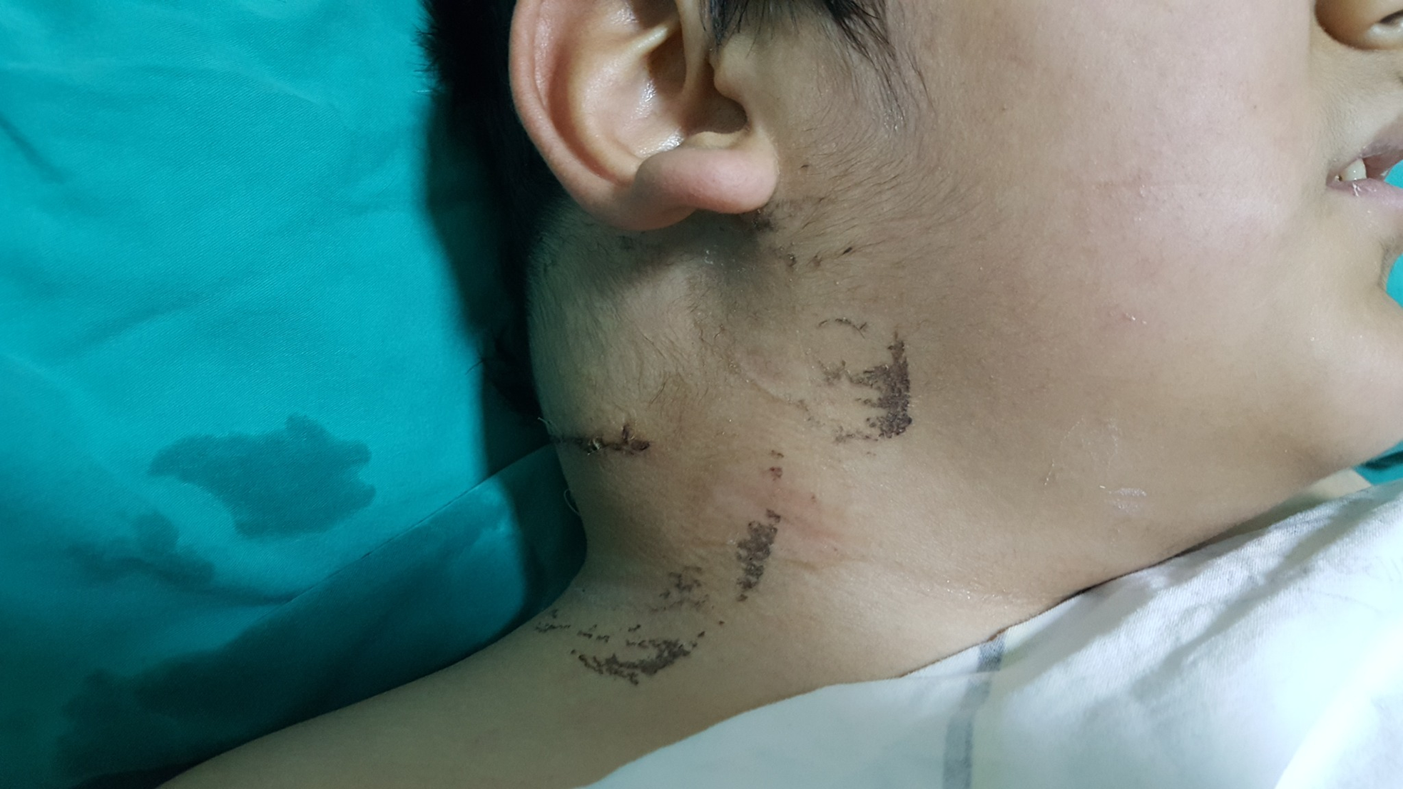
A 9-year-old boy was referred to our institution with a progressively enlarging swelling on the right side of his neck (Figure 1). The patient had previously been evaluated by an otolaryngologist. Surgical excision had initially been planned, and a skin incision was made; however, the procedure was abandoned because of the high risk of haemorrhage, and the incision was subsequently closed. Physical examination revealed a large, palpable, pulsatile mass in the right cervical region. A continuous vascular bruit was audible over the lesion on auscultation. The remainder of the physical examination was unremarkable.
Right-sided cervical swelling caused by the arteriovenous malformation at initial presentation.

Doppler ultrasonography demonstrated findings consistent with a high-flow vascular lesion. Contrast-enhanced CT of the neck revealed a 7 × 6 cm hypodense, intensely enhancing vascular lesion in the right suboccipital region. The lesion was supplied by branches of the right external carotid artery and the right subclavian artery. Digital subtraction angiography confirmed a high-flow arteriovenous malformation supplied by branches of the external carotid artery with early venous drainage (Video 1). Echocardiographic evaluation demonstrated no evidence of cardiac chamber enlargement or high-output cardiac failure. Based on the presence of pulsation and bruit without ulceration or cardiac failure, the lesion was classified as Schobinger stage II. According to the Yakes classification, it was categorised as type IIIb, characterised by multiple feeding arterioles shunting into an aneurysmal venous sac with multiple draining veins. Reference Schobinger1,Reference Yakes, Vogelzang, Ivancev, Yakes, Kim, Lee, Yakes and Do2
Considering the high-flow nature, large size, and progressive behaviour of the lesion, transcatheter embolisation was planned. Angiography demonstrated that the giant cervical arteriovenous malformation was supplied predominantly by a large feeding artery arising from the right external carotid artery, as well as two smaller feeding arteries originating from the right subclavian artery (Video 1).
The dominant feeding artery arising from the external carotid artery was selectively catheterised, and three detachable coils (two 5 × 15 mm and one 7 × 30 mm; Concerto™, Medtronic, Minneapolis, MN, USA) were deployed distal to its origin. During deployment, the coils were intentionally compacted using the catheter tip to create a stable coil framework. Completion angiography demonstrated complete occlusion of this feeding vessel (Video 2), although minimal residual flow persisted through the feeding artery arising from the right subclavian artery.
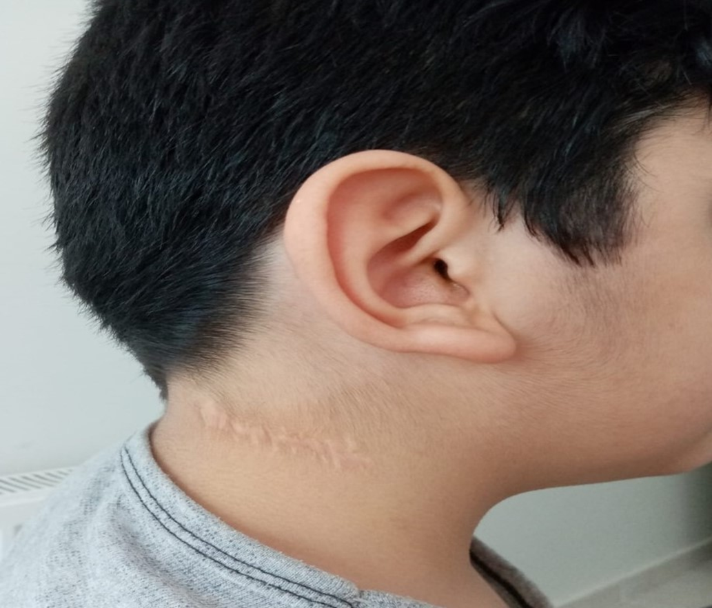
A staged embolisation strategy was intentionally adopted to minimise procedural duration and reduce anaesthesia exposure. Furthermore, the second intervention is scheduled for two months later so that we can observe any potential changes (such as permanent damage or complications). At repeat angiography performed two months later, the previously embolised external carotid artery feeder remained completely occluded, whereas the feeding artery arising from the right subclavian artery had become enlarged. This vessel was selectively embolised using one 9 × 30 mm and two 5 × 15 mm detachable coils (Concerto™, Medtronic, Minneapolis, MN, USA). Final angiography demonstrated complete elimination of the arteriovenous shunt. No procedural or post-procedural complications occurred. The patient was discharged the following day without complications. Progressive clinical improvement and marked reduction of the cervical swelling were observed, with sustained ultrasonographic evidence of complete occlusion during the 1-year follow-up period (Figure 2).
Clinical appearance one year after staged coil embolisation demonstrating marked regression of the cervical swelling.

Discussion
Although the optimal management of arteriovenous malformations remains controversial, a multidisciplinary treatment strategy is currently recommended. Therapeutic options depend on the location, size, flow characteristics, and extent of the lesion and include clinical observation, sclerotherapy, embolisation, ablative and coagulative techniques, and surgical resection. Reference Mattheis and Wanke3 While surgical excision may be curative for small, well-circumscribed arteriovenous malformations, complete resection is often not feasible in large or diffuse lesions. Moreover, incomplete excision is associated with high recurrence rates and may even stimulate aggressive lesion progression. Owing to these limitations and the potential morbidity of surgery, minimally invasive endovascular approaches have gained increasing acceptance. Various embolic materials, including sclerosants, absolute ethanol, liquid embolic agents, particulate embolic materials, and coils, have been used in the treatment of arteriovenous malformations. Reference Griauzde, Wilseck and Chaudhary4–Reference Funaki, Mori and Onozawa8
Although recurrence may occur following nonsurgical treatment, the risk of major complications is generally low. Reference Mattheis and Wanke3 Treatment success and recurrence rates vary according to the embolic agent used and the vascular architecture of the lesion. Le Fourn et al. Reference Le Fourn, Herbreteau and Papagiannaki9 performed 66 embolisation procedures for 32 arteriovenous malformations in 31 patients. Embolic agents included n-butyl cyanoacrylate, ethylene-vinyl alcohol copolymer, lauromacrogol, particulate embolic materials (collagen sponge and microspheres), and absolute ethanol. Cyanoacrylate glues were mixed with iodised oil in varying proportions to modify polymerisation and improve radiopacity. Among the eight patients with head and neck arteriovenous malformations, clinical improvement was achieved in five, one remained unchanged, and two experienced disease progression despite embolisation. Vascular coils have also been used as embolic devices in the endovascular management of arteriovenous malformations. These metallic devices produce mechanical vascular occlusion and are easily visualised under fluoroscopy. Coil embolisation may be particularly effective in arteriovenous malformations with a dominant feeding artery, a well-defined arteriovenous shunt, or a dominant draining vein. In cervical arteriovenous malformations, however, coils are most commonly used in combination with liquid embolic agents to reduce blood flow, prevent distal migration of liquid embolic materials, and improve control of venous outflow. Reference Dawkins, Motiwala and Peterson7,Reference Funaki, Mori and Onozawa8 The angioarchitecture defined by the Yakes classification also influenced our treatment strategy. Type IIIb lesions are characterised by multiple arteriolar feeders converging into a dilated venous sac with multiple draining veins. Although liquid embolic agents are commonly recommended for Yakes type IIIb lesions, the presence of a limited number of dominant feeding arteries in our patient allowed complete exclusion of the shunt using detachable coils alone.
Funaki et al. Reference Funaki, Mori and Onozawa8 reported a 10-year-old boy with a giant cervical arteriovenous malformation complicated by high-output cardiac failure who was successfully treated using detachable coils in combination with n-butyl cyanoacrylate, iodised oil, and ethanol, resulting in substantial clinical and echocardiographic improvement. In contrast, our patient had no evidence of high-output cardiac failure and was successfully treated using detachable coils alone. Gomes et al. Reference Gomes10 described two adult patients with arteriovenous malformations who underwent coil embolisation in combination with other embolic agents. Following coil migration into the pulmonary circulation in one patient who underwent venous embolisation, the author recommended selecting coils slightly larger than the target vessel diameter to reduce the risk of migration.
In the present case, detachable coils alone were selected because angiography demonstrated a limited number of dominant feeding arteries that could be selectively catheterised and completely occluded. The cervical location of the lesion further supported avoidance of liquid embolic agents because of the potential risk of non-target embolisation and cranial nerve ischaemia. Furthermore, detachable coils allowed controlled deployment with repositioning before release and stable anchoring within the target vessels. Coil diameters were intentionally selected slightly larger than the target vessel diameter to minimise the risk of migration, consistent with previous recommendations. To our knowledge, reports describing successful treatment of paediatric cervical arteriovenous malformations using detachable coils as the sole embolic agent remain extremely limited.
Conclusion
Coil-only embolisation may represent a safe and effective minimally invasive treatment strategy in carefully selected paediatric cervical AVMs with favourable angioarchitecture. Careful angiographic assessment may identify patients in whom coil-only embolisation can safely avoid the use of liquid embolic agents while maintaining excellent clinical outcomes. Long-term follow-up remains essential because recurrence may occur through collateral recruitment despite initial angiographic success.
Learning points
-
Cervical arteriovenous malformations are rare high-flow vascular anomalies that require detailed angiographic assessment for accurate classification and treatment planning.
-
In selected paediatric cervical arteriovenous malformations supplied by a limited number of dominant feeding arteries, detachable coil embolisation alone may achieve complete occlusion without the need for liquid embolic agents.
-
Careful angiographic evaluation is essential for identifying patients in whom coil-only embolisation may avoid the need for liquid embolic agents.
Supplementary material
The supplementary material for this article can be found at https://doi.org/10.1017/S1047951126124147.
Author contributions
All authors fulfil the criteria for authorship; Conceptualisation and design: AA, MAA. Data analysis and interpretation: AA, MAA Reviewing: AA, MAA Writing: AA. Editing: AA, MAA. Data collecting: AA.
Financial support
The authors received no financial support for this article’s research, authorship, and/or publication.
Competing interests
Concerning this paper’s research, authorship, and/or publication, the authors reported no potential conflicts of interest.
Consent for publication
Not applicable.


 Open access

