Introduction
Otitis media with effusion is defined as the presence of fluid in the middle ear, without signs or symptoms of acute infection.1 It is the most common cause of conductive hearing loss in children. Otitis media with effusion can lead to complications such as tympanic membrane atelectasis, retraction pocket formation and ossicular chain erosion. The more persistent the inflammatory condition, the greater the chance of these complications occurring, even if treated.
A normally functioning eustachian tube allows pressure regulation of the middle ear and protection from the nasopharynx.Reference Monsell and Harley2 Abnormal function of the eustachian tube has been implicated as one of the most important factors in the pathogenesis of all middle-ear disease.Reference Bluestone3 The placement of tympanostomy tubes provides an alternative means of middle-ear ventilation during periods of eustachian tube dysfunction. The tympanostomy tubes commonly used usually cease functioning because of extrusion, which occurs six to 18 months after insertion.Reference Weigel, Parker, Goldsmith, Postma and Pillsbury4, Reference Mandel, Rockette, Bluestone, Paradise and Nozza5 In many cases, this short-term ventilation is adequate for re-establishment of normal middle-ear aeration. However, 20–60 per cent of children will require repeated ventilation tube insertion.Reference Mandel, Rockette, Bluestone, Paradise and Nozza5–Reference Valtonen, Tuomilehto, Qvarnberg and Nuutinen7 Between 3 and 5 per cent of the adult population have evidence of persistent chronic otitis media; a larger proportion show signs of previous chronic otitis media.Reference Browning and Gatehouse8
A variety of transtympanic tubes have been designed to resist extrusion and provide long-term middle-ear aeration. However, the risk of complications (including otorrhoea, persistent perforation and cholesteatoma) is at least double that of conventional tympanostomy tubes.Reference Kay, Nelson and Rosenfeld9
T-tubes have been reported to have an average life span of 19 to 38 months.Reference Eliachar, Joachims, Goldsher and Golz10–Reference Van Heerbeek, De Saar and Mulder13 However, they have been associated with a significant risk of complications, notably persistent perforation after extrusion or removal. Rates of persistent perforation range from 10 to 33 per cent,Reference Eliachar, Joachims, Goldsher and Golz10–Reference Mangat, Morrison and Ganniwalla14 compared with a perforation rate of around 2 per cent for short-term ventilation tubes.Reference Kay, Nelson and Rosenfeld9 Otorrhoea, which can significantly impair quality of life,Reference Rosenfeld, Bhaya, Bower, Brookhouser, Casselbrant and Chan15 occurs in over 50 per cent of cases.Reference Strachan, Hope and Hussain12 Complication rates rise sharply if tubes remain in situ for longer than 36 months, leading some authors to suggest elective removal before 36 months.Reference Goode16 However, Mangat et al. Reference Mangat, Morrison and Ganniwalla14 have reported that surgical removal of tubes results in a much higher perforation rate than spontaneous extrusion (65 versus 19 per cent, respectively). Similar problems have been reported with other long-term transtympanic tubes such as the Per–Lee and Paparella II tubes, with complication rates of up to 70 per cent,Reference Per-Lee17 including perforation in 49 per cent of cases.Reference Kim, Moore and Rockley18 It may be argued that the high complication rate seen with long-term ventilation tubes is due to their use in patients who have failed prior treatment and thus constitute a more difficult group to treat. However, Strachan et al. Reference Strachan, Hope and Hussain12 and Mangat et al. Reference Mangat, Morrison and Ganniwalla14 have reported unacceptably high complication rates in patients with T-tubes inserted as a primary procedure.
Subannular placement of ventilation tubes via a tunnel beneath the skin of the external ear canal and fibrous annulus was first described by SimontonReference Simonton19 in 1968. This technique was used for atrophic and retracted tympanic membranes, in which there may not be sufficient fibrous material to retain a standard tympanostomy tube. A subannular ventilation tube may also be useful as an adjunct to tympanoplasty, as it avoids the placement of a tube through the grafted tympanic membrane.
One of the main reasons for extrusion of tympanostomy tubes is the centrifugal migration of the tympanic membrane epithelium towards the ear canal. Placement of a tube beneath the annulus may reduce the effect of these migratory forces and enable the tube to remain in situ for longer.Reference Jahn20 SilversteinReference Silverstein21 first reported the use of transosseous ventilation tube placement in 1970, describing placement of a silicone tube into the facial recess through a hole drilled in the posterior canal wall. The average lifespan of these tubes was 51 months, with some lasting longer than 10 years. However, 44 per cent of tubes became obstructed and required removal.Reference Haberkamp and Silverstein22 JahnReference Jahn20 further developed the transosseous technique by introducing a hydroxyapatite tube under the fibrous annulus into a groove drilled in the postero-inferior bony annulus. The advantage of this technique is osseous integration of the tube, resulting in bony fixation. There were no cases of spontaneous extrusion in Jahn's series. The overall success rate was 66 per cent, with tube blockage being the main complication.
Subannular ventilation tubes may provide long-term ventilation with few complications.Reference Martin-Hirsch, Woodhead and Vize23 However, a recent study has suggested that the risks of subannular ventilation tubes outweigh the benefits, compared with transtympanic T-tubes.Reference Jassar, Coatesworth and Strachan24
The current study examined the outcome of 57 consecutive subannular ventilation tube insertions in 45 patients at Dunedin, Invercargill and Mercy Hospitals, New Zealand, from 2000 to 2008. The results are discussed in relation to other reports of middle-ear ventilation.
Methods and materials
This was a retrospective review of 57 tube insertions in 45 consecutive patients who underwent subannular ventilation tube insertion, performed by or under the supervision of the senior author (PJD) between 2000 and 2008. The indications for subannular tube insertion were chronic otitis media with effusion and hearing loss (n = 54 cases), associated with adhesive otitis media (n = 7), tympanic membrane retraction (n = 17) or tympanic membrane perforation (n = 3).
Patients' case notes were examined for the following variables: pre- and post-operative pure tone audiometry threshold (averaged for 0.5, 1, 2 and 4 kHz); surgical complications; duration of tube placement; and post-operative complications, including perforation, infection, granulation, tube blockage, facial nerve damage and cholesteatoma. The extrusion time was taken as the first follow-up appointment at which the tube was no longer in situ. The results represent entries in the clinical notes up until the time of discharge, or for the most recent follow-up visit.
Surgical technique
All primary subannular ventilation tube insertions were performed under general anaesthesia, with the airway secured by either an endotracheal tube or laryngeal mask. The ear was prepared with Betadine® and draped in a sterile fashion.
The approach, determined at the time of surgery and by coincident procedures, was per-meatal, endaural or post-aural. Two per cent lignocaine with 1:80 000 adrenaline was infiltrated into the posterior ear canal and incision line. A tympanotomy incision was made in the posterior or inferior canal wall and a tympanomeatal flap elevated. Any adhesions between the tympanic membrane and the structures of the medial middle ear were separated. If necessary, the bony ear canal was widened. A groove was drilled in the postero-inferior bony annulus and adjacent canal wall. A ventilation tube was then positioned in the groove with the medial end in the hypotympanum and lower part of the facial recess. If necessary, the flange of the tube was trimmed to enable it to sit flush within the bony groove. The tympanomeatal flap was divided and the pedicles replaced below the neck of the tube.
Results
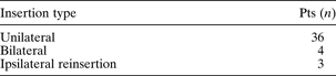
A total of 57 tubes were placed in 45 patients: 27 (60 per cent) males and 18 (40 per cent) females. Patients' mean age at surgery was 23 years (range, nine to 64 years). Thirty tubes (53 per cent) were placed in the right ear and 27 (47 per cent) in the left ear (Table I).
Tube insertions
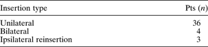
In addition, one patient had three insertions on the same side, and another had three insertions on the same side and a further insertion on the opposite side. Pts = patients
Thirty-five patients had previously been treated with a total of 59 tympanostomy tubes (mean number of previous tubes, two; range, one to seven), of which four (7 per cent) were subannular tubes. Four patients had no previous history of tympanostomy tube insertion. In six patients, we were unable to ascertain a history of previous tympanostomy tube insertion from the records. Two patients had undergone previous myringoplasties, with subsequent development of middle-ear effusions.
Forty-five ears (79 per cent) underwent a per-meatal approach, 10 (18 per cent) an endaural incision and two (4 per cent) a post-aural incision. Fifty (88 per cent) ears had Silverstein tubes inserted, four (7 per cent) Per-Lee tubes, two (4 per cent) long-term Armstrong tubes and one (2 per cent) a T-tube.
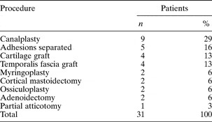
Table II shows additional procedures performed in 15 patients at the time of subannular tube insertion.

Twenty-nine tubes (51 per cent) remain in situ and 28 (49 per cent) were extruded or removed. The mean duration of ventilation for tubes still in situ was 22 months (range, one to 76 months; two patients were lost to follow up at one and two months, variously), and for tubes which were extruded or removed 23 months (range, one to 85 months). The mean follow up was 48 months (range, nine to 95 months).
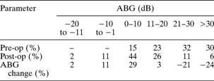
Fifty-three ears (93 per cent) had complete audiometric follow up (i.e. pure tone audiometry, averaged over 0.5, 1, 2 and 4 kHz). The mean pre-operative air–bone gap was 24 dB (range, 1–46 dB). The mean post-operative air–bone gap was 10 dB (range, −5 to 35 dB). The mean improvement in air–bone gap was 14 dB (range, −14 to 35 dB). One patient had worse hearing following intubation, attributable to scar tissue; this was corrected by revision surgery. The mean time to post-operative audiometry was six months (range, one to 40 months). Table III shows patients' pre- and post-operative air–bone gaps.
Patients' pre- vs post-operative air–bone gap*
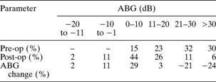
* For 53 ears. ABG = air–bone gap; pre-op = pre-operative; post-op = post-operative
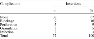
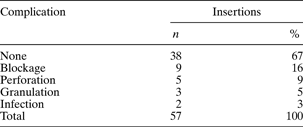
Table IV shows complications encountered following subannular ventilation tube insertion. In 38 ears (67 per cent), there were no complications. Nine tubes (16 per cent) became obstructed, of which three cases had spontaneous resolution, two blocked tubes resolved with topical ear drops, and one tube was unblocked using instrumentation. The final three blocked tubes were removed and replaced, and had no further complications. Granulation developed around the tube in three ears (5 per cent), which required cautery with silver nitrate in one. One patient developed granular myringitis, which is not uncommonly seen as a consequence of chronic otitis media. This was not considered to be caused by the subannular tube, as myringitis was also present in the contralateral ear. There were two episodes of post-operative infection (4 per cent) which cleared with topical antibiotic therapy. Five ears (9 per cent) had persistent perforation following extrusion or removal of the subannular tube. All perforations were small and in the region of the subannular tube insertion. None of these patients had infection. There were no cases of cholesteatoma formation or facial nerve injury.
Complications
Discussion
The maintenance of middle-ear aeration in patients with chronic eustachian tube dysfunction is an on-going problem for otologists. A variety of tubes have been designed to resist extrusion. However, meta-analysis has shown the risk of perforation, otorrhoea and cholesteatoma with long-term ventilation tubes to be at least double the risk of these complications with short-term tubes.Reference Kay, Nelson and Rosenfeld9 Subannular ventilation tubes have been described for use in atelectatic ears,Reference Simonton19 and may provide long-term ventilation with few complications.Reference Martin-Hirsch, Woodhead and Vize23
Martin-Hirsch et al. Reference Martin-Hirsch, Woodhead and Vize23 reported a series of 26 subannular tubes inserted via a tympanomeatal flap. Initial results were promising, with few complications and only one spontaneous extrusion. However, a subsequent study,Reference Jassar, Coatesworth and Strachan24 encompassing the original data set, found that 25 of 37 tubes were spontaneously extruded, and a further 10 had to be removed due to blockage or persistent infection. The mean time between insertion and extrusion was 29.6 months, with some tubes remaining in situ up to 74 months. There was a low perforation rate (8 per cent). However, Jassar et al. Reference Jassar, Coatesworth and Strachan24 questioned whether subannular placement could lead to erosion of the fibrous annulus, possibly resulting in marginal perforations, which are less likely to heal than central perforations. Overall, Jassar and colleagues reported that the use of subannular ventilation tubes did not compare favourably to T-tubes. Conversely, Al-Swadi et al. Reference Al-Swadi, Karlsmose, Gaihede, Henriksen and Rosberg25 reported that Per–Lee tubes placed in a groove drilled under the osseous annulus lasted significantly longer than T-tubes and had relatively few complications. The mean functioning time for tubes still in situ was 45 months, with a mean functioning time for all tubes of 17 months. The incidence of complications was comparable to that of standard tympanostomy tubes, with otorrhoea in 20 per cent of cases and perforation in 1 per cent.
O'Hare and GoebelReference O'Hare and Goebel26 described the anterior placement of a subannular tube in patients in whom the posterior canal wall or fibrous annulus was unsuitable for tube placement. After a mean follow up of 13.6 months, only one tube had been extruded. Complications included granulation tissue (26 per cent), acute infection (16 per cent) and persistent otorrhoea (5 per cent). In a longer term follow-up study of anterior subannular tubes (mean follow up, 26 months), 34 out of 38 ears had patent tubes and well aerated middle ears.Reference Elluru, Dhanda, Neely and Goebel27
More recently, Cloutier et al. Reference Cloutier, Arcand, Nartinez, Abela, Quintal and Guerguerian28 reported subannular ventilation tube insertion in 316 paediatric ears with a mean follow up of 53 months. Complications included otorrhoea (18 per cent), perforation (8 per cent), blockage (7 per cent) and cholesteatoma (2 per cent).
Our patients were predominantly either adolescents or adults, and presented primarily with a history of earlier ventilation tube insertion with persisting chronic otitis media with atelectasis, and/or middle-ear effusion. Two patients with a history of atelectasis and middle-ear effusion subsequently developed a perforation with hearing loss, and underwent subannular tube insertion to prevent subsequent recurrence of otitis media with effusion. There were 10 children aged between eight and 11 years who presented with persistent recurrence of otitis media with effusion, and for whom further short term intubation was considered inappropriate.
The patients included in our series represent the expected variety of individuals with persistent eustachian tube dysfunction for which ongoing middle-ear ventilation is required to maintain social hearing. Infection (4 per cent) and granulation (5 per cent) occurred less frequently than may be expected with long term transtympanic tubes. Persistent obstruction occurred in 5 per cent of patients. There were residual perforations in 9 per cent of the group, but in all cases these were small, situated lateral to the annulus, and associated with no tendency to discharge, with a small air–bone gap; that is, they functioned as an ongoing mode of middle-ear ventilation. The duration of ventilation was variable, and was less supportive of this approach to long-term middle-ear ventilation in that the tube life span was similar to that of transtympanic T-tubes (half the tubes were either extruded or removed, and one-quarter were in place for less than 18 months). In our patient group, the low rate of infective complications and the lack of complications associated with residual perforation argue in favour of subannular tubes as a means of promoting long-term, relatively trouble-free middle-ear ventilation.
The duration of ventilation and complication rates observed in this study are comparable to other reports.Reference Jassar, Coatesworth and Strachan24, Reference Al-Swadi, Karlsmose, Gaihede, Henriksen and Rosberg25, Reference Cloutier, Arcand, Nartinez, Abela, Quintal and Guerguerian28 Thus far, all studies of subannular ventilation tubes have been retrospective. Furthermore, the technique of subannular tube placement has varied. In our series, all patients had a postero-inferior bony groove drilled under the annulus, and division of the tympanomeatal flap with pedicle replacement around the neck of the tube. Whether this particular approach results in fewer complications or a longer duration of ventilation is uncertain at this stage. In three adult patients, drilling the groove subsequently allowed us to replace an extruded tube with a modified shortened T-tube under local anaesthetic in the out-patient department.
We report outcomes for a heterogeneous group of patients for whom long-term middle-ear ventilation was considered appropriate. While this allows us to comment on the outcome of our clinical practice, there are limitations to our conclusions. The patients predominantly had chronic otitis media with effusion and were symptomatic with hearing loss; others had retraction pocket disease and in some cases perforation consequent to breakdown of a thinned and retracted tympanic membrane. We are able to report on the duration of intubation and the clinical course following placement of the subannular tube; however, we are unable to state with confidence that this form of management was superior to either observation, hearing aid fitting or transtympanic tube placement. In order to fully assess the efficacy and safety of long-term middle-ear ventilation using subannular tubes, a large, prospective trial including a control group treated with standard T-tubes would be necessary. As subannular tubes have been reported to remain in place for over 10 years,Reference Silverstein21 long-term follow up would be required.
• Long-term tympanostomy tubes are associated with a significant incidence of complications, particularly persistent perforation
• Subannular ventilation tubes do not remain in situ longer than transtympanic T-tubes; however, associated complications, particularly persistent perforation after extrusion, are less common
• Subannular ventilation tubes provide an effective option for managing intractable middle-ear effusion and eustachian tube dysfunction
Further understanding of the mechanism by which subannular ventilation tubes extrude may be of use in improving their design. It is not known whether the tubes extrude due to inflammation, migration of the tympanic membrane epithelium, or erosion through the fibrous annulus. If there is a significant inflammatory component leading to extrusion, the use of biocompatible materials such as titanium may improve the longevity of the tube. The flange design may also contribute to duration of ventilation and rates of perforation after extrusion, as has been seen with transtympanic tubes.
Conclusion
Although subannular ventilation tubes do not remain in situ longer than transtympanic T-tubes, our series had fewer complications; in particular, persistent perforation after extrusion was less frequent. Subannular ventilation tube placement offers an alternative mode of providing long-term middle-ear ventilation for patients with persistent eustachian tube dysfunction. Whilst general anaesthesia and a tympanotomy are required for placement, the outcome, in terms of ventilation maintenance and infective complications, compares well with long-term transmyringeal ventilation.