Introduction
The detection rate of superficial pharyngeal legions has been increasing recently owing to surveillance endoscopy with narrow-band imaging. As a result, indications for transoral pharyngeal surgery have expanded widely.Reference Muto, Nakane, Katada, Sano, Ohtsu and Esumi1 Lesion grade and depth have been reported to be largely related to lesion size. Thus, lesions less than 5 mm tend to be low-grade intraepithelial neoplasias and do not progress to invasive carcinoma in the oesophagus.Reference Shimizu, Yoshida, Kato, Hirota, Ono and Nakagawa2 Takemura et al.Reference Takemura, Doyama, Nakanishi, Takeda, Kito and Ito3 reported that flat-type micro-lesions equal to or less than 5 mm in size in the orohypopharynx may be followed for up to 2 years without biopsy or endoscopic resection. Therefore, at our facility, micro-superficial lesions equal to or less than 5 mm in size in the pharyngeal region were observed without biopsy or endoscopic resection. No progression was detected in such cases initially.
However, we recently encountered a micro-superficial lesion measuring 5 mm that showed rapid growth. Detection of these small lesions is of great clinical significance in terms of treatment. To date, few reports have described characteristics of micro-superficial lesions equal to or less than 5 mm in size. The aim of this study was to identify the features of lesions equal to or less than 5 mm that become squamous cell carcinoma (SCC) or high-grade intraepithelial neoplasia. We also wanted to describe the characteristics of endoscopic findings of those micro-superficial lesions that are SCC or high-grade intraepithelial neoplasia in order to facilitate the administration of appropriate treatment while lesions are small.
Materials and methods
We conducted a retrospective cohort study in patients with pharyngeal lesions sized equal to or less than 5 mm who underwent endoscopic resection at Hiroshima University Hospital in Japan between April 2008 and June 2014. Eligible patients were aged equal to or more than 18 years at the time of resection. Patients who had a malignant tumour other than SCC on histopathological examination were not included in the study. The institutional review board of Hiroshima University Hospital approved this study (number: E-2039).
We retrospectively collected patient data on age, sex, smoking history, alcohol consumption, primary tumour location, gross findings on clinical examinations, endoscopic findings from electronic medical records and the pathological results post-resection.
Endoscopic examination
All examinations were performed by endoscopists with over 10 years of practical experience. A magnifying endoscope (GIF-H240Z, H260Z or H290Z; Olympus Medical Systems, Tokyo, Japan) was used. Tumour type classification was based on macroscopic findings, and the macroscopic characteristics of the lesion were classified in accordance with the Japanese Classification of Oesophageal Cancer (11th edition),4 General Rules for Clinical Studies on Head and Neck Cancer (6th edition) of the Japan Society for Head and Neck Cancer,5 and the Head and Neck Superficial Cancer Handling guidelines of the Japan Society for Head and Neck Cancer.6 Superficial-type lesions were categorised by the prefix 0 and were classified as follows: 0–I (superficial and protruding type), 0–II (superficial and flat type) and 0–III (superficial and excavated type). Type 0–II (superficial and flat type) was further classified as either 0–IIa (slightly elevated type: less than 1 mm in height), 0–IIb (true flat type) or 0–IIc (slightly depressed type).Reference Tateya, Morita, Muto, Miyamoto, Hayashi and Funakoshi7
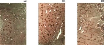
After white-light imaging assessment, magnifying endoscopy with narrow-band imaging was performed to evaluate the microvascular patterns of the lesions. The obtained microvascular patterns were classified according to the Japan Esophageal Society classification,4 which categorises vessels as either type A or B. Type A vessels showed mild or no intrapapillary capillary loop atypia (vessels with a diameter of 7–10 μm), whereas type B vessels showed obvious intrapapillary capillary loop changes. Type A and B vessels strongly indicated intraepithelial neoplasia and SCC, respectively.
Type B vessels were sub-classified into three groups as follows: type B1, dilated and tortuous vessels of various diameters and shapes with intact loop formation (dot-, spiral- or waist-thread-like loop vessels of 20–30 μm); type B2, multi-layered and irregularly and dendritically branched vessels with no loop formation; and type B3, vessels that were obviously thicker than the surrounding vessels (equal to or more than 3-fold thicker than B2 vessels (i.e. more than 60 μm in diameter)).
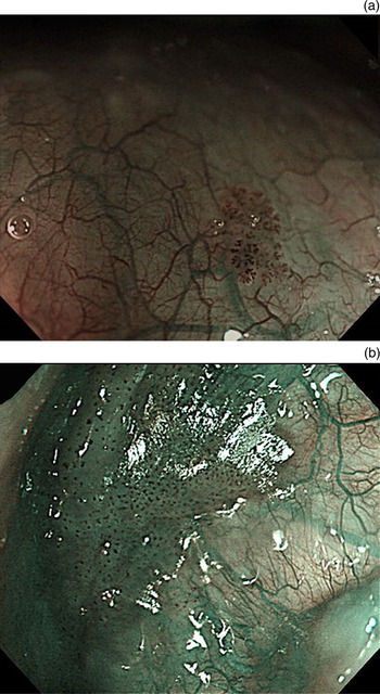
Typical cases of the typical type A, B1 and B2 type are shown in Figure 1. Background colouration was evaluated during lesion assessment with narrow-band imaging magnification. Cases showing a colour change in the epithelia between intrapapillary capillary loops were regarded as being positive for background colouration (Figure 2).
(a) Type A vessels showed mild or no atypia of the intrapapillary capillary loops (vessels with a diameter of 7–10 μm). (b) Type B1 dilated and tortuous vessels of various diameters and shapes with intact loop formation (dot-, spiral- or waist-thread-like loop vessels of 20–30 μm). (c) Type B2 multi-layered and irregularly and dendritically branched vessels with no loop formation.

(a) Negative for background colouration. The colour in the area between intrapapillary capillary loops is the same as the surrounding normal intrapapillary capillary loop area. (b) Positive for background colouration. The colour change in the epithelia between intrapapillary capillary loops is brownish compared with a normal intrapapillary capillary loop area.

Data analysis
Statistical differences in lesion characteristics were evaluated using the Mann–Whitney U test, in which the dependant variable was either ordinal or continuous. The Fisher's exact probability test was used to analyse the contingency table. The sensitivity, specificity, positive predictive value, negative predictive value and overall accuracy for identifying SCC or high-grade intraepithelial neoplasia and low-grade intraepithelial neoplasia or non-atypia cases were estimated of lesion characteristics, especially background colouration and intrapapillary capillary loop pattern classification. P-values less than 0.05 were considered statistically significant. All statistical analyses were performed using SPSS® (version 24) statistical analysis software.
Results
The study flow schema is shown in Figure 3. Among the 91 pharyngeal (oropharyngeal or hypopharyngeal) lesions from 69 patients who underwent endoscopic resection at our clinic between April 2008 and June 2014, 55 lesions measuring equal to or less than 5 mm from 45 patients were included in this study. The histological diagnoses included SCC (3 lesions), high-grade intraepithelial neoplasia (7 lesions), low-grade intraepithelial neoplasia (36 lesions) and non-atypia lesions (9 lesions; Figure 3).
Flow schema of the study. Eligible patients were equal to or more than 18 years of age at the initiation of the resection. Ninety-one lesions (69 patients) underwent endoscopic resection for oropharyngeal or hypopharyngeal lesions at our clinic between April 2008 and June 2014. SCC = squamous cell carcinoma; HGIN = high-grade intraepithelial neoplasia; LGIN = low-grade intraepithelial neoplasia.

The patients and characteristics are listed in Table 1. The median age at the time of treatment was 67 years (range, 40–84 years), and 39 patients (86.7 per cent) were male. The oropharynx (42 lesions, 76.4 per cent) was the most frequent primary region for lesions, followed by the hypopharynx (13 lesions, 23.6 per cent). Details on the subparts of the primary regions are presented in Table 1. Thirteen patients (28.9 per cent) were non-smokers, and 32 patients (71.1 per cent) were smokers. Seven patients (15.6 per cent) did not consume alcohol, 5 (11.1 per cent) were social drinkers and 33 (73.3 per cent) consumed alcohol regularly.
Patient characteristics

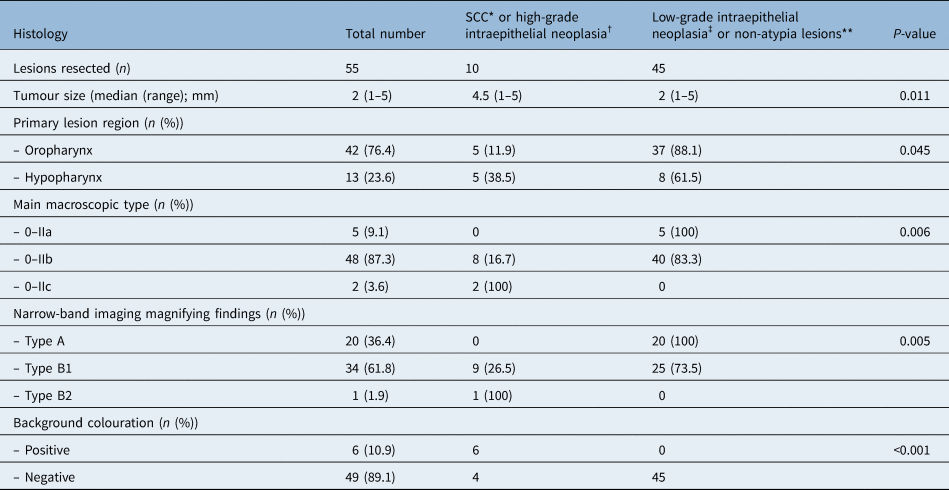
Eighteen patients (40.0 per cent) had oesophageal cancer and/or head and neck cancer synchronously or metachronously. Most of the patients were identified as having cancer by follow-up examinations for oesophageal or head and neck cancer. All cancers in the oesophagus and the head and neck region were treated with methods such as endoscopic resection, (chemo)radiotherapy or surgery. After physical examination, 27 patients (60.0 per cent) were referred for suspected pharyngeal cancer after endoscopy of the oesophagus, stomach or duodenum. Lesion characteristics are shown in Table 2. The histological diagnosis was SCC or high-grade intraepithelial neoplasia for 10 lesions (18.2 per cent) and low-grade intraepithelial neoplasia or non-atypia lesions for 45 lesions (81.8 per cent). Univariate analysis showed significant associations between the size of SCC or high-grade intraepithelial neoplasia and low-grade intraepithelial neoplasia or non-atypia lesions.
Lesion characteristics

*n = 3; †n = 7; ‡n = 36; **n = 9. SCC = squamous cell carcinoma
Lesion classification by macroscopic examination
Macroscopic categorisation according to the classification of oesophageal cancer published by the Japan Esophageal Society showed the following macroscopic types4: five lesions were 0–IIa; 48 lesions were 0–IIb and two lesions were 0–IIc.
In the 0–IIa category, all lesions were of low-grade intraepithelial neoplasia or non-atypia lesion histology. In the 0–IIb category, 8 lesions (16.7 per cent) were SCC or high-grade intraepithelial neoplasia, and 40 lesions (83.3 per cent) were low-grade intraepithelial neoplasia or non-atypia lesions. In the 0–IIc category, all lesions were SCC or high-grade intraepithelial neoplasia. Univariate analysis showed significant associations amongst the macroscopic types (Table 2; p = 0.006).
Narrow-band imaging findings
Numbers of lesions classified as intrapapillary capillary loop type A, B1 and B2 were 20 (36.4 per cent), 34 (61.8 per cent) and 1 (1.9 per cent), respectively. All type A lesions had low-grade intraepithelial neoplasia or non-atypia lesion histology. Among B1 lesions, 9 (26.5 per cent) were SCC or high-grade intraepithelial neoplasia and 25 (73.5 per cent) were low-grade intraepithelial neoplasia or non-atypia lesions. The histology of the type B2 lesion was SCC. Univariate analysis showed significant associations between SCC or high-grade intraepithelial neoplasia and magnifying endoscopy with narrow-band imaging findings (Table 2; p = 0.005).
Background colouration
Six lesions (10.9 per cent) were positive for background colouration, and 49 lesions (89.1 per cent) were negative for background colouration. All background colouration-positive lesions were SCC or high-grade intraepithelial neoplasia, and 45 (91.8 per cent) background colouration-negative lesions were of the low-grade intraepithelial neoplasia or non-atypia lesion type. Univariate analysis showed significant associations between SCC or high-grade intraepithelial neoplasia and background colouration positive status (Table 2; p < 0.001).
Performance characteristics of narrow-band imaging features
We analysed the ability of background colouration and intrapapillary capillary loop patterns to distinguish SCC or high-grade intraepithelial neoplasia from low-grade intraepithelial neoplasia or non-atypia lesions. Intrapapillary capillary loop pattern assessments by magnifying endoscopy with narrow-band imaging yielded negative findings for type A and positive findings for type B. The overall sensitivity, specificity, accuracy, positive predictive value and negative predictive value of background colouration for distinguishing SCC or high-grade intraepithelial neoplasia from low-grade intraepithelial neoplasia or non-atypia lesions was 60 per cent, 100 per cent, 92.7 per cent, 100 per cent and 91.8 per cent, respectively.
We analysed whether intrapapillary capillary loop patterns could be used to distinguish SCC or high-grade intraepithelial neoplasia from low-grade intraepithelial neoplasia or non-atypia lesions in 49 background colouration negative lesions.
The overall sensitivity, specificity, accuracy, positive predictive value and negative predictive value of magnifying endoscopy with narrow-band imaging for distinguishing between SCC or high-grade intraepithelial neoplasia and low-grade intraepithelial neoplasia or non-atypia lesions were 100 per cent, 44.4 per cent, 54.6 per cent, 28.6 per cent and 100 per cent, respectively. The overall sensitivity, specificity, accuracy, positive predictive value and negative predictive value of background colouration and intrapapillary capillary loop pattern together for distinguishing between SCC or high-grade intraepithelial neoplasia and low-grade intraepithelial neoplasia or non-atypia lesions were all 100 per cent (Table 3).
Sensitivities, specificities and accuracies of BGC and ME-NBI in distinguishing SCC or high-grade intraepithelial neoplasia from low-grade intraepithelial neoplasia or non-atypia lesions

BGC = background colouration; ME-NBI = magnifying endoscopy with narrow-band imaging; SCC = squamous cell carcinoma; CI = confidence interval
Discussion
Pharyngeal cancer is often detected after lesions have reached a large size. In such cases, the patient has a poor prognosis. Treatment of such advanced lesions with surgery or chemoradiotherapy may significantly deteriorate important functions such as swallowing, breathing, coughing and speech. Therefore, early detection of pharyngeal cancer improves prognosis and outcomes. Several studies have shown that narrow-band imaging improves the detection rate of superficial squamous cell carcinoma of the larynx and pharynx.Reference Yagishita, Fujii, Yano and Kaneko8,Reference Muto, Minashi, Yano, Saito, Oda and Nonaka9,Reference Popek, Bojanowska-Poźniak, Tomasik, Fendler, Jeruzal-Świątecka and Pietruszewska10 This study demonstrated that diagnosis based on background colouration and the intrapapillary capillary loop pattern on narrow-band imaging could improve differentiation of high-grade intraepithelial neoplasia and SCC from low-grade intraepithelial neoplasia or non-atypia lesions and so facilitate the identification and pathological examination of lesions measuring equal to or less than 5 mm.
Muto et al.Reference Muto, Satake, Yano, Minashi, Hayashi and Fujii11 reported that peroral organ-preserving endoscopic resection for superficial pharyngeal cancer is a feasible treatment option. In that study, no severe adverse events were reported, and patients had an extremely good prognosis.
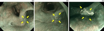
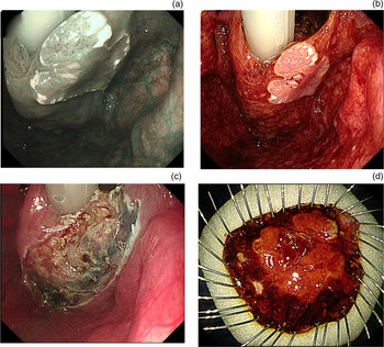
In our unit, micro-superficial lesions equal to or less than 5 mm in size in the pharyngeal region were observed without biopsy or endoscopic resection, and no progression was detected until recently, when we encountered a micro-superficial lesion that showed rapid growth. The patient was a 64-year-old man, with a 5-mm lesion in the right pyriform sinus that was initially diagnosed as a superficial lesion on endoscopic surveillance with narrow-band imaging. The lesion size was stable for 6 months after detection, but subsequently grew to 20 mm at 8 months later (Figure 4). The patient was referred to the Department of Otorhinolaryngology, Head and Neck Surgery, and he underwent endoscopic laryngopharyngeal surgery using a curved laryngoscope. Endoscopic laryngopharyngeal surgery is a hybrid of head and neck surgery and gastrointestinal endoscopic treatment. The concept is the same as that of endoscopic submucosal dissection in that both involve en bloc resection of a cancer lesion following submucosal injection; however, it differs from Endoscopic submucosal dissection in that the resection procedure is performed by a head and neck surgeon using both hands (Figure 5).Reference Tateya, Muto, Morita, Miyamoto, Hayashi and Funakoshi12 On the basis of this experience, we retrospectively investigated micro-superficial lesions equal to or less than 5 mm in size to clarify the difference between characteristics of SCC or high-grade intraepithelial neoplasia and low-grade intraepithelial neoplasia or non-atypia lesions.
Endoscopic findings of progression of a micro-superficial lesion in the right pyriform sinus. Macroscopic classification and narrow-band imaging categorised the lesion as 0–IIc and type B1, respectively, and the lesion showed positive findings for background colouration. (a) A 5-mm lesion was detected (yellow arrows). (b) After 6 months, the lesion was stable (yellow arrows). (c) After 14 months, the size of the lesion was 20 mm (yellow arrows).

Surgical procedure for superficial pharyngeal cancer with rapid growth in the right pyriform sinus. The histopathological examination showed squamous cell carcinoma in situ (600 μm) with lymphatic invasion (ly)0, blood vessel invasion (v)0, pathological horizontal margin (pHM)0 and pathological vertical margin (pVM)0. (a) Brownish area demonstrated using narrow-band imaging. (b) Tumour outlines were delineated by iodine staining. (c) The tumour was resected using the electric needle knife and curved forceps. (d) Resected specimen.

Nakamura et al.Reference Nakamura, Yano, Fujii, Kadota, Tomioka and Shinozaki13 reported that most superficial head and neck SCCs progressed in size naturally, suggesting that if permitted by the patient's condition, they should be treated using less invasive methods when small. Furthermore, the report suggested that superficial head and neck SCCs measuring equal to or more than 3 mm are significant lesions that require careful follow up or endoscopic intervention. Histological results obtained by Shimizu et al.Reference Shimizu, Kato, Yamamoto, Ono, Katsurada and Ono14 suggested that high-grade intraepithelial squamous neoplasia of the oesophagus show characteristics of carcinoma at a pre-invasive stage. That study also suggested that endoscopic mucosal resection should be performed for oesophageal lesions diagnosed as high-grade intraepithelial squamous neoplasia by endoscopic biopsy, not only because of their probable malignant potential but also because more than 30 per cent of such lesions are actually invasive carcinomas.
In our present study, the histological diagnosis indicated SCC and high-grade intraepithelial neoplasia histology in 3 and 7 lesions (18.2 per cent), respectively. Although these lesions measured equal to or less than 5 mm in size, they had the potential to grow. Thus, determination of characteristics of SCC or high-grade intraepithelial neoplasia via an endoscopic study before surgery may facilitate the identification of an appropriate surgical technique.
First, we examined whether the Japan Esophageal Society macroscopic classification was associated with a particular histological diagnosis. Results indicated that SCC or high-grade intraepithelial neoplasia tended to be larger than low-grade intraepithelial neoplasia or non-atypia lesions, and significant associations were observed among macroscopic types. This was attributed to the fact that in the 0–IIc group, all lesions were SCC or high-grade intraepithelial neoplasia, and in the 0–IIa group, all lesions were low-grade intraepithelial neoplasia or non-atypia lesions. A change in oesophageal micro-lesion morphology from type 0–IIb to type 0–IIc has been suggested to indicate cancerous potential. Thus, type 0–IIc lesions should be resected.Reference Kuwano15
Second, lesions classified on the basis of intrapapillary capillary loop patterns were evaluated by magnifying endoscopy with narrow-band imaging examinations. Narrow-band imaging has been reported to facilitate the detection of superficial cancers that are rarely identifiable by white-light imaging and thereby play an important role in the diagnosis of SCC of the oropharynx and hypopharynx.Reference Watanabe, Tsujie, Taniguchi, Hosokawa, Fujita and Sasaki16,Reference Cosway, Drinnan and Paleri17 In our study, all type A lesions had a low-grade intraepithelial neoplasia or non-atypia lesion histology, suggesting that type A lesions do not require resection. In contrast, the histology of type B2 lesion was SCC. We concluded that lesions with obviously multi-layered vessels and irregular and dendritic branching with no loop formation are SCC or high-grade intraepithelial neoplasia. The presence of type B vessels is evidence of oesophageal squamous cell carcinoma; however, it is not clear for the association between presence of type B vessels and pharyngeal cancer. On the other hand, Eguchi et al.Reference Eguchi, Matsui, Mukai and Sugimoto18 reported a significant correlation between the classification of type B vessels and tumour thickness in superficial pharyngeal cancer. In this study, 9 (26.5 per cent) of the B1 lesions were SCC or high-grade intraepithelial neoplasia and 25 (73.5 per cent) were low-grade intraepithelial neoplasia or non-atypia, indicating that the specificity of magnifying endoscopy with narrow-band imaging for cancer is very low. We thought that micro-superficial lesions of up to 5 mm in size might be difficult to diagnose as cancer based on being B1 alone.
Finally, background colouration was evaluated. All background colouration-positive lesions were SCC or high-grade intraepithelial neoplasia, and 45 background colouration negative lesions (91.8 per cent) had a low-grade intraepithelial neoplasia or non-atypia lesion histology. Background colouration indicates a colour change in the area between the intrapapillary capillary loops and can also be observed within the brownish area. Kanzaki et al.Reference Kanzaki, Ishihara, Ishiguro, Nagai, Matsui and Yamashina19 statistically analysed the cause of the colour change in background colouration and suggested that it may be related to the thinning of the keratinous layer, which is caused by neoplastic cell proliferation and thinning of the epithelium. A brownish epithelium was also closely associated with a diagnosis of high-grade intraepithelial neoplasia or cancer because it required the presence of abnormal cells in the upper half of the epithelium. Thus, a combination of vascular changes and brownish epithelium might predict the development and proliferation of neoplasia and facilitate the accurate diagnosis of oesophageal lesions. Additionally, narrow-band imaging reflects a wavelength that is specific to haemoglobin. Therefore, the colour change may be related to the extravascular haemoglobin component in the cancer area.
On the other hand, a pink colour sign may also reflect superficial oesophageal cancer in SCC or high-grade intraepithelial neoplasia. The presence of a pink colour sign was closely associated with the absence of a keratinous layer and was reported to be useful for the diagnosis of SCC or high-grade intraepithelial neoplasia.Reference Ishihara, Kanzaki, Iishi, Nagai, Matsui and Yamashina20 In this assessment, the discolouration of the iodine solution on the mucosal surface was evaluated. If a light-pink colouration appeared in the iodine-unstained area, the lesion was regarded as being pink colour sign positive. Takahashi et al.Reference Takahashi, Shimizu, Ono, Suzuki, Omori and Yoshida21 reported that the diagnosis of background colouration on narrow-band imaging is useful for differentiating high-grade intraepithelial neoplasia from low-grade intraepithelial neoplasia and may be an alternative means to diagnose many patients with oesophageal SCC based on pink colour sign. Notably, as it is difficult to stain the pharyngeal mucosa with iodine solution without general anaesthesia, it cannot be used for diagnosing SCC or high-grade intraepithelial neoplasia in an out-patient examination before surgery. Minami et al.Reference Minami, Inoue, Ikeda, Satodate, Hamatani and Nakao22 used background colouration to differentiate early SCC in the oesophagus from benign lesions, including inflammatory changes. They found that in combination with the intrapapillary capillary loop pattern classification, background colouration can provide additional information for the accurate discrimination of SCC or high-grade intraepithelial neoplasia from low-grade intraepithelial neoplasia or non-atypia lesions. Narrow-band imaging is a technique that exclusively identifies the wavelength of haemoglobin. Preliminary results reported by Minami et al. showed a significant correlation between haemoglobin immunopositivity and malignant pathology; in addition, background colouration and haemoglobin positivity were highly correlated.Reference Minami, Isomoto, Inoue, Akazawa, Yamaguchi and Ohnita23
• Early detection of pharyngeal cancer improves prognosis and outcomes
• This study aimed to elucidate features of malignant lesions equal to or less than 5 mm in size
• Diagnosis based on background colouration and the intrapapillary capillary loop pattern is useful for lesions equal to or less than 5 mm
In this study, univariate analysis showed significant associations between SCC or high-grade intraepithelial neoplasia and background colouration positivity, main macroscopic type and magnifying endoscopy with narrow-band imaging findings. However, a more accurate diagnosis may be needed to avoid the unnecessary resection of lesions less than 5 mm. Therefore, we investigated whether the combination of background colouration and the intrapapillary capillary loop pattern was useful for accurate discrimination of SCC or high-grade intraepithelial neoplasia from low-grade intraepithelial neoplasia or non-atypia lesions. Our results strongly suggested that diagnosis using background colouration and the intrapapillary capillary loop pattern together could differentiate high-grade intraepithelial neoplasia or SCC from low-grade intraepithelial neoplasia or non-atypia lesions. Our results suggested that diagnosis using background colouration and intrapapillary capillary loop patterns on narrow-band imaging is useful for differentiating high-grade intraepithelial neoplasia or SCC from low-grade intraepithelial neoplasia or non-atypia lesions. This data could facilitate decision-making regarding which lesions measuring 5 mm or less should be resected.
This study had several limitations. First, this was a retrospective study. Second, the study population was recruited from a single centre. Third, the number of patients included was relatively limited. Finally, all cases in this study had undergone resection; the natural history without resection is not known. However, it will be difficult to assess further cases because we do not actively resect lesions of equal to or less than 5 mm in size. Nevertheless, findings obtained for included cases appear to have diagnostic value. Further studies including further type B and background colouration positive lesions equal to or less than 5 mm in size are needed to validate our findings.
Acknowledgements
The authors acknowledge professor Hisham Mehanna (Chair of Head and Neck Surgery Institute for Head and Neck Studies and Education, University of Birmingham, UK) who commented on the study design and revised the paper. This study was supported by Japan Society for the Promotion of Science Kakenhi (grant number: 20K09713).
Competing interests
None declared