Introduction
During the development of an embryo from zygote to the blastocyst stage, the cytoplasmic volume and nuclear size reduced to different proportions following each cleavage, resulting in an increase in the nuclear to cytoplasmic (N/C) ratio in later embryo stages (Evsikov et al., Reference Evsikov, Morozoval and Solomko1990; Tsichlaki and FitzHarris, Reference Tsichlaki and FitzHarris2016). Whereas nucleus volume decreased approximately 10-fold during preimplantation development, cytoplasmic content is roughly halved after each cleavage, which can be translated into a c. 64-fold to 128-fold reduction in the same time course (Tsichlaki and FitzHarris, Reference Tsichlaki and FitzHarris2016). Based on this observation, it has been hypothesized that the initiation of compaction and blastocoel formation might be correlated with the increase in the N/C ratio. However, the experimental data supporting this hypothesis have been rather limited. Previous studies have indicated that a decrease in N/C ratio by enucleation (Kaufman and Sachs, Reference Kaufman and Sachs1976) or an increase in N/C ratio by microsurgical removal of some part of the zygote cytoplasm (Barton and Surani, Reference Barton and Surani1983) was found to have a negative effect on embryo development.
Conversely, the compaction and blastulation in preimplantation embryos are important events for successful implantation and pregnancy in mice (Kim et al., Reference Kim, Kim and Jun2017) and humans (Tao et al., Reference Tao, Tamis, Fink, Williams, Nelson-White and Craig2002; Skiadas et al., Reference Skiadas, Jackson and Racowsky2006). However, embryos with morphologically normal cleavage do not necessarily reach the morula and blastocyst stages at the appropriate time (Kim et al., Reference Kim, Kim and Jun2017). Time-lapse cinematography will facilitate the non-invasive tracking of cleavage and development of in vitro produced embryos, very useful in selecting good embryos for embryo transfer. Higher pregnancy rates were observed for animals in which transferred blastocysts had undergone normal cleavage, identified by the presence of two same-size blastomeres, compared with those with abnormal cleavage (Sugimura et al., Reference Sugimura, Akai, Somfai, Hirayama, Aikawa, Ohtake, Hattori, Kobayashi, Hashiyada, Konishi and Imai2010). In addition, in vitro technologies including somatic cell nuclear transfer (SCNT)-mediated gene transfer in pigs are very important for biomedical research, but the efficacy for offspring production is still low. Therefore, the improvement in cloned/transgenic embryo production systems in pigs is extremely necessary. Modification in starting materials including oocytes and donor cells have been made to improve the development of cloned embryos and understand the mechanism and factors that affect the progression of the nuclear reprogramming process. However, the effects of modification of N/C ratio on the initiation of morula compaction and blastocoel formation and on subsequent development in porcine embryos have not been reported until now.
The present study was conducted to examine whether N/C ratio has an influence on the timing of compaction and cavitation, and blastocyst quantity and quality, during early development of porcine embryos. In our study, N/C ratio was defined as volume of the nucleus, which contains DNA, to the volume of cytoplasm.
Materials and methods
Chemicals and media
All chemicals were purchased from Sigma-Aldrich (St. Louis, MO, USA), unless otherwise stated.
Oocyte collection and in vitro maturation (IVM)
Oocyte collection and IVM were conducted as described previously (Kikuchi et al., Reference Kikuchi, Onishi, Kashiwazaki, Iwamoto, Noguchi, Kaneko, Akita and Nagai2002). Ovaries were obtained from prepubertal crossbred gilts at a local slaughterhouse and transported to the laboratory at 35oC. Cumulus–oocyte complexes (COCs) were collected from follicles of 3–6 mm in a collection medium that was Medium 199 (with Hanks’ salts) supplemented with 10% (v/v) fetal bovine serum (Gibco, Thermo Fisher Scientific, MA, USA), 20 mM HEPES (Dojindo, Kumamoto, Japan), 100 IU/ml penicillin G potassium and 0.1 mg/ml streptomycin sulfate. Then the COCs were cultured in maturation medium in an incubator containing 5% CO2, 5% O2, and 90% N2 for 20–22 h in four-well dishes (Nunc, Thermo Fisher Scientific, MA, USA). The maturation medium was North Carolina State University (NCSU)-37 solution (Petters and Wells, Reference Petters and Wells1993) containing 10% (v/v) porcine follicular fluid, 0.6 mM cysteine, 50 mM β-mercaptoethanol, 1 mM dibutyl cAMP, 10 IU/ml eCG (Serotropin; ASKA Pharmaceutical Co. Ltd, Tokyo, Japan), and 10 IU/ml hCG (Puberogen 500 units, Novartis Animal Health, Tokyo, Japan). The COCs were subsequently cultured for 24 h in the maturation medium without dibutyl cAMP and hormones. After IVM, the cumulus cells were removed from the oocytes by repeated pipetting in collection medium supplemented with 150 IU/ml hyaluronidase. Denuded oocytes were treated with sucrose 0.2 M (Dang-Nguyen et al., Reference Dang-Nguyen, Hiep, Somfai, David, Men, Viet-Linh, Noguchi, Kaneko, Kikuchi and Nagai2018) to select the high-quality oocytes with the first polar body (PB) under a stereomicroscope. After treatment with sucrose, oocytes were washed three times in collection medium to completely remove sucrose and used for further experiments.
Ooplasm micromanipulation
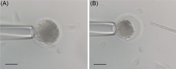
Oocyte micromanipulation was conducted with the aid of micromanipulators (Narishige, Tokyo, Japan) on an inverted microscope (Olympus, Tokyo, Japan). Handling medium was collection medium (Men et al., Reference Men, Kikuchi, Nakai, Fukuda, Tanihara, Noguchi, Kaneko, Linh, Nguyen, Nagai and Tajima2013) supplemented with 1 µg/ml cytochalasin D (CD). Injection solution was IVC-PyrLac supplemented with 20 mM HEPES and 4% (w/v) polyvinyl pyrolidone (Nakai et al., Reference Nakai, Kashiwazaski, Takizawa, Hayashi, Nakatsukasa, Fuchimoto, Shino and Kikuchi2003). Approximately 30 matured oocytes were transferred to a 20-µl droplet of handling medium. All droplets were covered with paraffin oil (Paraffin Liquid; Nacalai Tesque, Inc., Kyoto, Japan). The injection pipette was washed three times in injection solution, then used to make a hole in the zona pellucida using a piezo drill-assisted micromanipulator (PMAS-CT150; Prime Tech Ltd, Tsuchiura, Japan) and approximately one-third of the ooplasm was removed (Fig. 1). The PB was orientated at either 6 or 12 o’clock to avoid aspirating/damaging the meiotic spindle. The aspiration was gently conducted to reduce/avoid oolemma damage. The degenerated oocytes after micromanipulation were excluded. Before in vitro fertilization (IVF), the ooplasm-reduced oocytes were treated with 1% pronase that was dissolved in collection medium to remove the zona pellucida (from this point forwards referred to as the reduced cytoplasm (RC) group).
Removal of one-third of cytoplasm volume by micromanipulation in porcine oocytes. (A) A matured oocyte was held before micromanipulation. (B) The same oocyte after micromanipulation. Scale bar, 50 µm.

Oocyte enucleation, fusion and activation
After IVM, sucrose-treated oocytes with the first PB were enucleated in HEPES-buffered Medium 199 supplemented with 20% FCS and 5 µg/ml CD. Enucleated oocytes were subjected to fuse with non-enucleated oocytes. Before fusion, both enucleated oocytes and non-enucleated oocytes were freed from the zona pellucida by a short treatment with pronase 1%. The oocytes were washed three times in collection medium and then transferred into droplets of collection medium supplemented with 3 mg/ml phytohemagglutinin (PHA) in pairs for 10 min to facilitate the adhesion prior to fusion. The agglutinated oocytes were transferred into a fusion chamber (Super Electro Cell Fusion Generator, Nepagene, Chiba, Japan) and were automatically aligned by applying an alternating current (AC) field of 50 V/cm for 4 s, and fused using a single direct current (DC) pulse of 2 kV/cm for 20 µs. The fusion/activation buffer was 0.28 M d-mannitol supplemented with 0.05 mM CaCl2, 0.1 mM MgSO4 and 0.01% (w/v) bovine serum albumin (BSA). Only fused oocytes were washed and transferred to droplets of collection medium with CD for 30 min before being subjected to IVF (from this point forwards referred to as the doubled cytoplasm DC group). The normal IVM oocytes without ooplasm micromanipulation and without enucleation were also freed from the zona pellucida and used as the control group.
In vitro fertilization (IVF) and in vitro culture (IVC)
IVF was conducted according to Kikuchi et al. (Reference Kikuchi, Onishi, Kashiwazaki, Iwamoto, Noguchi, Kaneko, Akita and Nagai2002). Frozen epididymal sperm of a Meishan boar were thawed in pre-warmed TCM199 (with Earls salts, Gibco; pH adjusted to 7.8), centrifuged for 2 min at 750 g and the supernatant was removed. Pelleted sperm were re-suspended in the same medium and capacitated for 15 min at 37oC before co-incubation with oocytes. The medium for IVF was a modified Pig-FM supplemented with 2 mM sodium pyruvate, 2 mM caffeine, and 5 mg/ml BSA (Suzuki et al., Reference Suzuki, Asano, Eriksson, Niwa, Nagai and Rodriguez-Martinez2002). The final sperm concentration was adjusted to 1 × 10⁵/ml. Sperm and oocytes were co-incubated for 30 min at 5% CO2, 5% O2, and 90% N2. Then sperm-binding oocytes were gently transferred to fresh IVF droplets for another 2.5 h under the same conditions.
Parthenogenetic activation
The zona pellucida was removed from IVM oocytes by a short treatment with 1% pronase. Then they were fused in pairs in a chamber with the same conditions as described above. The fused oocytes were parthenogenetically activated by a single DC pulse of 1.5 kV/cm for 99 μs using the same mannitol buffer as for fusion. After stimulation, the fused oocytes were randomly divided into two groups and incubated in culture medium with or without CD.
Evaluation of pseudopronucleus (PPN) formation after electric stimulation and CD treatment
To confirm the number of pseudopronucleus(i) in parthenogenetically activated embryos, embryos from four groups were collected at 10 h after electrical stimulation, mounted onto slides, fixed in ethanol:acetic acid (3:1 w/v) for several days and then stained with aceto-orcein. The number of activated oocytes with different numbers of PPN (1PPN, 2PPNs, 3PPNs or 4PPNs) and nonactivated (no PPN) oocytes were recorded among experimental groups.
Analysis of compaction and cavitation of embryos in time-lapse system
After 6 d of IVC, onset of compaction and cavitation of embryos and numbers of blastocysts were recorded based on a time-lapse record. Each individual blastocyst was separately fixed and stained to examine total cell numbers (Somfai et al., Reference Somfai, Yoshioka, Tanihara, Kaneko, Noguchi, Kashiwazaki, Nagai and Kikuchi2014). Firstly, they were washed in phosphate-buffered saline without Ca2+ and Mg2+ (PBS(−)) and treated for 5 min with 99.5% ethanol supplemented with 50 μg/ml Hoechst 33342. Then they were washed in PBS(−) supplemented with 0.3% polyvinylpyrrolidone and individually mounted in glycerol on a glass slide covered with a coverslip. A digital image of each blastocyst was taken using an epi-fluorescence microscope (BX51, Olympus, Tokyo, Japan) under UV light and then total cell numbers were counted using ImageJ software (v.1.41; National Institutes of Health, Bethesda, MD, USA).
Experimental design
Experiment 1: In vitro development of IVF embryos after ooplasm manipulation
After IVM, high-quality oocytes with the first PB were divided into three groups: RC group, in which approximately one-third of ooplasm was removed from the oocyte (Fig. 1); doubled cytoplasm (DC group) in which one enucleated oocyte was fused with one non-enucleated oocyte; and the control group. The oocytes from the three groups were freed from the zona pellucida and subjected to IVF and IVC. Then two-cell embryos from three experimental groups were selected at 24 h to 27 h after IVF and transferred into 16 microwell culture dishes (Vitrolife, Tokyo, Japan) for tracking by time-lapse. After 6 d of IVC, the time-lapse recording was finalized and the following data were recorded: the onset of compaction and cavitation, the number of blastocysts; and total cell number per blastocyst were determined by Hoechst staining.
Experiment 2: In vitro development of parthenogenetically activated embryos
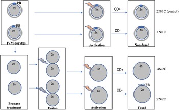
After IVM, high-quality oocytes with the first PB were treated with pronase to remove the zona pellucida. Then they were fused in pairs using PHA. The fused and non-fused oocytes were parthenogenetically activated by a single DC pulse of 1.5kV/cm for 99 µs. After electric stimulation, the oocytes were incubated in IVC medium with or without CD depending on groups. In this experiment, four experimental groups were designated: (1) non-fused oocytes with CD treatment (referred to as the diploid, 2N/1C – control group); (2) non-fused oocytes without CD treatment (haploid, 1N/1C group); (3) fused oocytes with CD treatment (tetraploid, 4N/2C group); and (4) fused oocytes without CD treatment (diploid, 2N/2C group) (Fig. 2). Two-cell embryos from the four experimental groups were selected at 24 h to 27 h after electric stimulation and transferred into 16 microwell culture dishes (Vitrolife, Tokyo, Japan) for tracking by time lapse. After 6 d of IVC, time-lapse system was finalized, and the onset of compaction and cavitation was analyzed. Blastocysts formation rate was determined, and blastocysts were staining separately to count total cell numbers.
Embryo production with different ploidies by parthenogenetic activation. Metaphase II (MII) oocytes have a 2n set of chromosomes. After electric stimulation and CD treatment to block the 2nd PB extrusion, oocytes still have a 2n set of chromosomes (2N/1C – control group); without CD, the second PB will extrude and oocytes have only a 1n set of chromosomes (1N/1C group). Oocytes were freed from zona pellucida and agglutinated in pairs and then automatically aligned by applying an alternating current (AC) field of 50 V/cm for 4 s, then fused by using a single DC pulse of 2 kV/cm for 20 µs. These fused oocytes have a 4n set of chromosomes, after electric activation and CD treatment, oocytes still have a 4n set of chromosomes (4N/2C group); without CD, the second PB will extrude and oocytes have a 2n set of chromosomes (2N/2C group). CD+/−: with/without cytochalasin D treatment; N/C: nuclear to cytoplasmic ratio. PB: polar body.

In a separate experiment, we also examined oocyte activation status after electrical stimulation by counting the number of formed PPN(s) in activated oocytes that may be useful to provide further information about the association between PPN(s) and ploidy. At 10 h after electric stimulation, embryos in each group were mounted on slides and fixed in ethanol:acetic acid (3:1 w/v) for several days and then stained with 1% (w/v) aceto-orcein solution and examined for nuclear status under a phase contrast microscope. The embryos with pseudopronuclear formation (from 1PPN to 4PPNs) were considered activated and the embryos without PPN were nonactivated embryos.
Statistical analysis
The data on pseudopronucleus formation, onset of compaction and cavitation, blastocyst formation rate and total cell number were expressed as mean ± standard error of the mean (SEM) and analyzed by one-way analysis of variance (ANOVA) following by Bonferroni correction using Stata/SE 15.0 software package (StataCorp., College Station, TX, USA). Data expressed as percentages were transformed by arcsine before being subjected to ANOVA test. Differences were considered statistically significant at P < 0.05.
Results
Experiment 1: In vitro development of IVF embryos after ooplasm manipulation
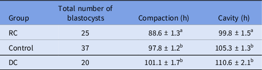
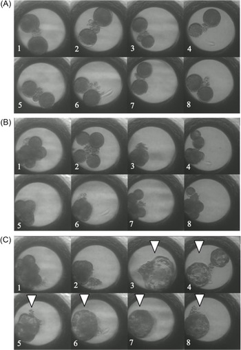
Compaction in the RC group started c. 88.6 ± 1.3 h after IVF, significantly earlier compared with the control group (97.8 ± 1.2 h) and DC group (101.1 ± 1.7 h), (P < 0.001). Similarly, the onset of cavitation in the RC (99.8 ± 1.5 h) group was significantly earlier than those of the control (105.3 ± 1.3 h; P < 0.05) and DC (110.6 ± 2.1 h; P < 0.001) groups (Table 1, Fig. 3).
Onset of compaction and cavitation in IVF embryos after ooplasm manipulation

DC: doubled cytoplasm; RC: reduced cytoplasm.
Data were expressed as mean ± standard error of the mean (SEM). Five replicates were carried out.
a,bValues with different superscripts within the same column indicate significant difference (P < 0.05).
Cell division from two-cell stage (A) to the blastocyst stage (C) in IVF embryos by time-lapse system. DC group (A1, A2, B1, B2, C1, C2), RC group (A3, B3, C3), Control group (A 4–A8, B4–B8, C4–C8). An embryo from the RC group (B3) showed compaction earlier than the others. Arrowheads showed blastocysts in (C).

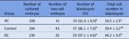
As shown in Table 2, the rates of blastocyst formation in RC and DC group were not significantly different (61.0 ± 6.9% and 57.1 ± 4.8%, respectively) but were significantly lower than that of control group (86.1 ± 7.0%; P < 0.05). In addition, total cell numbers of blastocysts obtaining from RC group was significantly (P < 0.05) lower than the DC group but not different from the control group.
Blastocyst formation rate and quality of IVF embryos after ooplasm manipulation

DC: doubled cytoplasm; RC: reduced cytoplasm.
Data were expressed as mean ± standard error of the mean (SEM). Five replicates were carried out.
a,bValues with different superscripts within the same column indicate significant difference (P < 0.05).
Experiment 2: In vitro development of parthenogenetically activated embryos
The embryos with PPN formation (from 1 to 4PPNs) at 10 h after electric stimulation were classified as activated and the embryos without PPN as nonactivated. As shown in Table 3, the embryos showed 4PPNs occupy 25.3% of activated embryos in the 4N/2C group, significantly higher than that of the remaining groups (P < 0.05). Similarly, the embryos showed that 3PPNs occupy 33.7% of activated embryos in the 4N/2C group, significantly higher than that of the remaining groups (P < 0.05). The rates of embryos showed that 2PPNs in the 2N/2C and 2N/1C groups were 67.7% and 59.2% of activated embryos, respectively, significantly higher than that of the 1N/1C group. The rate of 1 PPN embryos in the 1N/1C group (77.1% of activated embryos) was significantly higher compared with the 2N/2C and 4N/2C groups (P < 0.05).
Pseudopronucleus formation in porcine parthenogenetic embryos after electrical stimulation and CD treatment

N/C: nuclear to cytoplasmic ratio; 1PPN to 4PPN: one to four pseudopronucleus(i).
Data were expressed as mean ± standard error of the mean (SEM). Four replicates were carried out.
a,bValues with different superscripts within the same column indicate significant difference (P < 0.05).
* Percentage of 1PPN to 4PPN was based on the number of activated oocytes.
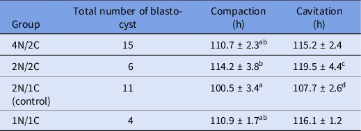
By tracking in vitro embryo development from the two-cell stage to the blastocyst stage using a time-lapse system, our data (Table 4) indicated that the start of compaction in the 2N/2C group was significantly delayed compared with that of the 2N/1C (control) group (P < 0.05). In addition, the start of blastocoel formation in this group also tended to be slower compared with the control group (P = 0.064). Conversely, there was no significant difference in the initiation of compaction and cavitation between the 4N/2C and 2N/1C (control) groups that had a N/C ratio considered equivalent.
Onset of compaction and cavitation of parthenogenetically activated embryos tracking by time-lapse cinematography

N/C: nuclear to cytoplasmic ratio.
Data were expressed as mean ± standard error of the mean (SEM). Five replications were carried out.
a,bValues with different superscripts within the same column indicate significant difference (P < 0.05).
c,dValues with different superscripts within the same column indicate tendency difference (P = 0.064).
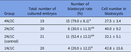
After 6 day of IVC, blastocyst formation rate in the 4N/2C group was significantly higher than that of the 1N/1C group (P < 0.05), but not significantly different from the remaining groups. However, there were no significant differences in total cell numbers of blastocysts in all groups (Table 5).
Parthenogenetic development of IVM porcine oocytes after electric activation and CD treatment observed by time-lapse cinematography

N/C: nuclear to cytoplasmic ratio.
Data were expressed as mean ± standard error of the mean (SEM). Five replications were carried out.
Embryos with a clear blastocoel during time-lapse recording were defined as blastocysts after 6 d of IVC. Blastocysts rate was calculated by division of number of the blastocyst to number of two-cell embryos used for time-lapse system.
a,bValues with different superscripts within the same column indicate significant difference (P < 0.05).
Discussion
The aim of the present study was to investigate the effects of modification of N/C ratio on the initiation of compaction and cavitation and on subsequent development in porcine embryos using two different approaches. Firstly, we conducted ooplasm manipulation including reducing cytoplasm and doubling cytoplasm before IVF to see how the modified N/C ratios affected the initiation of compaction and cavitation in preimplantation embryos (Experiment 1). Then, using oocyte fusion, activation and CD treatment, we generated and compared four experimental groups with different N/C ratios and ploidies (Experiment 2). In both experiments we compared the embryos with different N/C ratios in terms of onset of compaction and cavitation, blastocyst quality and quantity. In this study, only equally cleaved two-cell embryos at 24 h to 27 h after IVF or electric stimulation were selected for tracking by a time-lapse system. Embryos with unequal blastomeres were removed from the experiments as this type of cleavage pattern was reported to be correlated with aneuploidy and multinucleation of embryos (Hardarson et al., Reference Hardarson, Hanson, Sjögren and Lundin2001). In addition, the embryos with more than two cells were also not selected at this time point, as embryos that underwent a direct cleavage from 1 cell to 3–4 cells in the first cell division might be generated by fragmentation (for parthenogenetic activation) or polyspermic fertilization (for IVF) (Han et al., Reference Han, Wang, Abeydeera, Petersen, Kim, Murphy, Day and Prather1999) and are likely to be linked to abnormal chromosome numbers (Somfai et al., Reference Somfai, Inaba, Aikawa, Ohtake, Kobayashi, Konishi and Imai2010; Sugimura et al., Reference Sugimura, Akai and Imai2017).
In Experiment 1, we were aiming to remove roughly half of the cytoplasm content as Wakayama and Yanagimachi (Reference Wakayama and Yanagimachi1998) reported that a mouse oocyte can develop to full-term even after losing about half of its cytoplasm. However, our preliminary data suggested that removal of this amount of cytoplasm was technically challenging and often resulted in low developmental competence of the generated embryos, suggesting species difference (data not shown). We ended up taking out approximately one-third of the volume of cytoplasm. Removal of cytoplasm resulted in an increase in the N/C ratio, and we hypothesized that embryos derived from this RC group would have earlier initiation of compaction and cavitation. As expected, the initiation of compaction and cavitation was happening significantly earlier in RC group compared with control and DC group. However, although the initiation of compaction and cavitation in DC groups occurred later than in the control group, there were no significant differences. As reported in mice, nucleus volume scales with the size of the cytoplasm during the preimplantation development and following manipulation (Novakova et al., Reference Novakova, Kovacovicova, Dang Nguyen, Sodek, Skuultety and Anger2016; Tsichlaki and FitzHarris, Reference Tsichlaki and FitzHarris2016). This phenomenon might also occur in pigs and caused nucleus upscaling in DC group embryos, which in turn affected the onset of compaction and cavitation of such embryos. This hypothesis, however, should be confirmed by experimental data in a further study. In the previous study by Mita (Reference Mita1983) using starfish egg, cytoplasm was reduced to half by cutting eggs before fertilization. As a result, half eggs underwent nine synchronous divisions in contrast with control eggs with 10 synchronous cleavages. The author suggested that the N/C ratio may serve as a mechanism for the timing of initiation of asynchronous cleavages (or midblastula transition). Although the effect of modification of N/C ratio on cell cleavage has been reported in many organisms, the mechanisms by which the embryo senses the N/C ratio to regulate the cell cycle has remained unknown until now.
In present study, the effects of modified N/C ratios on blastocyst quantity and quality were also evaluated. We found that the percentage of blastocysts in groups in which the oocytes were subjected to manipulation (RC and DC groups) was significantly lower than that of the control group. Probably, damage triggered by cytoplasm removal or enucleation process may explain the lower developmental potential of embryos obtained from these groups. Our results were consistent with previous studies showing a decrease in the N/C ratio by enucleation (Kaufman and Sachs, Reference Kaufman and Sachs1976) or an increase of N/C ratio by microsurgical removal of some part of the zygote cytoplasm (Barton and Surani, Reference Barton and Surani1983) which was found to have a negative effect on embryo development. Total cell numbers in blastocyst of RC group were significantly lower compared with the DC group but not different from the control group. This means that blastocyst quality was not much affected when the porcine oocytes lost about one-third of its cytoplasm. Moreover, double-sized oocytes generated by fusion of pairs of non-enucleated and enucleated oocytes resulted in good quality blastocysts after IVF. In mice, Wakayama et al. (Reference Wakayama, Kishigami, Nguyen, Ohta, Hikichi, Mizutani, Bui, Miyake and Wakayama2008) succeeded in obtaining normal offspring from double-sized oocytes that were produced by fusion of pairs of non-enucleated and enucleated oocytes after ICSI.
In Experiment 2, embryos in the 2N/2C and 1N/1C groups were artificially manipulated so that their N/C ratio reduced to approximately half compared with the control groups (4N/2C and 2N/1C), and therefore are expected to show delays in initiation of compaction and cavity (Fig. 2). Indeed, the initiation of compaction in the 2N/2C group was significantly delayed compared with the 2N/1C (control) group (P < 0.05), and the initiation of cavitation also showed a delayed tendency compared with the control group (P = 0.064). In other words, these results confirmed that the decrease in the N/C ratio delayed the initiation of compaction and cavitation compared with the control group.
Markedly, the initiation of compaction and cavitation in the 4N/2C group was similar to that of the 2N/2C and 1N/1C groups rather than the 2N/1C (control) group, although the 4N/2C group had a N/C ratio comparable with the 2N/1C group. The exact reason is still unclear but there were only about 25% of activated embryos in the 4N/2C group that had 4 PPNs (Table 1), which were expected to develop to tetraploid embryos. Although occasionally two sets of chromosomes merge and form one diploid PPN (Somfai et al., Reference Somfai, Ozawa, Noguchi, Kaneko, Ohnuma, Karja, Fahrudin, Maedomari, Dinnyés, Nagai and Kikuchi2006), the number of PPNs generally reflects the ploidy of an embryo. Therefore, the N/C ratio in embryos in the 4N/2C group was probably not as high as in the 2N/1C group, as expected, but alternatively ranged somewhere between the 2N/1C and 2N/2C groups, and may explain the delayed initiation in compaction and cavitation in this group. We also confirmed that the majority of embryos from other groups showed the expected ploidy: about 68% and 60% of embryos in the 2N/2C and 2N/1C groups had 2PPNs, respectively, whereas over 77% of embryos in the 1N/1C group had 1PPN.
Interestingly, the blastocyst formation rate was the highest in the 4N/2C group in which embryos were produced by the fusion of two zona-free oocytes. Wu et al. (Reference Wu, Lai, Mao, McCauley, Caamaño, Cantley, Rieke, Murphy, Prather, Didion and Day2004) reported that zona-free porcine oocytes can be fertilized and developed not only to the blastocyst stage but also to piglets after embryo transfer to recipients, suggesting that the loss of the zona pellucida had no negative effect on subsequent development of porcine oocytes. Regarding the total cell numbers in blastocysts, it was reported that tetraploid embryos obtained from fertilized oocytes had fewer cells than diploid blastocyst, although they were morphologically normal blastocysts in mice (Snow, Reference Snow1975; Koizumi and Fukuta, Reference Koizumi and Fukuta1995) and in pigs (He et al., Reference He, Kong, Shi, Xie, Jiao, Huang, Guo, Hu and Liu2013). Similarly, total cell numbers of tetraploid blastocysts obtained from parthenogenetically activated oocytes were significantly lower than those of diploid blastocysts (Sembon et al., 2011). In contrast, our results showed that there were no significant differences in total cell numbers in blastocysts between the 4N/2C and 2N/1C groups. Various methods in generating tetraploid embryos might be responsible for the differences in results from these studies. Several studies have generated tetraploid embryos by fusion of two blastomeres of two-cell stage embryos in cattle (Iwasaki et al., Reference Iwasaki, Kono, Fukatsu and Nakahara1989), mice (Kubiak and Tarkowski Reference Kubiak and Tarkowski1985, Kaufman and Webb Reference Kaufman and Webb1990) and pigs (He et al., Reference He, Kong, Shi, Xie, Jiao, Huang, Guo, Hu and Liu2013) or using cytochalasin B or D to inhibit the PB extrusion in mice (Koizumi and Fukuta Reference Koizumi and Fukuta1995) and pigs (Sembon et al., 2011). Sembon et al., (2011) used a single oocyte to produce tetraploid parthenogenetic embryos by two treatments with CB: during IVM and after electric stimulation to inhibit the extrusion of a PB, whereas our study performed the fusion of two IVM oocytes combined with one CD treatment, expecting to obtain tetraploid parthenogenetic embryos. However, it also should be noted that only roughly 25% of embryos in the 4N/2C group in the present study were supposedly tetraploid (with 4PPN), the remaining were diploid and triploid (32.5% and 33.7%, respectively), explaining this discrepancy.
In this study, the time-lapse video system allowed us to observe the expansion, collapse or re-expansion of some blastocysts even several times during IVC. The collapse of blastocysts is caused by an efflux of blastocoel fluid because of loose binding in the trophectoderm. These were low-grade blastocysts that originated from poor-quality oocytes or sperm or suboptimal culture conditions (Marcos et al., Reference Marcos, Pérez-Albalá, Mifsud, Molla, Landeras and Meseguer2015). Indeed, in our study, the blastocysts that showed collapse and re-expansion several times had low cell numbers or degenerated at Day 6 of IVC (Fig. 3).
In conclusion, this is the first study that demonstrated that the increase in N/C ratio (in the RC group) accelerated compaction and blastocoel formation in embryos compared with the control group after IVF in pigs. In contrast, the decrease in the N/C ratio delayed the initiation of compaction and blastocoel formation in parthenogenetically activated porcine embryos.
Acknowledgements
We would like to thank Ms. Nagai for technical assistance.
Conflict of interest
None
Financial Support
This research received no specific grant from any funding agency, commercial or not-for-profit sectors.
Ethical Standards
Not applicable.