INTRODUCTION
Histomonas meleagridis is the causative agent of histomonosis (histomoniasis or blackhead disease or enterohepatitis) affecting gallinaceous birds, predominantly turkeys. Inflammation and ulceration of the caeca filled with sulphur-coloured exudates and necrotic liver lesions are considered to be pathognomonic (McDougald, Reference McDougald2005). Since the ban of all effective control measures (Anonymous, 1995, 2001, 2002), there are, at present, no therapeutic or prophylactic drugs available against H. meleagridis, resulting in an increasing number of fatal outbreaks in the poultry industry, particularly in turkey farms (McDougald, Reference McDougald2005; Callait-Cardinal et al. Reference Callait-Cardinal, Leroux, Venereau, Chauve, Le Pottier and Zenner2007). The current situation urges the need for basic knowledge on H. meleagridis. Although Tyzzer (Reference Tyzzer1920) renamed the protozoon parasite observed by Smith (Reference Smith1895) as H. meleagridis and established its causal nature of this disease, many conflicting reports on the cause of enterohepatitis had been reported (Delappe, Reference Delappe1957). Based on frequent observations of other organisms, besides H. meleagridis, presumptions of coccidian protozoa, trichomonad species e.g. Tetratrichomonas gallinarum (Delappe, Reference Delappe1957), and moulds e.g. Candida albicans (Kemp and Reid, Reference Kemp and Reid1966b) as the etiological agent of blackhead disease had been made (Delappe, Reference Delappe1957). Further the co-infection of different protozoa in caeca, mainly T. gallinarum and Blastocystis spp., interferes and impedes the propagation of cultured H. meleagridis isolated from field outbreaks. In the past, many authors highlighted the necessity of cultural investigations. Many reports dealt with in vitro isolation of H. meleagridis from embryonated eggs of the Heterakis gallinarum nematode (Ruff et al. Reference Ruff, McDougald and Hansen1970), from caecal contents (DeVolt, Reference DeVolt1943; Dwyer, Reference Dwyer1970; McDougald and Galloway, Reference McDougald and Galloway1973), from caecal discharges (Delappe, Reference Delappe1952, Reference Delappe1953; Delappe and Pierce, Reference Delappe and Pierce1953), from feces (Drbohlav, Reference Drbohlav1924; Bishop, Reference Bishop1938) and from liver lesions (Bayon and Bishop, Reference Bayon and Bishop1937; Delappe, Reference Delappe1957). However, few data on growth behaviour and life-time of the cultured parasites were reported. Therefore, it is not clear whether other host-derived pathogens were present in the in vitro isolates of H. meleagridis. Exclusion of interfering organisms such as Blastocystis spp. and T. gallinarum is essential for the maintenance of H. meleagridis cultures. So far, a clonal H. meleagridis culture from caecal content was established through micromanipulation (Hess et al. Reference Hess, Kolbe, Grabensteiner and Prosl2006b). This technique depends on precise microscopic recognition and selection of a single H. meleagridis parasite complicated by the morphological similarity to other host organisms such as T. gallinarum (Allen, Reference Allen1936; Harrison et al. Reference Harrison, Hansen, DeVolt, Holst and Tromba1954; Delappe, Reference Delappe1957) and Blastocystis spp. (Delappe, Reference Delappe1952; Harrison et al. Reference Harrison, Hansen, DeVolt, Holst and Tromba1954) commonly present in caecal content of birds. The development of the mono-eukaryotic H. meleagridis culture from a number of cells mimics the field isolate, as opposed to cloning, and offers a key tool for future in-depth studies of H. meleagridis. With this perspective, the objective of this study was to develop an efficient technique to isolate and set up H. meleagridis cultures free of other avian protozoa starting from mixed infections of H. meleagridis, T. gallinarum and Blastocystis spp. Following the successful in vitro propagation of the isolated H. meleagridis parasites, their sustained pathogenicity was verified in vivo in the turkey model.
MATERIALS AND METHODS
Histomonas meleagridis mixed strain: origin and storage of the stock
The virulent field strain H. meleagridis/Turkey/France/HNA.C2.L2/06 was isolated from the caeca and liver of diseased birds from a clinical outbreak at a French commercial breeder in June 2006. The H. meleagridis-infected liver and caeca were collected at autopsy and macerated in warm PBS. For long-term storage in liquid nitrogen 8% heat-inactivated horse serum (Gibco TM, Invitrogen) and 8% dimethylsulfoxide (Sigma Aldrich, Germany) were added to the suspension and frozen to −80 °C under controlled conditions (±1 °C/min). The presence of H. meleagridis and other host protozoa in the suspension was verified with an inverted light microscope at 200–400× magnification and confirmed by PCR amplication of the small subunit rRNA gene of H. meleagridis (Bleyen et al. Reference Bleyen, De Gussem, De Gussem and Goddeeris2007) of T. gallinarum and of Blastocystis spp. (Grabensteiner and Hess, Reference Grabensteiner and Hess2006). For further ease of reference this suspension will be referred to as ‘mixed strain’.
Comparison of the growth kinetics of H. meleagridis with T. gallinarum and Blastocystis spp. in cultures of the mixed strain
The mixed strain was resuscitated in culture. Hereby, the cryostabilate stored in the liquid nitrogen was defrosted as fast as possible in 37 °C warm water. The mixed strain was carefully transferred into a culture flask containing culture medium. This culture medium consisted of 90% M199 medium supplemented with Hanks salts, L-glutamine, 25 mm HEPES and L-amino acids (Gibco TM, Invitrogen), 10% heat-inactivated horse serum (Gibco TM, Invitrogen) and 12 mg of rice starch (Gibco TM, Invitrogen). The medium was inoculated with one loop of the turkey's caecal bacterial culture grown on a Columbia agar plate with 5% sheep blood (Biotrading, Belgium). The culture was incubated at 40 °C under anaerobic conditions sealing the culture flasks tightly. Parasite growth was examined with an inverted light microscope at 200–400× magnification. For the relative kinetic growth studies of different cultured protozoa, the cryostabilate of the mixed strain and culture samples from different incubation times (3, 4, 5, 6, 12 and 24 h) were processed for transmission electron microscopic (TEM) analysis. The cell suspensions were centrifuged and the cell pellets were treated for TEM analysis of ultra-fine sections as described by Mast et al. (Reference Mast, Nanbru, van den Berg and Meulemans2005). Briefly, cells were fixated in a cacodylate buffer containing 2·5% glutaraldehyde and 2% paraformaldehyde as active compounds and subsequently in 1% (wt/vol) osmium tetroxide. Thereafter the samples were fixated with 2% (wt/vol) uranylacetate in distilled water and dehydrated in ethanol. The cells were embedded in Epon-Spurr (1 : 1) medium. Ultrafine sections were cut with a Leica Ultracut ultramicrotome (Leica) and were stained with Reynolds lead citrate and uranyl acetate. Twenty to 100 sections of culture sample of each incubation time were analysed with a Technai Spirit transmission electron microscope, thereby taking digital pictures with a Eagle 4*4 camera (FEI).
Birds/poults
Commercial male B.U.T. 9 turkeys (British United Turkeys; Claeys, Kruishoutem, Belgium) and broiler chickens (Ross; Belgabroed NV, Merksplas, Belgium) were housed in a disinfected stable. At arrival, the one-day-old birds were kept together in a floor pen. Water and feed were supplied ad libitum. The experiments were approved by the Ethical Committee for Animal Experiments of the KU Leuven with licence number P029/2011 according to international regulations.
The mono-eukaryotic H. meleagridis culture
To establish a H. meleagridis culture free of other host intestinal protozoa, 5-week-old B.U.T. 9 turkeys and 3-week-old chickens were cloacally inoculated with a cryostabilate of H. meleagridis/Turkey/France/HNA.C2.L2/06. Ten days after inoculation the birds were euthanized by cranial dislocation. When the liver showed very small off-white foci, these starting-necrotic liver lesions were collected while still warm. They were chopped into small pieces and put into 9 mL culture medium. The culture medium was prepared and inoculated with caecal bacteria one day before and incubated at 40 °C under anaerobic conditions. Cultures were microscopically examined daily, and were maintained according to Hauck et al. (Reference Hauck, Armstrong and McDougald2010a). Every second or third day, 1 mL of the culture was transferred into a new culture flask containing 9 mL culture medium. The parasites were identified as H. meleagridis by the diagnostic Hime-PCR test for the detection of the H. meleagridis 18S rRNA gene (Bleyen et al. Reference Bleyen, De Gussem, De Gussem and Goddeeris2007). Furthermore, to exclude the presence of other protozoa, PCR-amplification of the small subunit rRNA gene of T. gallinarum and of Blastocystis spp. was performed (Grabensteiner and Hess, Reference Grabensteiner and Hess2006). TEM analysis was performed to ascertain the identity of the cultured protozoa, as histomonads show morphological similarity to other intestinal protozoa under light microscopic conditions (Kemp and Reid, Reference Kemp and Reid1966a).
Confirmation of the sustained pathogenicity of the mono-eukaryotic H. meleagridis culture in turkeys
Forty birds were randomly allocated to the infected group (IG) and the uninfected control group (UC) at a 1 : 1 ratio. At the age of 17 days, the birds of the IG were intracloacally inoculated with 1·7 × 105H. meleagridis per animal (culture passage 8). The amount of cultured H. meleagridis was determined using a Neubauer counting chamber and inverted light microscopy at 200–400× magnification. Subsequently, the suspensions were diluted in culture medium to obtain the required concentration for inoculation. During the study all birds were examined daily and mortality was recorded. At 14 days post infection (dpi), all surviving birds were euthanized by cranial dislocation for investigation of the macroscopic histomonosis caecal and liver lesions applying the adapted 0–4 lesion score system of McDougald and Hu (2001). Normal caeca with dark-coloured content were scored as 0, whereas caecal lesions were scored as follows: 1=caecal content is yellow, slimy and foamy to fluid and little or no thickening of the caecal wall with some small petechiae or few scattered, small lesions on the mucosa; 2=thickening of the caecal wall with some bleeding or inflammation of mucosa, caecal content is yellow, foamy to fluid with some fibrinous exudates; 3=caeca enlarged, thickening of caecal wall, bleeding and/or necrotic confluent lesions on mucosa, beginning of ulcers visible, caeca empty or yellow, foamy to fluid content with blood and/or caeseous core; 4=death from histomonosis, severe thickening of caecal wall, distended caeca with severe necrotic lesions or ulcers on the mucosa, caeca empty or filled with yellow caseous material. Macroscopic liver lesions were scored as follows: 0=normal; 1=a few very small foci; 2=lesions covering up to 50% of liver surface, lesions are off-white and variable in appearance; 3=more than 50% of the liver surface covered with necrotic lesions; 4=death from histomonosis, at least 70% of the liver show coalescing huge necrotic lesions with craters or huge amount of smaller necrotic spots.
RESULTS
Comparison of the growth kinetics of H. meleagridis with T. gallinarum and Blastocystis spp. in cultures of the mixed strain
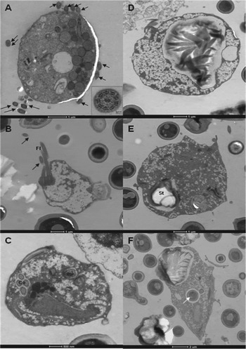
The culture of the mixed strain was not pure. Besides H. meleagridis, presence of at least two other protozoa was detected by microscopic examination and PCR analysis. Based on their characteristic morphology, T. gallinarum and Blastocystis spp. were identified. The kinetic TEM study showed the relative amounts of H. meleagridis, T. gallinarum and Blastocystis spp. (Fig. 1). In the cryostabilate of the mixed strain as well as in the 3-h incubated culture predominantly T. gallinarum were observed (Fig. 1A). After the incubation time of 4 and 5 h equal amounts of Blastocystis spp. (Fig. 1D) and T. gallinarum (Fig. 1B) were present in the culture samples. After 6 h of incubation T. gallinarum were still observed (Fig. 1C), however most of the cultured protozoa were identified as Blastocystis spp. (Fig. 1E) and after 12 and 24 h the culture consisted of only Blastocystis spp. (Fig. 1F), no other protozoa were observed. Throughout the study, the relative amount of H. meleagridis remained relatively low: only one H. meleagridis could be demonstrated on all examined sections of the TEM analysis. Obviously, the culture of the mixed strain contained several bacterial species originating from the host's caeca. The bacterial population shifted from Gram-negative to Gram-positive bacteria and, interestingly, these Gram-positive bacteria could be consumed by the only remaining Blastocystis spp. (Fig. 1F).
TEM analysis at several incubation time points to study the relative amount of Histomonas meleagridis to Tetratrichomonas gallinarum and Blastocystis spp. in a culture of caecal content originated from turkeys suffering from histomonosis. T. gallinarum were identified based on the presence of 4 flagella (black arrow) outside the cell or 4 costas (Co) in the cell at incubation time of 0 (A), 4 (B), and 6 (C) hours. Blastocystis spp. were identified at incubation time of 4 (D), 6 (E), 24 (F) hours. The black arrows show transversal sections of the flagellum (Fl). On the insert of the picture (A) the cross-section of the axenoma with the characteristic 9*2 plus 2 (9 fused pairs of microtubule doublets surrounding 2 central ones) organization of microtubules is shown. Gram-positive bacteria with the characteristic thick peptidoglycan layer were observed in the environment of the protozoal cells but also inside Blastocystis spp. (white arrow in F).

The mono-eukaryotic H. meleagridis culture
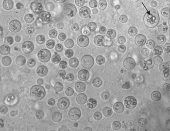
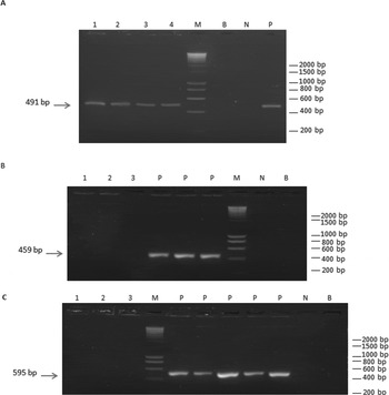
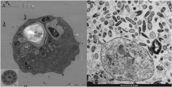
At post-mortem examination of the euthanized birds (10 dpi), liver and caeca of the infected turkeys and chickens displayed typical histomonosis lesions. The caecal lesions were scored as 2 to 3. Score 2 was assigned to the turkey liver lesions and score 1 for the chicken liver lesions. Overall, the liver lesions were still small and were off-white coloured. Twenty-four hours after inoculation of the cultures with turkey liver material no H. meleagridis could be detected. However, after 48 h H. meleagridis protozoa were numerous so 10 mL fresh culture medium was added. In order to maintain the parasite's proliferation in vitro for more than 1 year, every 2 or 3 days at growth peak (±106H. meleagridis mL−1) the culture needs to be passaged (1/10 dilution). No other protozoa could be detected by light microscopic examination (Fig. 2) and specific PCR analysis as shown in Fig. 3. Irrespective of the passage number of the culture originating from liver lesions of H. meleagridis infected turkeys, only the protozoon H. meleagridis was identified in the culture (Fig. 3A). No other protozoon such as Blastocystis spp. (Fig. 3B) and T. gallinarum (Fig. 3C) were detected in the culture, whereas the PCR controls with genomic DNA from protozoa frequently occurring in caeca of poultry were positive. Through electron microscopic examination the cultured parasites were unequivocally identified as H. meleagridis (Fig. 4A). Remarkably, a high number of Gram-negative bacteria surrounding H. meleagridis was observed (Fig. 4B). Similar observations resulted from examination of the mono-eukaryotic H. meleagridis culture originating from liver lesions of chickens after infection with the H. meleagridis mixed strain (data not shown).
Light microscopic examination (400×) of the cultured protozoa confirmed the presence of Histomonas meleagridis and the absence of any other protozoa in the mono-eukaryotic culture originating from turkey liver lesions. The parasites showed phagocytosis of rice starch granules (black arrow).

Diagnostic PCR analysis detected Histomonas meleagridis but no other protozoa were detected in the mono-eukaryotic culture originating from turkey liver lesions. To amplify the small subunit rRNA gene of each protozoon species, specific primers for Histomonas meleagridis (A), Blastocystis spp. (B) and Tetratrichomonas gallinarum (C) were used in the mono-eukaryotic culture at passage 6 (lane 1), passage 34 (lane 2), passage 49 (lane 3) and passage 52 (lane 4). M: molecular size marker (Smartladder SF, Eurogentec); B: blanco PCR control without DNA template; N: negative PCR control; P: positive PCR controls, respectively for Histomonas meleagridis (A), Blastocystis spp. (B) and Tetratrichomonas gallinarum (C).

TEM examination identified unequivocally the cultured protozoon to be the flagellated Histomonas meleagridis. Large part of the cytoplasma consists of organelles surrounded by membranes as hydrogenosomes (H), food vesicles containing bacterial debris (FV) or phagocytosed bacteria (B) or rice starch granules (ST). Black arrows show transversal sections of the flagellum with the typical microtubule organization presented in the inserted picture (A). Very high number of Gram-negative bacteria and rice starch granules (ST) were observed in the mono-eukaryotic H. meleagridis culture (passage 3) (B).

Confirmation of the sustained pathogenicity of the mono-eukaryotic H. meleagridis culture in turkeys
Experimental infection of turkeys with the isolated H. meleagridis culture resulted in the cumulative mortality rate and mean lesion scores of liver and caeca of birds presented in Table 1. One bird of the IG group was excluded from the data as it was decapitated by other birds. Since 18 of 19 birds of the group IG deceased due to histomonosis, from 7 dpi to 14 dpi, score 4 was assigned to the caecal and liver lesions. The caecal wall was thickened and showed necrotic to caseous exudates. Strong necrotic lesions with ulceration were noticed on the mucosa of the caeca and on the liver. The one remaining bird was killed at termination of the study and its caecal and liver lesion scores were 1 and 0 respectively. The caecal lesion score was 1 based on the observation of the aspecific yellow and foamy content. The mean caecal and liver lesion scores for the group IG resulted in 3·84 and 3·79, respectively. Meanwhile, for the group UC lesion scores of 0·2 and 0·0 were assigned to the caeca and the liver, respectively. Seventeen non-infected birds had normal caecal content whereas in 3 birds of the UC group the content was yellow and foamy. During the experiment none of the non-infected birds died due to histomonosis.
Confirmation of the sustained pathogenicity of the cultured Histomonas meleagridis originated from turkey liver lesions in turkeys intracloacally infected with the mono-eukaryotic H. meleagridis culture passage 8

a UC: uninfected group; IG: infected group.
b Total number of birds in each group.
c Except for UC group, all birds of IG were intracloacally infected at 17 days of age.
d The mean lesion scores of the caeca and the liver of each group, assigned at day of autopsy (14 dpi).
Birds that died from histomonosis during the study were given a lesion score of 4 per organ.
DISCUSSION
Since the first in vitro isolation of H. meleagridis by Drbohlav from feces (Drbohlav, Reference Drbohlav1924), a wide range of culture media and conditions have been studied to optimize growth of H. meleagridis (DeVolt, Reference DeVolt1943; Dwyer, Reference Dwyer1970; van der Heijden et al. Reference van der Heijden, McDougald and Landman2005; Hauck et al. Reference Hauck, Armstrong and McDougald2010a). In the current investigation and in accordance with Tyzzer's observations (Tyzzer, Reference Tyzzer1920), besides H. meleagridis other protozoa such as T. gallinarum and Blastocystis spp. were identified in cultures inoculated with caecal contents of birds from field outbreaks of histomonosis. Moreover, the frequent contamination of field isolates with other protozoa such as T. gallinarum and Blastocystis spp. hampers the propagation and maintenance of cultured H. meleagridis. The deleterious effect of T. gallinarum (Delappe, Reference Delappe1957) and Blastocystis spp. on H. meleagridis has been mentioned without specific details (Delappe, Reference Delappe1952; Tyzzer, Reference Tyzzer1934). Taking into account these perspectives, an efficient approach to isolate and propagate H. meleagridis free of other host's protozoa was described in the present study. Thereby, the contamination of H. meleagridis with T. gallinarum and Blastocystis spp. was analysed by TEM at different culture incubation times. The parasite population shifted from mainly T. gallinarum to only Blastocystis spp. Concomitantly, the main bacterial populations also shifted from Gram-negative to Gram-positive bacteria. Possibly, the inhibitory power of T. gallinarum and Blastocystis spp. in contaminated cultures could be attributed to their rapid growth driven by their selective bacterial preference as a food source, in agreement with Delappe's findings (Delappe, Reference Delappe1952). Conversely, in our pure H. meleagridis culture pre-inoculated with caecal bacteria, predominantly Gram-negative bacteria were present. Unfortunately, the underlying explanation for the shift from Gram-negative to Gram-positive bacteria when cultures were overgrown by Blastocystis spp. needs further investigation.
Furthermore, the successful establishment of mono-eukaryotic H. meleagridis cultures from liver was possible by pre-incubation of the culture medium with caecal bacteria one day before, highlighting the indispensable addition of bacteria. Previously, many attempts to culture H. meleagridis in an axenical manner failed (Bishop, Reference Bishop1938; Delappe, Reference Delappe1952; Lesser, Reference Lesser1961; Hauck et al. Reference Hauck, Armstrong and McDougald2010a). Although underlying reasons remain unsolved, it is generally assumed that the co-cultured caecal bacteria are able to produce an anaerobic environment (Delappe and Pierce, Reference Delappe and Pierce1953) suitable for H. meleagridis, a facultative anaerobic flagellate (McDougald, Reference McDougald2005), and serve as food for the cultured protozoa as observed in vacuoles (Munsch et al. Reference Munsch, Lotfi, Hafez, Al-Quraishy and Mehlhorn2009). Possibly, the inclusion of host's caecal bacteria promotes the parasite to switch from its amoebic tissue form to the flagellated stage increasing the success rate of establishing mono-eukaryotic H. meleagridis cultures from bird liver lesions. As previously described by Lee et al. (Reference Lee, Long, Millard and Bradley1969), the flagellated form in the caecal lumen consumes bacteria by phagocytosis while the non-flagellated tissue form ingests dissolved host liver by pinocytosis. In our study, flagellated H. meleagridis could be detected microscopically 48 h after inoculation of the cultures. However, flagellated parasites were already observed 24 h after establishment of H. meleagridis cultures from liver lesions of hen or turkey (Bayon and Bishop, Reference Bayon and Bishop1937; Goedbloed and Bool, Reference Goedbloed and Bool1962). Unfortunately, specific identification of cultured organisms, growth behaviour and effective propagation of the cultures in those previous studies were not documented in detail, impeding interpretation of their findings. The only in-depth defined H. meleagridis culture was established through micromanipulation by picking up one single cell (Hess et al. Reference Hess, Kolbe, Grabensteiner and Prosl2006b) rather than from a population of cells as performed in our study. The propagation of the latter cultures allows maintaining the characteristic nature of a field isolate which enables their use for in vitro screening of candidate antihistomonal compounds. Long-term cultivation of H. meleagridis however, might narrow the genetic variation between cells.
In the present study, the ultra-structural characteristics of the parasites from our pure cultures matched the descriptions and observations of H. meleagridis in the literature (Schuster, Reference Schuster1968; Rybicka et al. Reference Rybicka, Honigberg and Holt1972; Mielewczik et al. Reference Mielewczik, Mehlhorn, Al-Quraishy, Grabensteiner and Hess2008). In our study, only H. meleagridis could be isolated from the liver of birds which had been inoculated with a contaminated stabilate, whereas in other studies liver of H. meleagridis-infected birds appeared to be contaminated with other organisms including bacteria and protozoa (Harrison et al. Reference Harrison, Hansen, DeVolt, Holst and Tromba1954; Delappe, Reference Delappe1957; Goedbloed and Bool, Reference Goedbloed and Bool1962; Grabensteiner and Hess, Reference Grabensteiner and Hess2006; Hauck et al. Reference Hauck, Balczulat and Hafez2010b).
Investigations using PCR amplification of the SSrRNA-gene (Grabensteiner and Hess, Reference Grabensteiner and Hess2006; Hauck et al. Reference Hauck, Balczulat and Hafez2010b) ascertained the microscopic identification of Blastocystis spp. (Delappe, Reference Delappe1952) and T. gallinarum (Delappe, Reference Delappe1957) in the necrotic liver lesions of birds, besides H. meleagridis. Although they are regarded as apathogenic possibly due to subtype differences, contradictions remain about the pathogenic potential of Blastocystis spp. (Stensvold et al. Reference Stensvold, Lewis, Hammerum, Porsbo, Nielsen, Olsen, Arendrup, Nielsen and Mølbak2009) and T. gallinarum (Amin et al. Reference Amin, Neubauer, Liebhart, Grabensteiner and Hess2010). In addition, the amount of detected bacteria in liver samples of turkeys killed during the course of the disease was much lower than in birds which died from histomonosis (Harrison et al. Reference Harrison, Hansen, DeVolt, Holst and Tromba1954). In this regard, H. meleagridis causes extensive damage of the caecal tissue allowing dissemination of other host's organisms as postulated by Delappe (Reference Delappe1957). So, it is worth mentioning to precisely determine the moment of isolating liver lesions for obtaining the mono-eukaryotic H. meleagridis culture as H. meleagridis might play the first invader role causing liver lesions in birds subjected to a mixed infection. Clearly, development of H. meleagridis cultures free of growth-impeding protozoa such as T. gallinarum and Blastocystis spp. is an important tool to characterize this protozoon parasite.
Next to the successfully acquired mono-eukaryotic H. meleagridis culture, an infection study in turkeys was performed to confirm the sustained pathogenicity of this H. meleagridis culture. In accordance with reports about mortality and development of histomonosis regardless of the several variable factors (Hess et al. Reference Hess, Grabensteiner and Liebhart2006a), the mortality due to histomonosis reached 95% by 14 dpi. At autopsy severe inflammation with necrosis in the caeca and liver displaying the maximum lesion score 4 were seen, while in the IC control group no clinical signs or death due to histomonosis were observed. So, the mono-eukaryotic H. meleagridis cultures were still considered as very pathogenic.
In conclusion, it was possible to establish mono-eukaryotic H. meleagridis cultures in an efficient and successful way from liver lesions of birds infected with a contaminated H. meleagridis strain. Hereby, the co-cultivation of caecal bacterial flora is essential for the in vitro multiplication of H. meleagridis. As the pathogenicity of the parasite was not altered by propagation in vitro, this method of producing pure H. meleagridis cultures provides an important tool to gain more in-depth information about the protozoon H. meleagridis itself. Moreover, the mono-eukaryotic culture could reveal questions about the interactions between H. meleagridis and its bacterial environment in the culture or in the host.
ACKNOWLEDGEMENTS
We would like to thank Marcel Samain, Nele Steen and Sharon Beernaert for their technical assistance. Special thanks to Ward Heggermont MD for his careful reading of the manuscript.
FINANCIAL SUPPORT
This work was financially supported by the ‘Agency for Innovation by Science and Technology’ of Belgium (IWT grant number SB-71506).