Introduction
Larval stages of ascarid nematodes elicit severe tissue damage when they invade hosts which are not normally the definitive host. The racoon roundworm, Baylisascaris procyonis, is particularly prone to cause a fatal neurological disturbance (Huff et al., Reference Huff, Neafie, Binder, De Leon, Brown and Kazacos1984; Kuchle et al., Reference Kuchle, Knorr, Medenblik-Frysch, Weber, Bauer and Naumann1993; Moertel et al., Reference Moertel, Kazacos, Butterfield, Kita, Watterson and Gleich2001; Wise et al., Reference Wise, Sorvillo, Shafir, Ash and Berlin2005). The dog roundworm, Toxocara canis, and the cat roundworm, T. cati, are also responsible for the visceral larva migrans syndrome (VLM) in humans (Glickman & Magnaval, Reference Glickman and Magnaval1993; Fisher, Reference Fisher2003). The VLM caused by these two roundworms is commonly known as toxocariasis, which is considered a disease of infants and children, although adults are also infected (Glickman et al., Reference Glickman, Magnaval, Domanski, Shofer, Lauria, Gottstein and Brochier1987; Aragane et al., Reference Aragane, Akao, Matsuyama, Sugita, Natsuaki and Kitada1999; Yoshida et al., Reference Yoshida, Shirao, Asai, Nagase, Nakamura, Okazawa, Kondo, Takayanagi, Fujita and Akao1999). Moreover, outbreaks of VLM due to the pig roundworm, Ascaris suum, have been reported from the southern part of Japan (Maruyama et al., Reference Maruyama, Nawa, Noda, Mimori and Choi1996; Sakakibara et al., Reference Sakakibara, Baba, Niwa, Yagi, Wakayama, Yoshida, Kobayashi, Yokoi, Hara, Itoh and Kimura2002). Meningitis can occur when Ascaris larvae migrate into the central nervous system (Osoegawa et al., Reference Osoegawa, Matsumoto, Ochi, Yamasaki, Horiuchi, Kira, Ishiwata, Nakamura-Uchiyama and Nawa2001). However, a precise diagnosis of these animal-borne ascarid infections is not always possible and an adequate anthelminthic therapy against these infections was not available. The lack of a comprehensive study using animal models for ascarid infections has also prevented progress in this field.
The Mongolian gerbil Meriones unguiculatus is known to be susceptible to a variety of parasites including Brugia pahangi (Ash & Riley, Reference Ash and Riley1970), Strongyloides stercoralis (Nolan et al., Reference Nolan, Megyeri, Bhopale and Schad1993), Nippostrongylus brasiliensis (Horii et al., Reference Horii, Khan and Nawa1993), and Entamoeba histolytica (Chadee & Meerovitch, Reference Chadee and Meerovitch1984). Akao et al. (Reference Akao, Takayanagi, Suzuki, Tsukidate and Fujita2000) and Takayanagi et al. (Reference Takayanagi, Akao, Suzuki, Tomoda, Tsukidate and Fujita1999) demonstrated that gerbils could serve as an animal model for ocular toxocariasis due to both T. canis and T. cati. The occurrence of retinal haemorrhages including larval invasion into the retina was found to be quite high compared with that in mice after oral inoculation of infective eggs. However, no information is available on the migration route, the final site of infection, or on the pathogenesis of ascarid larvae in gerbils. Here, we present our findings on the characteristic features of ascarid infections in gerbils as they relate to the pathogenesis of VLM in humans.
Materials and methods
Mongolian gerbils Meriones unguiculatus ranging between 2 and 3 months of age, were raised in the Animal Centre of Nippon Medical University and were maintained under pathogen-free conditions. Only male gerbils with black hair were used and all experiments were carried in accordance with the guidelines of the Institutional Animal Care and Use Committee of Tokyo Medical and Dental University.
Recovery of ascarid eggs
Eggs of T. canis were obtained from the uteri of adult worms collected from faeces following the administration of anthelmintics to naturally infected puppies. Worms of B. procyonis were recovered from the intestine of infected racoons (Sato et al., Reference Sato, Furuoka and Kamiya2002), and those of B. transfuga collected from the faeces of infected bears. Worms of B. transfuga were naturally expelled from bears during their fasting period just prior to hibernation. Adult worms of A. suum were obtained from slaughtered pigs and A. lumbricoides were collected after treatment with anthelmintics from naturally infected humans. Female worms of each species were isolated, and matured embryonated eggs were prepared following the method of Oshima (Reference Oshima1961).
Fully embryonated eggs were treated with 50% hypochlorous acid for 10 min to remove their proteinous membranes. After repetitive washing with distilled water, 1000 eggs of each ascarid were inoculated into each gerbil through a gastric tube under light anaesthesia.
Recovery of larvae
After ophthalmological observations (Takayanagi et al., Reference Takayanagi, Akao, Suzuki, Tomoda, Tsukidate and Fujita1999), four gerbils from each group were sacrificed using sodium pentobarbital at predetermined intervals. One gerbil from each group was examined histopathologically and the remainder were used for larval recovery from the gastrointestinal tract, liver, lungs with heart, and skeletal muscle including bone and genital organs except for the skin. The contents of the gastrointestinal tract were collected in a conical tube at 6 and 12 h after inoculation. Each organ was minced and digested with artificial gastric juice (0.5% 1:10,000 pepsin, 0.7% hydrochloric acid) for 2 h at 37°C along with vigorous agitation. After digestion, the fluids were sieved with a tea strainer, and centrifuged at 320 × g for 5 min. The supernatant was discarded and a small amount of distilled water was added to the tube. The sediment was then spread out on glass, and larvae in the fluid were counted using a stereoscopic microscope. Examination of the brain tissue was performed as follows: each brain including the olfactory bulb, cerebrum, cerebellum, and pons was enucleated and minced into small pieces (approximately 2 mm3) on a slide glass using forceps, and these were then covered with another slide glass. Migrating larvae were then counted using a microscope with eight or nine slide glasses being examined in each sample. Recovery rates were calculated from a mean of three gerbils at each period.
Histopathology
Tissue samples of liver, lungs, femoral muscle, brain, and gastrointestinal tract including the stomach, duodenum, ileum, caecum and rectum were fixed in 10% neutral formalin solution. Serial sections were prepared and stained with haematoxylin and eosin or periodic acid Schiff haematoxylin.
Results
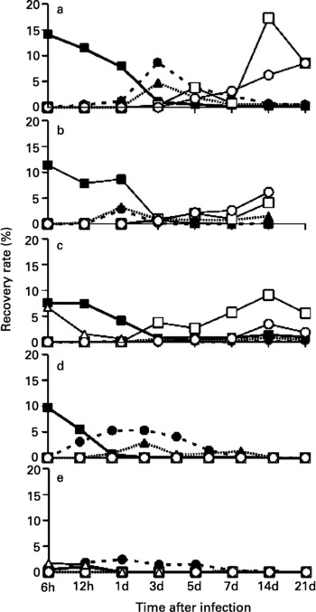
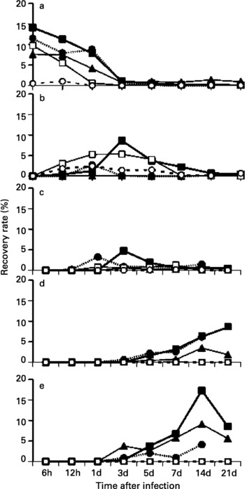
Changes in the recovery rates of larvae from various organs after oral inoculation of the five ascarid species were recorded (fig. 1) and recovery rates arranged in the order of the migration route shown in fig. 2. In Toxocara canis-infected gerbils, almost all larvae were recovered from the intestinal wall up to 24 h after infection. Thereafter, larvae began to appear in the liver and lungs (by day 3), and then in the skeletal muscle and brain. The number of larvae in the muscle and brain were approximately equal. Macroscopically, haemorrhagic lesions which were observed in the lung 3, 5, 7 and 14 days after infection gradually disappeared. Ophthalmoscopically, a motile larva was observed in the retina 14 days after infection.
Recovery rates (%) of larvae from various organs in gerbils after oral inoculation with five ascarid species up to day 21 post-infection. a, Toxocara canis; b, Baylisascaris procyonis; c, B. transfuga; d, Ascaris suum; e, A. lumbricoides; ■, gastrointestinal tract; ●, liver; ▲, lung and heart; □, muscles; ○, brain; △, intestinal contents.

Recovery rates (%) of larvae in gerbils after oral inoculation with five ascarid species to show the route of migration. ■, Toxocara canis; ●, Baylisascaris procyonis; ▲, B. transfuga; □, Ascaris suum; ○, A. lumbricoides; a, gastrointestinal tract; b, liver; c, lung and heart; d, brain; e, muscles.

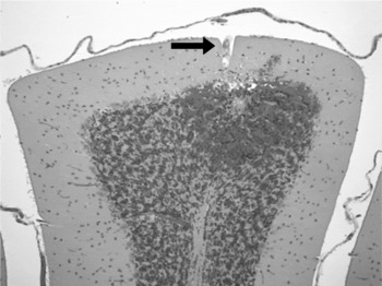
In B. procyonis- and B. transfuga-infected gerbils, the recovery rates of larvae were significantly lower than that in the case of T. canis. Both species of larvae had migrated into the lungs through the liver within 24 h after infection, and some had already arrived in the brain by this time, resulting in small haemorrhagic foci in the brain (fig. 3). With B. procyonis, ocular invasion by the larvae was observed and neurological disorders such as rotational and involuntary movement or paraplegia emerged, resulting in the mortality of gerbils between days 15 and 20 post-infection. Macroscopically, the gerbils exhibited fresh petechial haemorrhages of the lung 1 day after infection, but no new haemorrhagic lesions were evident beyond that time. Ophthalmic examination demonstrated motile larvae in the retina and severe chorioretinitis 7 days after infection. The number of migrating larvae of B. procyonis in the brain was higher than that of B. transfuga-infected gerbils at all times after the infection. By day 3 post-infection an average of 6.3 larvae (range 6–7) in B. procyonis-infected gerbils and 1.7 larvae (range 1–2) in B. transfuga-infected gerbils were found. By day 7, an average of 26.0 larvae (range 23–31) in B. procyonis-infected gerbils and 6.7 larvae (range 5–8) in B. transfuga-infected gerbils were recovered. Ophthalmic and neurological abnormalities were less severe than those observed with B. procyonis, although gait difficulty and circulatory movements in the same direction were presented by day 10 post-infection. The number of B. transfuga larvae in the skeletal muscle was higher than that in the brain; i.e. an average number of larvae recovered were 37.7 (range 21–67) in muscle and 1.7 (range 1–2) in brain at the day 3 post-infection, and 91.0 (range 95–100) in muscle and 34.7 (range 24–49) in brain at the day 14 post-infection.
A fresh haemorrhagic lesion in the granular layer of the cerebellum in gerbils 24 h after infection with Baylisascaris procyonis; larva (arrowed) in the molecular layer of cerebellum immediately beneath the pia mater.

With A. lumbricoides, the average recovery rate of larvae after 6 h of infection was 2.3% (range 1.7–2.6%) compared with about 10% or higher in other ascarid parasites (14.1% in T. canis, 11.4% in B. procyonis, 16.3% in B. transfuga and 9.7% in A. suum; fig. 1a). Migrating larvae were present in the liver 12 h after infection, and remained there for up to 5 days after infection in the case of A. lumbricoides and 7 days in the case of A. suum. On day 7, haemorrhagic lesions in the lungs were the most prominent feature in gerbils infected with both species, although these lesions gradually disappeared and no larvae were recovered thereafter from any organ.
As far as the migration route is concerned, the highest recovery rate in the gastrointestinal wall occurred in T. canis-infected gerbils followed by B. procyonis-, A. suum-, and B. transfuga-infected gerbils 6 h after infection. Larvae of both species of Ascaris immediately migrated away from the intestinal wall to the liver within 24 h after infection, with Toxocara and Baylisascaris larvae remaining there until the end of experiment. Ascaris lumbricoides larvae were minimally recovered from not only the gastrointestinal tract but also from other organs throughout the experiment. The recovery rate of ascarid larvae from the brain was high in the case of T. canis (3.1%) and B. procyonis (2.6%) at day 7 post-infection as compared with B. transfuga (0.7%), even though B. procyonis-infected gerbils did not survive until the end of the experiment. On the other hand, no A. lumbricoides larvae were found and only one A. suum larva was observed on day 7 post-infection. The recovery rate from skeletal muscles was high in the case of T. canis and B. transfuga, although the number of muscle stage larvae of B. transfuga was always higher than that in B. procyonis.
Discussion
Takayanagi et al. (Reference Takayanagi, Akao, Suzuki, Tomoda, Tsukidate and Fujita1999) demonstrated that the Mongolian gerbil is a suitable animal model for ocular toxocariasis because of the high incidence of ocular invasion by the larvae. However, little is known about the migratory behaviour or pathogenesis of ascarid larvae in gerbils. In the present study, T. canis larvae migrated to the liver within 3 days after infection, and were thereafter distributed equally in skeletal muscles and the brain. These results are similar to those of Olson (Reference Olson1962) and Sprent (Reference Sprent1952), suggesting that the migration route and final site of infection have little influence on the development of ocular toxocariasis in gerbils.
In the present study, B. procyonis larvae more so than B. transfuga were likely to accumulate in the brain and all gerbils infected with B. procyonis died from severe neurological disturbances within 2 weeks after infection. On the other hand, gerbils infected with B. trasfuga survived throughout the duration of the experiment, despite exhibiting neurological disorders. The number of B. transfuga muscle stage larvae was always higher than in B. procyonis-infected gerbils. Sato et al. (Reference Sato, Matsuo, Osanai, Kamiya, Akao, Owaki and Furuoka2004) reported that the B. procyonis and B. transfuga larvae that had migrated into the brain of gerbils were larger than those of T. canis; however, no significant differences in larval size were observed between B. procyonis and B. transfuga. These results suggest that severe neurological disorders caused by B. procyonis could be attributed to the total amount of larvae in the brain. Additionally, these findings suggest that B. procyonis larvae may have a neurotropism, whereas B. transfuga larvae may have an affinity for muscular tissue. Further studies are needed to better understand the pathogenetic differences between B. procyonis and B. transfuga larvae in the brain of infected gerbils. Ophthalmologically, the lesions elicited by both species closely resembled each other although the incidence was extremely low in B. transfuga-infected gerbils. These results indicate that B. transfuga should not be used as an alternative parasite for studying diffuse unilateral subacute neuroretinitis induced by B. procyonis in gerbils (Akao et al., Reference Akao, Hayashi, Sato, Fujita and Furuoka2003).
In the present study, the infectivity of A. suum and A. lumbricoides in gerbils was very low, with migration to the central nervous system being minimal and no ophthalmological changes were found. Therefore, A. suum and A. lumbricoides are considered inappropriate parasites for studying ophthalmological and neurological disorders in gerbils. Severe to mild pulmonary haemorrhagic lesions were common in infected gerbils, although a complete healing of these lesions occurred in the case of A. suum and A. lumbricoides. Interestingly, no larvae were recovered from any organs of these gerbils beyond 14 days post-infection. Mouse models have shown a similar pattern (Slotved et al., Reference Slotved, Eriksen, Murrell and Nansen1997, Reference Slotved, Eriksen, Murrell and Nansen1998). To further document the migratory behaviour of A. suum and A. lumbricoides larvae in gerbils after 7 days of infection, the contents of the gastrointestinal tract were examined daily between days 8 and 13 post-infection because we assumed that the larvae might return to the intestine via the larynx and pharynx. However, no larvae were detected (data not shown), suggesting their rapid expulsion from infected gerbils.
Further studies are needed to more fully elucidate the migration behaviour and pathogenesis of T. cati so that we may potentially improve the therapy against this important zoonotic parasite of human VLM (Akao et al., Reference Akao, Takayanagi, Suzuki, Tsukidate and Fujita2000; Fisher, Reference Fisher2003).
Acknowledgements
The authors are grateful to Drs Masanori Kawanaka, Hiroshi Sugiyama, and Yasuyuki Morishima for providing some of the B. procyonis eggs in this study.