INTRODUCTION
Schistosoma mansoni, the digenean parasite causing human intestinal schistosomiasis in sub-Saharan Africa and South America, uses the freshwater snail Biomphalaria glabrata as an intermediate host during its life-cycle. Although it is known that complex host-parasite interplay takes place between snails and schistosomes (Yoshino et al. Reference Yoshino, Boyle and Humphries2001; Coppin et al. Reference Coppin, Lefebvre, Caby, Cocquerelle, Vicogne, Coustau and Dissous2003; Lockyer et al. Reference Lockyer, Jones, Noble and Rollinson2004; Walker, Reference Walker2006; Guillou et al. Reference Guillou, Roger, Moné, Rognon, Grunau, Théron, Mitta, Coustau and Gourbal2007; Lockyer et al. Reference Lockyer, Spinks, Noble, Rollinson and Jones2007), knowledge of molecular events that are modulated in schistosomes by snail host factors, or through reciprocal interactions, is poor. Analysis of cell signalling in the host and/or parasite during such interplay represents a useful means of exploring interactive effects at the cellular level.
Protein tyrosine phosphorylation, mediated by receptor and non-receptor protein tyrosine kinases (PTKs), plays a pivotal role in many cellular signal transduction pathways in multicellular organisms (Hunter, Reference Hunter1995). Such phosphorylation has been shown to be modulated in host-parasite systems during processes as diverse as host cell invasion (Neira et al. Reference Neira, Ferreira and Yoshida2002) and growth factor cross-talk (Vicogne et al. Reference Vicogne, Cailliau, Tulasne, Browaeys, Yan, Fafeur, Vilain, Legrand, Trolet and Dissous2004), and in response to other host-derived molecular cues (Smith et al. Reference Smith, Selkirk and Gounaris2000; Marcila et al. Reference Marcilla, De la Rubia, Espert, Carpena, Esteban and Teledo2004). Although relatively few signalling enzymes have been characterized in schistosomes, recent studies have identified several receptor and non-receptor PTKs in S. mansoni. These include the receptor PTKs SER (Ramachandran et al. Reference Ramachandran, Skelly and Shoemaker1996) and SmRTK1 (Vicogne et al. Reference Vicogne, Pin, Lardans, Capron, Noel and Dissous2003), and the non-receptor PTKs TK3 (Kapp et al. Reference Kapp, Knobloch, Schussler, Sroka, Lammers, Kunz and Grevelding2004), TK4 (Knobloch et al. Reference Knobloch, Winnen, Quack, Kunz and Grevelding2002), TK5 (Kapp et al. Reference Kapp, Schussler, Kunz and Grevelding2001) and SmFes (Bahia et al. Reference Bahia, Andrade, Ludolf, Mortara and Oliveira2006). Although the cellular locations (e.g. muscle for SER) of these PTKs provide clues to their potential function, precise roles for these schistosome PTKs remain to be discovered.
Much of our recent research has focused on cell signalling in snail haemocytes (e.g. Lacchini et al. Reference Lacchini, Davies, Mackintosh and Walker2006; Plows et al. Reference Plows, Cook, Davies and Walker2006; Walker, Reference Walker2006) to facilitate studies into the effects of schistosome components on host defence signalling mechanisms (Plows et al. Reference Plows, Cook, Davies and Walker2005). Here, in complementary work, we report the effects of snail host factors on cell signalling in S. mansoni, and reveal a striking difference in PTK signalling in miracidia following exposure to haemolymph from snails that are susceptible or resistant to schistosome infection. Moreover, we show that the PTK inhibitor, genistein, significantly impairs the in vitro transformation of S. mansoni miracidia into primary sporocysts.
MATERIALS AND METHODS
Snails and haemolymph extraction
Resistant (NHM accession number 3017, derived from BS 90 (Paraense and Correa, Reference Paraense and Correa1963)) and susceptible (NHM accession number 1742) B. glabrata strains were used. Adult snails were washed with distilled water, dried, and the haemolymph from 15 snails of each strain was extracted by head-foot retraction (Sminia and Barendsen, Reference Sminia and Barendsen1980). Haemolymph was then pooled on ice and mixed with 2 vols of sterile Chernin's balanced salt solution, pH 7·2 (Chernin, Reference Chernin1963) containing glucose and trehalose (1 g/l each; CBSS+).
Haemolymph-miracidia co-culture and protein extraction
Miracidia were hatched from S. mansoni (Belo Horizonte strain) eggs in spring water, collected, and washed 3 times in each of spring water and CBSS+. Miracidia were then concentrated using an Express Stericup filter unit (Millipore) with a 0·22 μm membrane. Diluted haemolymph (150 μl in CBSS+, above) from resistant or susceptible snails was added to individual wells of a 96-well microtitre plate and the haemocytes were allowed to attach to the bottoms of the wells for 60 min at 26°C. After this period, cell culture inserts (Nunc; 0·2 μm Anopore membrane) were placed in the wells containing haemolymph and 800 miracidia (concentrated into 60 μl CBSS+) were added to each insert and the co-culture incubated at 26°C. Miracidia were therefore exposed to haemolymph and haemocyte exudates, but were not in direct contact with the haemocytes themselves. At various time-points (10–60 min) miracidia were quickly removed from the inserts, placed in 15 μl of 5× SDS-PAGE sample buffer and immediately boiled for 5 min. Control (0 min) miracidia were processed at the start of the experiment without being exposed to haemolymph. Phosphatase and protease inhibitor cocktails (Sigma) were then added to the cooled samples at the manufacturer's recommended concentrations and the samples were stored at −20°C for subsequent electrophoresis. In a parallel set of experiments, the tyrosine kinase inhibitor, genistein (final concentration 50 μm), or vehicle (DMSO; 0·05%), were pre-incubated with the concentrated miracidia for 30 min prior to adding the miracidia to the cell culture inserts. This dose of genistein was used as it reduces protein tyrosine phosphorylation in Fasciola hepatica (see Marcilla et al. Reference Marcilla, De la Rubia, Espert, Carpena, Esteban and Teledo2004).
SDS-PAGE and Western blotting
Protein samples were loaded onto discontinuous SDS-PAGE slab gels, containing 10% acrylamide in the resolving gel. After electrophoresis, proteins were electrophoretically transferred onto a Hybond nitrocellulose membrane (0·45 μm; Amersham Bioscience) using a semi-dry transfer unit, and homogeneous transfer was confirmed by staining with Ponceau S. Membranes were then blocked for 1 h at room temperature with 1% (w/v) bovine serum albumin (BSA) in Tris-buffered saline (TBS) containing 0·1% (v/v) Tween-20 (TTBS). After blocking, membranes were incubated with anti-phosphotyrosine mouse monoclonal primary antibodies (P-Tyr-100, Cell Signalling Technology; 1/2000 in TTBS containing 1% (w/v) BSA) overnight at 4°C and subsequently washed 3 times in TTBS. After exposure to anti-mouse horseradish peroxidase (HRP)-conjugated secondary antibodies (1/5000 in TTBS; Sigma) for 1 h at room temperature and further washing, signal was developed using enhanced chemiluminescence (ECL) detection reagent (PerBioscience) and ECL film (Amersham Bioscience). Blots were then stripped using Restore Western blot stripping buffer (PerBioscience), washed and incubated with anti-actin rabbit polyclonal antibodies (1/2500 in TTBS; Sigma) for 1 h at room temperature prior to detection with anti-rabbit HRP-conjugated secondary antibodies (1/5000 in TTBS; Sigma) and ECL. In all cases, no signal was detected when membranes were incubated in secondary antibody alone. The intensities of the immunoreactive signals on replicate blots from 5 independent experiments were then analysed using Kodak 1 D Image analysis software. Values were calculated as the net difference in tyrosine phosphorylation following exposure, compared to the control value for each replicate; thus each control value was assigned a standardized value of 1.
Confocal microscopy
Following exposure of miracidia to haemolymph from either resistant or susceptible snails (or CBSS+, control) for 60 min at 26°C, miracidia were removed and immediately fixed in acetone on ice for 15 min; they were then pelleted by a brief centrifugation (~5 s) at 800 g. Most of the supernatant was discarded and the isolated miracidia were applied to cover-slips which were then left to air-dry overnight at 4°C. Specimens were blocked in 1% (w/v) BSA in phosphate-buffered saline (PBS) containing 0·3% (v/v) Triton X-100 for 30 min and then exposed to anti-p-Tyr-100 antibodies (1/100 in PBS containing 1% (w/v) BSA) for 3 h in a humid chamber at room temperature. After 4 washes in PBS and subsequent incubation in anti-mouse FITC-conjugated secondary antibodies (1/100 in PBS; Sigma), covers-slips were washed a further 4 times with PBS, mounted in Vectorshield anti-bleaching medium (Vector laboratories), and sealed with clear nail varnish. Slides from 3 independent experiments were examined on a Leica TCS SP2 AOBS confocal laser scanning microscope using a 40× immersion objective; the laser settings were kept constant for all observations.
Effect of genistein on motility of miracidia and miracidia to sporocyst transformation
The effects of the tyrosine kinase inhibitor, genistein, on the ability of miracidia to swim and to transform into sporocysts were determined. Miracidia were collected, washed as described above with CBSS+ containing penicillin and streptomycin (100 U/ml of each), and were treated either with 50 μm genistein or vehicle (DMSO, 0·05%). Motility of miracidia was then assessed under a dissecting microscope by recording the speed at which they swam across a blackened watch glass; 20 miracidia were observed for each treatment. The remaining miracidia were then maintained at 26°C in 6-well tissue culture plates (Nunc). After 48 h, the cultures were observed with an inverted microscope and the number of fully transformed miracidia (sporocysts lacking epidermal plates) determined as a proportion of the total number of miracidia/sporocysts alive (those in which flame cell movement was visible). Two independent experiments were performed, each being done in duplicate.
Statistical analysis
Where appropriate, results were analysed using one-way analysis of variance (ANOVA) and post-hoc multiple comparison tests (Tukey) using the statistical software package MINITAB.
RESULTS
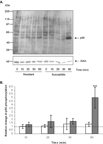
When proteins extracted from freshly hatched S. mansoni miracidia were probed with the anti-phosphotyrosine (p-Tyr-100) antibody, a number of immunoreactive bands were observed (Fig. 1A); these included 3 relatively strong bands representing tyrosine phosphorylated proteins with approximate molecular weights of 56, 60 and 68 kDa. The phosphorylated proteins appear to be derived from the miracidia and not the haemolymph because in the absence of haemolymph (0 min lanes, Fig. 1A) a similar overall banding pattern was observed. Moreover, no tyrosine-phosphorylated proteins were revealed on Western blots containing medium (CBSS+ plus haemolymph molecules) from cell culture inserts lacking miracidia (data not shown). The effect of B. glabrata haemolymph on the phosphorylation status of miracidia proteins was then explored. Exposure of miracidia to haemolymph from a resistant (NHM 3017) B. glabrata strain had limited effects on the tyrosine phosphorylation levels of any of the proteins observed over 60 min (Fig. 1A). In stark contrast, exposure of miracidia to haemolymph from susceptible (NHM 1742) snails caused a substantial increase in the phosphotyrosine content of the 56 kDa protein (p56) (Fig. 1A) after 60 min. Image analysis of immunoblots and ANOVA revealed that the effect of susceptible snail haemolymph on the tyrosine phosphorylation of p56 was highly significant (P⩽0·001), and that after 60 min a 5-fold increase in mean tyrosine phosphorylation levels were observed when compared to the control (Fig. 1B; P⩽0·001).
Protein tyrosine phosphorylation in Schistosoma mansoni miracidia is differentially affected by haemolymph from resistant and susceptible strains of Biomphalaria glabrata. (A) Miracidia were exposed to snail haemolymph for 0–60 min. Miracidia proteins were extracted and global protein tyrosine phosphorylation was assessed by Western blotting using anti-p-Tyr-100 antibodies (upper panel); actin was probed as a loading control (lower panel). Blots shown are representative of 5 independent experiments. (B) Immunoreactive bands corresponding to phospho-p56 were analysed for their relative intensities and the mean fold-change (±s.d.; n=5) in p56 tyrosine phosphorylation following exposure of miracidia to haemolymph from resistant (□) or susceptible ( ) snails was calculated. *** P⩽0·001 when compared to the control (0 min) value represented by the broken line.

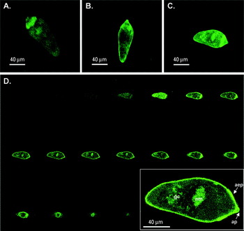
In an attempt to define the location of tyrosine-phosphorylated proteins in intact S. mansoni miracidia we used the p-Tyr-100 antibody combined with fluorescence confocal microscopy. It proved difficult to detect increased tyrosine phosphorylation in any particular region of whole miracidia following exposure to haemolymph from susceptible snails. However, these miracidia often displayed a greater overall fluorescence intensity than miracidia exposed to CBSS+ or haemolymph from resistant snails (see for example, Fig. 2A–C). Analysis of serial optical z-sections of miracidia revealed that tyrosine-phosphorylated proteins were associated with the whole tegument, with intense staining present in the anterior epidermal plates (Fig. 2D; a miracidium exposed to haemolymph from susceptible B. glabrata is shown). In addition, tyrosine-phosphorylated proteins were particularly evident in the region corresponding to the neural mass (Fig. 2D; see Bruckner and Voge (Reference Bruckner and Voge1974) for detailed description of the S. mansoni nervous system); this staining was observed in most specimens and, when absent (e.g. in Fig. 2B), there was no obvious correlation with the exposure conditions. Finally, diffuse phosphotyrosine staining was regularly seen in the region containing the germinal cells (Fig. 2D).
Protein tyrosine phosphorylation in intact Schistosoma mansoni miracidia. Miracidia were exposed to (A) CBSS+ (control) or haemolymph from either (B) a resistant or (C) a susceptible strain of Biomphalaria glabrata and were processed for confocal microscopy using anti-p-Tyr-100 antibodies; images show Z-axis projections displayed in average pixel brightness mode. (D) Serial optical Z-sections of (C) show the distribution of proteins phosphorylated on tyrosine residues. The inset shows an enlargement of Z-section no. 8, labelled to indicate the location of the anterior epidermal plates (aep), the germinal cells (gc), the neural mass (nm), and the apical papillae (ap). No fluorescence was observed when miracidia were incubated with the secondary antibody alone.

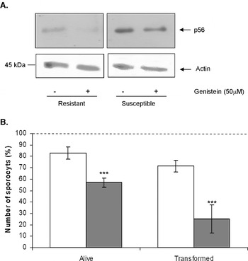
When miracidia were treated with the tyrosine kinase inhibitor genistein (50 μm) prior to being exposed to haemolymph from resistant or susceptible snails for 60 min, a decrease in p56 tyrosine phosphorylation was seen using Western blotting (Fig. 3A); no other tyrosine-phosphorylated proteins were visible on blots following treatment of miracidia with genistein (data not shown). Analysis of immunoblots revealed that genistein (50 μm) reduced mean p56 phosphotyrosine levels by 41% (s.d.±12%; n=3) and 50% (s.d.±16%; n=3) in miracidia exposed to resistant and susceptible snail haemolymph, respectively. Next, the effects of this inhibitor on the motility of S. mansoni miracidia, the viability of the larval stages (miracidia and sporocysts), and the transformation efficiency of miracidia to sporocysts were explored. When genistein was initially added to the miracidia cultures, within a very short period (5 min) an effect on miracidial motility was observed, with swimming speeds being impaired considerably (by about 50%) in all experiments; moreover, this effect appeared to last for several hours. After 48 h in culture, 50 μm genistein significantly reduced the mean viability of larvae when compared to the DMSO-treated controls (P⩽0·001; Fig. 3B); mean mortality rates of 43% and 17% were recorded for genistein and DMSO-treated larvae, respectively (Fig. 3B). Transformation efficiency was also impaired in miracidia treated with genistein. Whereas 71% of the miracidia transformed in control groups, inhibition of PTKs by genistein significantly reduced transformation to 25% (P⩽0·001; Fig. 3B). Thus, whereas the majority (85%) of surviving larvae had transformed in the control groups, only 37% transformed in the genistein-treated group.
The PTK inhibitor, genistein, reduces miracidia p56 phosphotyrosine levels and inhibits miracidia to sporocyst transformation. (A) Miracidia were treated with genistein (or vehicle, DMSO) for 30 min prior to exposure to haemolymph from either resistant or susceptible Biomphalaria glabrata. Proteins were extracted and miracidia protein tyrosine phosphorylation assessed by Western blotting using anti-p-Tyr-100 antibodies (upper panel). After genistein treatment only phospho-p56 was apparent on blots; thus only p56 is shown. Actin was probed as a loading control (lower panel) and blots shown are representative of 3 independent experiments. (B) Miracidia were cultured for 48 h with genistein () or DMSO ((□); control) and the mean number of viable larvae (miracidia/sporocysts), and transformation rates calculated (±s.d.; n=140 from 4 experiments). *** P⩽0·001 when compared to the control group.

DISCUSSION
Our interest in snail-schistosome interactions prompted us to explore the effects of B. glabrata haemolymph on signalling processes in S. mansoni miracidia; we chose to study protein tyrosine phosphorylation mediated by PTKs because this conserved process is central to many cellular signal transduction pathways in eukaryotic organisms (Hunter, Reference Hunter1995). The most striking finding of the present research was that an S. mansoni miracidia protein with a molecular weight of approximately 56 kDa was substantially phosphorylated on tyrosine residues following 60 min exposure to haemolymph from a susceptible B. glabrata strain (NHM 1742); this response could be blunted by pre-incubation of miracidia with the PTK inhibitor genistein. To our knowledge, this is the first demonstration of snail host-derived factors altering specific signalling processes in schistosome larvae. Importantly, this effect was not observed when miracidia were exposed to haemolymph from a resistant B. glabrata strain (NHM 3017). This differential effect suggests that p56 might be important in the biological responses of schistosomes to the host environment and opens up opportunities to study the potential involvement of phospho-p56 and upstream PTKs in the success or failure of infection.
Although this study is the first to report effects of host factors on specific cell signalling mechanisms in snail-schistosome interactions, molecular interplay at the level of kinase signal transduction has been reported in other host-parasite systems. For example, bovine bile induces specific tyrosine phosphorylation of 29 and 45 kDa proteins in F. hepatica adults (Marcilla et al. Reference Marcilla, De la Rubia, Espert, Carpena, Esteban and Teledo2004). In Trichinella spiralis, bile exposure, a treatment which renders the parasite infective for intestinal epithelia, alters the serine/threonine and tyrosine phosphorylation status of a number of proteins, particularly that of a 45 kDa protein phosphorylated by a serine/threonine kinase (Smith et al. Reference Smith, Selkirk and Gounaris2000). Studies with Trypanosoma cruzi trypomastigotes revealed that a soluble factor present in extracts from susceptible HeLa cells substantially enhances the phosphotyrosine levels of a T. cruzi 175 kDa protein; interestingly, this effect was not observed when trypomasigotes were treated with a soluble extract of resistant (K562) cells (Favoreto et al. Reference Favoreto, Dorta and Yoshida1998; Neira et al. Reference Neira, Ferreira and Yoshida2002).
Haemolymph of resistant snails sometimes contains more haemocytes than that of susceptible snails; protein composition of the two haemolymphs is also likely to be different. We did not equalize the number of haemocytes (or the haemolymph protein concentrations) in our experiments to compensate for this, instead we used similar volumes of haemolymph from each snail strain. This approach was taken as we wished to determine effects in a physiologically-relevant context. It is possible that the differential effects of haemolymph on the tyrosine phosphorylation of p56 are due to the presence of a stimulatory factor in the haemolymph of susceptible snails, or an inhibitory factor in haemolymph of resistant snails; concentration-dependent effects also cannot be excluded. We are also unable to conclude from the present work whether the haemolymph factor originally derives from haemocytes or other cells, it is, however, a soluble haemolymph factor able to diffuse across the 0·2 μm membrane present in the cell culture inserts. We are only aware of one other report in which host factors have been shown to influence the tyrosine phosphorylation of schistosome proteins (Vicogne et al. Reference Vicogne, Cailliau, Tulasne, Browaeys, Yan, Fafeur, Vilain, Legrand, Trolet and Dissous2004); in this study, SER, an S. mansoni epidermal growth factor (EGF)-like molecule was found to be phosphorylated on tyrosine residues when adult worm membranes were treated with human EGF. Given that EGF-like molecules have been characterized in snails (Hermann et al. Reference Hermann, van Kesteren, Wildering, Painter, Reno, Smith, Kumar, Geraerts, Ericsson, Smit, Bulloch and Nagle2000), studies into the effects of the conserved EGF signature on signal transduction in miracidia and sporocysts would likely provide useful insights into molecular aspects of snail-schistosome interplay.
Fluorescence confocal laser scanning microscopy revealed that tyrosine-phosphorylated proteins existed predominantly in the miracidium tegument, the region of the schistosome that directly contacts the snail host milieu. In addition, there was a tendency for miracidia exposed to haemolymph from susceptible snails to possess higher levels of general fluorescence, which is reflected in the increase in the intensity of tyrosine phosphorylated bands seen in Western blotting. Although a specific location for phospho-p56 was not defined, p56 might be associated with the tegument region, tempting speculation that it might be involved in early signalling processes that enable the parasite to develop successfully in the susceptible snail host. Knowledge of PTK signalling in the schistosome tegument is poor; however, recent work by Bahia et al. (Reference Bahia, Mortara, Kusel, Andrade, Ludolf, Kuser, Avelar, Trolet, Dissous, Pierce and Oliveira2007) has identified an ~ 143 kDa Fes-like tyrosine kinase (SmFes) in all life-cycle stages and has been located to the tegument and the region (terebratorium) containing the apical papillae of the miracidium, thus implicating SmFes in host penetration. In addition to the tegument, tyrosine-phosphorylated proteins were observed in regions corresponding to the neural mass and, sometimes, the germinal cells. Previous research has not focused on the presence of PTKs in these regions. However, PTKs are known to be crucial mediators of neuronal signalling (Ohnishi et al. Reference Ohnishi, Yamamori, Ono, Aoyagi, Kondo and Takahashi2001) and development (Birchmeier, Reference Birchmeier, Sonnenberg, Weidner and Walter1993; see below).
Functional experiments conducted with the inhibitor, genistein, demonstrated that PTKs plays an important role in the swimming of S. mansoni miracidia, the survival of miracidia, and in the miracidia to sporocyst transformation. This inhibitor has previously been shown to reduce motility of human sperm (Bajpal and Doncel, Reference Bajpal and Doncel2003) and to abolish adaptive swimming behaviour leading to chemoattraction in Tetrahymena thermophila (Leick et al. Reference Leick, Iversen, Kemp and Christensen1997). In the transformation experiments reported in the present study, the proportion of surviving larvae that transformed into sporocysts (85%) in the presence of DMSO (control group) was only slightly lower than that (90%) reported elsewhere (Boyle et al. Reference Boyle, Wu, Dhoemaker and Yoshino2003) for transformation in CBSS+ alone. Notably, the dramatic reduction in larval transformation observed in the presence of genistein confirms a role for PTKs as important mediators of development in schistosomes. In this context, studies with adult S. mansoni have demonstrated that the PTK inhibitor, herbimycin A, reduces protein tyrosine phosphorylation, and blocks mitotic activity and egg production in paired females (Knobloch et al. Reference Knobloch, Kunz and Grevelding2006). Moreover, an S. mansoni Syk-family PTK, TK4, has been identified that may play a role in germ cell development (Knobloch et al. Reference Knobloch, Winnen, Quack, Kunz and Grevelding2002). The importance of PTKs to schistosome development is not surprising given that these signalling enzymes regulate multiple physiological processes, including differentiation, in animals; indeed, the importance of protein kinases to development is highlighted by the recent finding that 85% of sea urchin signalling kinases are expressed in the developing embryo (Bradham et al. Reference Bradham, Foltz, Beane, Arnone, Rizzo, Coffman, Mushegian, Goel, Morales, Geneviere, Lapraz, Robertson, Kelkar, Loza-Coll, Townley, Raisch, Roux, Lepage, Gache, McClay and Manning2006). Genistein blocks the activity of multiple PTKs in S. mansoni miracidia (as demonstrated here by p-Tyr-100 blotting) so the effects observed in the present work are unlikely specific to a single PTK. We anticipate that further characterization of tyrosine kinases in S. mansoni will facilitate important inhibitor- and/or RNAi-based studies into the role of specific receptor and non-receptor PTKs during larval transformation.
The work described here represents the first study on tyrosine phosphorylation and PTK signalling in schistosome miracidia. Although we are presently unable to ascribe a distinct function to S. mansoni phospho-p56, future studies will focus on the potential of this protein to mediate biological responses in miracidia that are crucial to establishing a successful infection in B. glabrata. Ultimately, a thorough analysis of cell signalling pathways that drive immediate functional responses as well as those that mediate changes in gene expression in the snail (Walker, Reference Walker2006; Lockyer et al. Reference Lockyer, Spinks, Noble, Rollinson and Jones2007) and schistosome (Jolly et al. Reference Jolly, Chin, Miller, Bahgat, Lim, DeRisi and McKerrow2007), is crucial to gaining a comprehensive understanding of both unidirectional and reciprocal snail-schistosome interactions.
This work was carried out while A. J. W. was in receipt of a Higher Education Funding Council for England (HEFCE) Promising Researcher Fellowship. We would like to thank Jayne King and Mike Anderson of the Natural History Museum for culturing the parasite.