Connecting the hepatic veins to the azygos venous system so as to relieve arteriovenous fistulas subsequent to a Kawashima operation can be cumbersome. It has previously been shown that this procedure is particularly difficult when performed through a redo-median sternotomy, and when using extracorporeal cardiopulmonary bypass with the need for deep hypothermia.Reference Baskett, Ross, Warren, Sharratt and Murphy1, Reference Kaneko, Murakami, Miyamoto and Takamoto2 Mindful of these difficulties, we sought an alternative means of connecting the hepatic veins to the azygos venous system, approaching through a lateral thoracotomy and without the use of extracorporeal circulation and hypothermia. We have now used this approach in 3 patients, all with isomerism of the left atrial appendages and interruption of the inferior caval vein, and all having undergone some form of the Kawashima procedure.
Materials and methods
Operative technique
Access to the hepatic veins is obtained through a postero-lateral thoracotomy just cranial to the diaphragmatic dome. The pericardium is opened, anterior to the phrenic nerve, and the hepatic veins are encircled by a vessel loop. The inferior pulmonary ligament is divided in order to retract the lung cranially. The azygos vein, strictly a hemiazygos vein in the setting of left isomerism, runs paravertebrally and is identified easily, whereafter it is encircled by another vessel loop. Within the pericardium, and anteromedial to the azygos vein, the hepatic venous confluence enters the atrium on that side of the body. Due to the distance between the veins, a direct anastomosis between them is likely to kink, so we prefer to insert a short vascular prosthesis. We place a side-biting Satinsky clamp on the hepatic venous confluence, and anastomose the vascular prosthesis to it in end-to-side fashion. After measurement of the prosthetic length, another Satinsky clamp is placed on the azygos vein, and a similar end-to-side anastomosis is constructed. After de-airing of the prosthesis, and removal of the clamps, the opening of the hepatic venous confluence to the atrium is closed easily by means of a vascular stapler.
Results
We have performed the procedure in 3 female patients, all with isomerism of the left atrial appendages, interruption of the inferior caval vein, and continuation of the inferior caval venous return through the azygos system. The patients also had atrioventricular septal defect with common atrioventricular junction, valvar and subvalvar pulmonary stenosis, and in all of them one of the ventricles was hypoplastic.
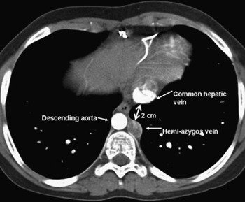
The first patient was born in 1990 with mirror-imaged arrangement of the abdominal organs, and double outlet right ventricle with anterior aorta. She was in a reasonable haemodynamic balance until she was 6 years old, at which stage we constructed a semi-total bilateral bidirectional cavo-pulmonary connection, placing an atrial pacemaker because of sinus nodal dysfunction. The hepatic veins were left draining into the left-sided atrium. The immediate postoperative saturations of oxygen ranged between 85% and 90%. By 2002, the saturation of oxygen had dropped to 70% at exercise. Echocardiography and contrast injected computed tomography revealed arterio-venous fistulas in the upper lobe of the left lung. At a second operation in 2002, her left-sided hepatic vein was redirected surgically to the azygos venous system as described above through a left lateral thoracotomy. The azygos and hepatic veins both had diameters of 20 millimetres. We used a Gore-tex® vascular prosthesis of 16 millimetres diameter, and with a length of 2 centimetres, to make the connection. (Table 1, Fig. 1). There were no complications, and postoperatively her saturations of oxygen increased to 98% within one month. At her most recent follow-up visit, in 2006, her saturation of oxygen measured transcutaneously was 100%, and she was in an excellent physical condition.
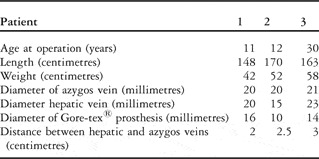
The Table shows the age, length and weight of our patients, and the diameters of the vessels and vascular prosthesis used during the operative procedure.

Computed tomographic scan showing the distance between the common hepatic vein and azygos vein in our first patient.

The second patient, shortly after her birth in 1991, required construction of a left-sided modified Blalock-Taussig shunt, followed in 1992 by construction of a right-sided shunt. In 1994, we created a semi-total cavopulmonary connection by connecting the left superior caval vein end-to-side to the left pulmonary artery. The hepatic veins were left draining into the left-sided atrium, the Blalock-Taussig shunts were divided, and the restrictive interatrial communication was enlarged. Her postoperative saturations of oxygen rose immediately to 90%. Over a period of three years, the saturation dropped again to between 55% and 77% at rest. Lung perfusion scans showed a right-to-left shunt in the lungs, indicating the existence of arterio-venous fistulas, that were more prominent in the right than in the left lung.
In 1998, her left-sided hepatic veins were connected to the right pulmonary artery, interposing an extracardiac Gore-tex® ringed vascular prosthesis of 10 millimetres diameter, without the use of extracorporeal circulation. One year postoperatively her saturation of oxygen has increased to 90% at rest, and 75% during exercise.
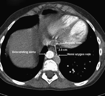
In the following years, her saturations of oxygen gradually decreased again to 56% at rest, and a lung perfusion scan at this time showed arteriovenous fistulas remaining in the left lung, with preferential flow of the hepatic veins to the right lung. In 2003, at the age of 11 years, a computed tomographic scan showed the diameter of her hemi-azygos vein to be 20 millimetres, that of the common hepatic vein to be 15 millimetres, and the distance between the two veins 2.5 centimetres. Therefore, in 2004, at the age of 12 years, she underwent a further operation (Table 1, Fig. 2). In this operation, her unilateral left-sided hepatic vein was redirected to the azygos venous system via a left lateral thoracotomy, using Gore-tex® vascular prosthesis of 10 millimetres diameter, and without the use of extracorporeal circulation. The previously inserted vascular prosthesis was closed. The small size of the vascular prosthesis was dictated by the length of the common hepatic vein and the previous operation. There were no complications. Two months postoperatively her saturations were measured at 94 to 95%, and they have remained at this level until now.
Computed tomographic scan showing the distance between the common hepatic vein and azygos vein in our second patient.

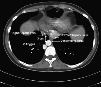
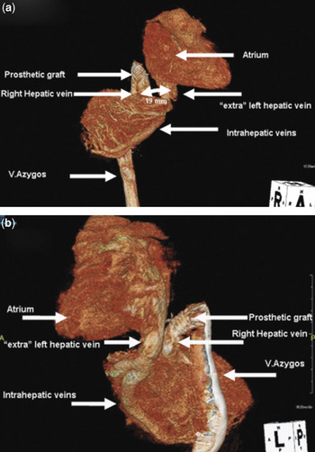
The third patient, born in 1973, came from abroad at the age of 25 years. At presentation, when she had undergone no previous surgery, her saturation of oxygen at rest was 86%, dropping to 54% during maximal exercise. In 1998, we constructed a semi-total cavopulmonary connection, leaving the hepatic veins draining into the atrium. Postoperative exercise testing showed an improved saturation of oxygen at rest of 97%, which dropped at maximal exercise to 89%. She returned five years later with overt cyanosis, a saturation of oxygen of 80%, and polycythaemia. Pulmonary angiography and contrast echocardiography established the presence of pulmonary arterio-venous fistulas. A computed tomographic scan in 2003 showed the diameter of her azygos vein to be 21 millimetres, that of the common hepatic vein to be 23 millimetres, and the distance between the two veins to be 3 centimetres. Shortly thereafter, we redirected her hepatic veins to the azygos vein through a right lateral thoracotomy, without the use of extracorporeal circulation, using a Gore-tex® prosthesis of 15 millimetres diameter (Table 1, Fig. 3). The hepatic venous connection to the right atrium was closed by means of a vascular stapler. One month postoperatively, exercise testing showed the saturation of oxygen at rest to be 87%, falling at maximal exercise to 60%. After an interval of 3 years, in 2006, she returned with relapsing cyanosis and polycythaemia. Catheterisation and computed tomographic scanning at this time showed an additional left-sided hepatic vein not previously recognised. This vein permitted right-to-left retrograde shunting from the azygos vein via the vascular prosthesis into the right-sided hepatic vein, passing through the hepatic parenchyma to enter the left-sided atrium (Fig. 4). This situation also precluded hepatic venous blood flowing directly into the lungs, thus causing arteriovenous fistulas. Through a left thoracotomy, we closed this large left-sided intrapericardial hepatic vein, whence the saturation increased immediately to 98%, and remained at this level postoperatively. In retrospect it proved possible to see the bilateral hepatic veins in the computed tomographic scan performed in 2003, albeit that they were not recognised as such, nor were they recognised during the operations. The distance between the hepatic veins was 19 millimetres.
Computed tomographic scan showing the distance between the right hepatic vein and azygos vein in our third patient. Note the bilateral hepatic veins.

Three-dimensional reconstruction of the computed tomographic scan in our third patient after injection of contrast into the right femoral vein. It shows the course of the dye from the azygos vein through the vascular prosthesis, through part of the hepatic parenchyma, to the left-sided hepatic vein and thence into the atrium. Figure 4a is taken in right anterior oblique projection, while Figure 4b shows the left posterior oblique projection.

Discussion
Patients with continuation of the interrupted inferior caval venous return through the azygos venous system can well have intracardiac morphology that dictates creation of the Fontan circulation. This combination of anomalies usually exists in the setting of isomerism of the left atrial appendages.Reference Uemura, Yagihara, Hattori, Kawahira, Tsukano and Watanabe3 Kawashima and colleagues described some time agoReference Kawashima, Kitamura, Matsuda, Shimazaki, Nakano and Hirose4 how it was possible, in the setting, to connect the superior caval vein surgically to the right pulmonary artery, and divide or ligate the pulmonary trunk. This operative manoeuvre produces a close to total cavopulmonary connexion, with only hepatic and coronary venous blood returning to the atrium and producing a physiological right-to-left shunt at atrial level. The volume of this shunt increases the preload on the left ventricle similar to the effect of a fenestration in a lateral tunnel, although it has no function in limiting central venous pressure.
Cyanosis after this surgical palliation can occur over time, due to development of diffuse bilateral pulmonary arteriovenous fistulas. These fistulas develop gradually, allegedly because of absence of a substance as yet unidentified in the hepatic venous blood that either has an extremely short half-life, or else is rapidly inactivated in the systemic circulation. It has been demonstrated that rerouting of hepatic venous blood towards the lungs reverses the development of these arteriovenous fistulas, and thus diminishes cyanosis.Reference Srivastava, Preminger and Lock5, Reference Duncan and Desai6 The situation is similar to the pathogenesis of the so-called hepato-pulmonary syndrome, where pulmonary arteriovenous fistulas develop as a result of reduced synthesis of the elusive hepatic factor due to hepatic dysfunction. Technically, the hepatic veins can also be connected to the pulmonary arteries by means of an extracardiac vascular prosthesis.Reference Shah, Rychik, Fogel, Murphy and Jacobs7 Construction of such a veno-arterial connection has the disadvantages of requiring resternotomy, and often the need for extracorporeal circulation. Even then, our second patient, in whom this was the adopted procedure, shows that the distribution of hepatic venous blood may well be unbalanced, so that fistulas may well disappear in one lung, whilst they remain or develop in the other lung. The potential physiologic disadvantage of this procedure is precluded by connecting the hepatic veins to the azygos venous system, which reasonably guarantees balanced distribution over both lungs. Thrombosis in extracardiac conduits placed between the hepatic veins and the pulmonary arteries can be a second potential disadvantage.Reference Konstantinov, Puga and Alexi-Meskishvili8 Connecting the hepatic veins to the pulmonary arteries by means of a vascular prosthesis, therefore, appears only to have disadvantages.
Several investigatorsReference Baskett, Ross, Warren, Sharratt and Murphy1, Reference Kaneko, Murakami, Miyamoto and Takamoto2, Reference Amodeo, Di Carlo, Grigioni, De Santis and Di Donato9 have described solitary cases where the hepatic veins were directly connected to the azygos vein. All used deep hypothermic circulatory arrest, which has the well-known disadvantages of providing a limited period of operative exposure and the risk of neurological sequels. Kaneko and his groupReference Kaneko, Murakami, Miyamoto and Takamoto2 used an autologous pericardial roll to make the hepatic veins-azygos connection. We chose Gore-tex® conduits, since all our patients had undergone one or more previous operations, producing scarring of the pericardium, which was hence less suitable for use as a conduit. A potential limitation of using Gore-tex® is the need of postoperative anticoagulation, but all our patients were already anticoagulated prior to their operations. In 2002, Steinberg et al.Reference Steinberg, Alfieris and Brandt10 used an extracardiac conduit to connect multiple hepatic veins to the azygos venous system in the setting of interruption of the inferior caval vein, albeit through a median sternotomy and with the use of cardiopulmonary bypass.Reference Steinberg, Alfieris and Brandt10
Bilateral hepatic veins exist in one-third of patients with isomerism of the left atrial appendages,Reference Uemura, Ho, Devine, Kilpatrick and Anderson11 so it is not surprising that one of our 3 patients has this anatomy. The possibility of bilateral hepatic veins could be regarded as an academic oddity when the hepatic veins are left to drain to the atriums, such as frequently the case when using the Kawashima operation. When the hepatic veins need to be redirected towards the pulmonary circulation, such a bilateral arrangement achieves paramount technical importance. As we have demonstrated, one of the hepatic veins can readily be connected to the azygos venous system on the side of that vein. We closed the hepatic vein on the other side in our third patient, at the cost of a tendency of retention of fluid, which could be treated with diuretics. This was done after our discovery of a transhepatic shunt subsequent to connection of only the one hepatic vein to the azygos vein. It is doubtful whether the second hepatic vein can be closed as a primary procedure. Such a process is likely to produce severe hepatic congestion, with parenchymal damage as a result. In retrospect, we could have connected both hepatic veins to the azygos vein, as described by Steinberg et al.Reference Steinberg, Alfieris and Brandt10 Judging from the computed tomographic scan, such a manoeuvre is certainly technically feasible (Figs. 3 and 4). The distance between the two hepatic veins in our third patient is so small that it should be possible to fit another vascular prosthesis between them.
Thus, we have shown that, through a lateral thoracotomy, it is readily possible to connect together the hepatic and azygos venous systems by using a short vascular prosthesis. This manoeuvre carries that added advantage of not having to touch the heart, which may well result in deteriorated function, with the possible need of instituting extracorporeal circulation. The approach also avoids an extended operative time, and the bleeding associated with resternotomy. The accessibility of the venous structures precludes the need for extracorporeal circulation. Care should be taken, nonetheless, to identify multiple hepato-atrial connections at the time of redirection surgery, as they may cause recurrent cyanosis when missed. All 3 of our patients achieved excellent results as judged by postoperative saturations of oxygen. The only disadvantages of the approach are the need for a new incision, and thus an extra scar.