INTRODUCTION
Microsporidia are widespread in aquatic environments. Almost half of the 190 genera described to date infect aquatic hosts, with over 20 in fish, 50 in aquatic arthropods and more than 20 in non-arthropod invertebrates, protists, and as hyperparasites of other taxa infecting aquatic animals (Stentiford et al. Reference Stentiford, Bateman, Feist, Stone and Dunn2013). Microsporidian hyperparasites infect myxozoans (myxosporean and actinosporean stages) and acanthocephalans in fish (Diamant and Paperna, Reference Diamant and Paperna1985; de Buron et al. Reference de Buron, Loubès and Maurand1990; Morris and Freeman, Reference Morris and Freeman2010), digeneans in fish, crustaceans and molluscs (Canning and Nicholas, Reference Canning and Nicholas1974; Levron et al. Reference Levron, Ternengo, Toguebaye and Marchand2004, Reference Levron, Ternengo, Toguebaye and Marchand2005), monogeneans in toads (Cable and Tinsley, Reference Cable and Tinsley1992), cestodes in snails and fish (Canning et al. Reference Canning, Lai and Lie1974; Sene et al. Reference Sene, Ba, Marchand and Toguebaye1997; Ba et al. Reference Ba, Toguebaye and Marchand2007), dicyemidans in squid (Czaker, Reference Czaker1997) and gregarines in polychaetes and nemerteans (Caullery and Mesnil, Reference Caullery and Mesnil1897, Reference Caullery and Mesnil1914; Vinckier, Reference Vinckier1975). Their reported ability to infect parasitic and free-living protists in the Gregarinasina (see above and Larsson, Reference Larsson2000; Larsson and Koie, Reference Larsson and Koie2006), Amoebozoa (Scheid, Reference Scheid2007) and Ciliophora (Foissner and Foissner, Reference Foissner and Foissner1995; Fokin et al. Reference Fokin, Di Giuseppe, Erra and Dini2008) probably vastly under represents their true potential to interact with protistan hosts within aquatic and terrestrial habitats (Stentiford et al. Reference Stentiford, Bateman, Feist, Stone and Dunn2013).
The Paramyxida is an order of parasitic protists within the class Ascetosporea (supergroup Rhizaria) (Ward et al. Reference Ward, Bennett, Bateman, Stentiford, Feist, Williams, Berney and Bass2016). Paramyxids are related to haplosporidians, paradinids and mikrocytids (Bass et al. Reference Bass, Chao, Nikolaev, Yabuki, Ishida, Berney, Pakzad, Wylezich and Cavalier-Smith2009; Hartikainen et al. Reference Hartikainen, Ashford, Berney, Okamura, Feist, Baker-Austin, Stentiford and Bass2014a , Reference Hartikainen, Stentiford, Bateman, Berney, Feist, Longshaw, Okamura, Stone, Ward, Wood and Bass b ), although the evolutionary relationships among the five ascetosporean orders is currently unresolved. All characterized ascetosporeans are parasites of invertebrate hosts (Chatton, Reference Chatton1911; Haskin et al. Reference Haskin, Stauber and Mackin1966; Grizel et al. Reference Grizel, Comps, Bonami, Cousserans, Duthoit and Le Pennec1974; Larsson, Reference Larsson1987; Feist et al. Reference Feist, Hine, Bateman, Stentiford and Longshaw2009). Recent work utilizing genome sequence data from representative lineages within the Rhizaria have revealed how certain groups (e.g. the mikrocytids) are more diverse and wide spread in aquatic habitats than previously thought (Burki et al. Reference Burki, Corradi, Sierra, Pawlowski, Meyer, Abbott and Keeling2013; Hartikainen et al. Reference Hartikainen, Stentiford, Bateman, Berney, Feist, Longshaw, Okamura, Stone, Ward, Wood and Bass2014b ). Despite this, the rhizarians, and their constitutive taxa, remain one of the most understudied groups of eukaryotes. Representatives of the genus Marteilia are perhaps the best-studied paramyxids, largely due to their impact on cultured oyster stocks, and their listing in international legislative frameworks (OIE, 2014).
Several examples of microsporidian hyperparasitism of paramyxean hosts are reported in the literature. Based upon morphological data, Comps et al. (Reference Comps, Pichot and Deltreil1979) described Nosema ormieresi infecting Marteilia refringens, in turn parasitizing the oyster Ostrea edulis. The taxonomic placement of the hyperparasitic microsporidian within Nosema was based upon the presence of a diplokaryon in all life stages; now considered a plesiomorphic trait (see Stentiford et al. Reference Stentiford, Bateman, Small, Moss, Shields, Reece and Tuck2010) and therefore likely to be erroneous. Furthermore, a microsporidian hyperparasite was also reported infecting M. refringens in mussel Mytilus galloprovincialis (Villalba et al. Reference Villalba, Mourelle, Carballal and Lopez1997) and recently, infecting the sister paramyxid Marteilia cochillia in cockles (Villalba et al. Reference Villalba, Iglesias, Ramilo, Darriba, Parada, No, Abollo, Molares and Carballal2014). In the latter case, the authors proposed that the microsporidian hyperparasite was likely synonymous with that previously described infecting the cockle digestive gland by Comps et al. (Reference Comps, Grizel, Tigé and Duthoit1975). An additional complication in this case, however, is the co-infection of cockles by digenean trematodes which can also play host to a microsporidian hyperparasite, Unikaryon legeri (Canning and Nicholas, Reference Canning and Nicholas1974).
Whilst paramyxid parasites are best known for their devastating effects on cultured and wild molluscan populations, they can also parasitize crustacean hosts. There is strong evidence that M. refringens may require passage through crustacean hosts to complete its lifecycle (Audemard et al. Reference Audemard, Le, Barnaud, Collins, Sautour, Sauria, de, Coustau, Combes and Berthe2002; Arzul et al. Reference Arzul, Chollet, Boyer, Bonnet, Gaillard, Baldi, Robert, Joly, Garcia and Bouchoucha2014). Furthermore, representatives of the understudied genus Paramarteilia, a sister taxon to Marteilia, are known to infect amphipod (Ginsburger-Vogel and Desportes, Reference Ginsburger-Vogel and Desportes1979) and decapod (Feist et al. Reference Feist, Hine, Bateman, Stentiford and Longshaw2009) hosts. In amphipods, Paramarteilia orchestiae infections commonly co-occur with microsporidian pathogens (again, incorrectly attributed to the genus Nosema) (Ginsburger-Vogel and Desportes, Reference Ginsburger-Vogel and Desportes1979). Recent work has shown that paramyxean pathogens may at least be partly responsible for the elevated rates of feminization and other sexual disruption noted in some amphipod populations – a consequence previously assigned only to infection by microsporidians (Ginsburger-Vogel, Reference Ginsburger-Vogel1991; Short et al. Reference Short, Guler, Yang, Kille and Ford2012). These paramyxeans, and co-infecting microsporidians of the genus Dictyocoela can be vertically transmitted to amphipod eggs and larvae (Short et al. Reference Short, Guler, Yang, Kille and Ford2012), possibly offering a route of re-infection to molluscan hosts. Taken together, these observations provide an intriguing insight in to potential for microsporidian pathogens to exploit the invasion and pathogenic strategies of paramyxean pathogens, and their potential to ‘hitch-hike’ between hosts of differing trophic positions (Lefèvre et al. Reference Lefèvre, Lebarbenchon, Gauthier-Clerc, Missé, Poulin and Thomas2008; Thomas et al. Reference Thomas, Renaud and Poulin2008). Furthermore, the observations pose interesting questions about the taxonomic status of microsporidian pathogens infecting paramyxean protist hosts. Are they primitive associations or, perhaps more recent acquisitions, thus taking advantage of an existing parasitic strategy of their protistan hosts? (Stentiford et al. Reference Stentiford, Bateman, Feist, Stone and Dunn2013).
We have previously stated that of the known microsporidian genera infecting aquatic organisms, phylogenetic data pertaining to the ssuRNA gene are available for approximately 35 arthropod-infecting taxa, 17 fish-infecting taxa, and six taxa associated with non-arthropod invertebrates. To date, little sequence data exist for microsporidian parasites infecting protists or hyperparasites (Stentiford et al. Reference Stentiford, Bateman, Feist, Stone and Dunn2013). In particular, the absence of data from apparently primitive genera, such as Metchnikovella which infect gregarine hyperparasites of polychaetes (Sokolova et al. Reference Sokolova, Paskerova, Rotari, Nassonova and Smirnov2013) provides a notable example. In the current study, we investigated the phylogeny of two microsporidian hyperparasites infecting the paramyxean protist M. cochillia and the digenean trematode Meiogymnophallus minutus, both occurring as co-infections in cockles. We show that rather than representing basally branching taxa, these hyperparasites align with other known microsporidian parasites infecting marine crustaceans. In doing so, we erect a new genus and species (Hyperspora aquatica n. gn., n.sp.) to contain the hyperparasite of Marteilia spp. and propose that such hyperparasitism may provide a strategy for the vectoring of microsporidian pathogens between hosts of different trophic status in aquatic systems.
MATERIALS AND METHODS
Sampling, histology and electron microscopy
Cockles (Cerastoderma edule) were hand-collected from shellfish beds of O Sarrido and Lombos do Ulla in the Ría de Arousa, Galicia, NW Spain in June 2012. For histology, a transverse section of each individual containing gill, visceral mass, mantle lobe and foot muscle, plus portions of the adductor muscle and siphon were fixed in Davidson's sea water fixative (Hopwood, Reference Hopwood, Bamcroft and Stevens1996) for 24 h. Following fixation, samples were dehydrated in an ethanol series and embedded in paraffin according to standard protocols. Histological sections (c. 5 µ m) were mounted to glass slides before staining with Harris’ haematoxylin and eosin (H&E) prior to examination under light microscopy for detection of two microsporidian hyperparasites of M. cochillia and metacercariae of the digenean M. minutus, infecting cockles. Smears of cockle digestive glands infected with M. cochillia were fixed in ethanol and stained with Panoptic rapid stain (a modified Romanowsky stain). Digital images were obtained using a Nikon E800 light microscope and the Lucia™ Screen Measurement System (Nikon). For electron microscopy, 2 mm3 blocks of cockle digestive gland were fixed in a solution containing 2·5% glutaraldehyde in filtered seawater, post-fixed in 1% osmium tetroxide in 0·2 m sodium cacodylate solution and embedded in Epon resin. Ultrathin sections (50–70 nm) were stained with uranyl acetate and lead citrate and examined in a JEM-1010 transmission electron microscope (TEM, JEOL).
DNA extraction, PCR amplification and sequencing
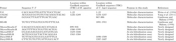
Corresponding pieces of digestive gland and mantle from individual cockles were preserved in 96% ethanol at the time of dissection. DNA extractions were performed using the Wizard Genomic DNA Purification Kit (Promega) according to the manufacturer's protocol to isolate genomic DNA. DNA quality and quantity was checked using a Nanodrop® ND-1000 spectrophotometer (Nanodrop technologies, Inc.). A set of previously published primers and additional primer designs were utilized in this study (Table 1). Primers were designed to amplify overlapping fragments of the small subunit rRNA gene (SSU rRNA), the internal spacer transcribed (ITS) and the large subunit rRNA gene (LSU rRNA) of the microsporidian pathogens infecting M. cochillia and M. minutus in cockle hosts. The whole SSU region of the two microsporidian hyperparasites was amplified by PCR using the primers V1f/1342AC (Weiss et al. Reference Weiss, Zhu, Cali, Tanowitz and Wittner1994; Yang et al. Reference Yang, Short, Kille and Ford2011). To amplify the end of the SSU, the ITS, and the end of the LSU rRNA gene of the microsporidian hyperparasites of M. minutus, HG4F/HG4R primers were used (Gatehouse and Malone, Reference Gatehouse and Malone1998). To amplify the end of the SSU, the ITS, and the end of the LSU rRNA gene of the hyperparasite of M. cochillia, a new primer set, defined as Micros_Mcochi_F/Micros_Mcochi_R was used. To do so, SSU rRNA sequences obtained for this microsporidian, and for another six microsporidian sequences deposited in GenBank were aligned using the programme Clustal W. These microsporidians were Dictyocoela berillonum (AJ438957), Microsporidia sp. (GQ246188), Microsporidium sp. (AJ438962), Microsporidium sp. (HM800853), Potaspora morhaphis (EU534408) and Thelohania butleri (DQ417114). Subsequently, new primers were designed using the Primer 3 program (Rozen and Skaletsky Reference Rozen, Skaletsky, Krawetz and Misener2000).
Primer sets used for PCR and ISH detection of Hyperspora aquatica n. gen., n. sp. and Unikaryon legeri

All PCR assays were performed in a total volume of 25 µL containing 1 µL of genomic DNA (200 ng), PCR buffer at 1× concentration, 1·5 mm MgCl2, 0·2 mm nucleotides (Roche Applied Science), 0·3 µ m of each pair of primers and 0·025 U µL−1 Taq DNA polymerase (Roche Applied Science). A negative control (no DNA) was used in each PCR assay. The PCR assays were carried out in a T gradient thermocycler (Biometra), under the following reaction parameters: 94 °C for 5 min, 35 cycles at a melting temperature of 94 °C for 1 min, an annealing temperature of 62 °C for primers V1f/1342AC1 and 50 °C for primers HG4F/HG4R and Micros_Mcochi_F/Micros_Mcochi_R, for 1 min, an extension temperature of 72 °C for 2 min, followed by a final extension period of 72 °C for 10 min. After PCR, 10 µL of amplified DNA was separated by electrophoresis on 2% agarose gels, in 1% Tris acetate EDTA buffer, stained with ethidium bromide and scanned in a GelDoc XR documentation system (BioRad). PCR products were cleaned for sequencing using ExoSap-It (USB Corporation) for 15 min at 37 °C, followed by inactivation for 15 min at 80 °C. Sequencing was performed by the company Secugen (Madrid) and the chromatograms were analysed using ChromasPro v.1.41 (Technelysium Pty Ltd.). All generated sequences were assessed for similarity against known sequences using BLAST (Basic Local Alignment Search Tool) of the National Center for Biotechnology Information (NCBI, USA).
Phylogenetic analyses
Sequence alignments were made in mafft v.7 e-ins-i algorithm (Katoh and Standley, Reference Katoh and Standley2013). The alignment was refined by eye and analysed in RAxML BlackBox v.8 (Stamatakis, Reference Stamatakis2014) (GTR model with CAT approximation (all parameters estimated from the data); average of 10 000 bootstrap values was mapped onto the tree with the highest likelihood value). A Bayesian consensus tree was constructed using MrBayes v.3.2.5 (Ronquist et al. Reference Ronquist, Teslenko, van der Mark, Ayres, Darling, Höhna, Larget, Liu, Suchard and Huelsenbeck2012). Two separate MC3 runs with randomly generated starting trees were carried out for 2 M generations each with one cold and three heated chains. The evolutionary model applied included a GTR substitution matrix, a four-category autocorrelated gamma correction and the covarion model. All parameters were estimated from the data. Trees were sampled every 1000 generations. A total of 500 000 generations were discarded as ‘burn-in’ (trees sampled before the likelihood plots reached a plateau) and a consensus tree was constructed from the returning sample. All phylogenetic analyses were performed via the Cipres Science Gateway (Miller et al. Reference Miller, Pfeiffer and Schwartz2010).
In situ hybridization (ISH) assays
ISH assays were performed in order to confirm that sequences obtained from PCR co-located to microsporidian hyperparasites of M. cochillia and M. minutus in cockles observed in histology sections. Two digoxigenin polynucleotide probes were used: (1) MicrosMarteiliaISH probe designed based on the LSU sequence of the microsporidian hyperparasites of M. cochillia and another Microsporidium spp. downloaded from the GenBank database; and (2) MicrosMetacercariaISH probe designed based on the LSU sequence of the microsporidian hyperparasite of M. minutus, and other Dictyocoela spp. obtained from GenBank. The PCR DIG ProbeSynthesis kit (Roche Applied Science) was used to amplify the fragment of LSU of the hyperparasite of M. cochillia and M. minutus using the primers MicrosISH_F/MicrosISH_R and DictyISH_F/DictyISH_R, respectively (Table 1). Both sets were labelled with digoxigenin by incorporation of DIG-dUTP during the PCR process. Three serial tissue sections (about 5 µ m) were produced from each paraffin block to allow for testing of the MicrosMarteiliaISH and MicrosMetacercariaISH probes, and negative control (without probe), respectively. A further serial section was stained with Harris’ H&E in order to identify and localize the paramyxean, digenean and hyperparasites of interest. Sections for ISH assays were placed on to silanized slides (Silane-prep slides; Sigma) following which assays were performed as detailed in Ramilo et al. (Reference Ramilo, Abollo and Villalba2014). Slides were viewed using light microscopy as above.
RESULTS
Light microscopy and ultrastructure of hyperparasitism
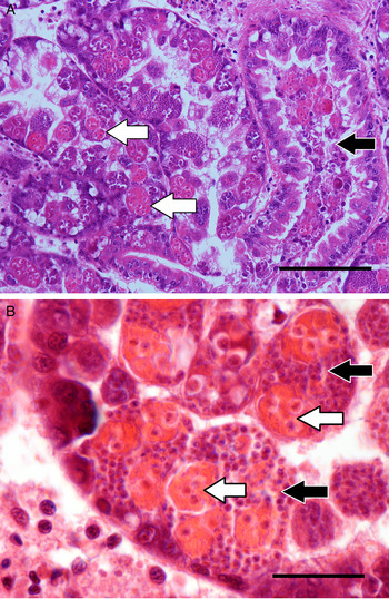
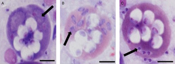
The microsporidian stages infect the cytoplasm of the primary cell of M. cochillia which itself initially located within the epithelial cells of the cockle digestive gland (Fig. 1A). Although early M. cochillia stages harbour the microsporidian parasite, the infection is most prominent in more advanced larger stages within which sporonts are also present (Fig. 1B). Sporonts and developing spores do not appear to be infected with the microsporidian hyperparasite and development of M. cochillia spores proceeds normally. Early development of the microsporidian can be visualized at the light microscope level in stained impression smears of infected cockle digestive gland. A sequence of merogonic development to mature spore formation is seen in Fig. 2. Released spores are also visible but we were unable to detect examples of M. cochillia in the process of spore release which is likely to be an uncontrolled event on rupture of primary cells within the lumen of the digestive tubule.
Microsporidian hyperparasitism of Marteilia cochillia infecting cockles (Cerastoderma edule). (A) The digestive gland tubules are significantly altered during advanced M. cochillia infection. Marteilia cells are clearly visible as large oeosinophilic bodies within tubule epithelial cells (white arrows). Hyperparasitized M. cochilla are shed to the lumen of infected tubules (black arrow). Scale 100 µ m; (B) M. cochilla (white arrows) and microsporidian hyperparasite (black arrows) within tubule epithelial cells and tubule lumen. Scale 25 µ m H&E histology.

Cockle digestive gland impression smears showing microsporidian hyperparasitism of Marteilia cochillia primary cells. (A) Infected primary cell containing six developing spores and putative Hyperspora aquatica meronts appearing as relatively pale staining regions (arrow); (B) Multiple H. aquatica sporonts with dark basophilic cytoplasm (arrow); (C) Intracellular spores of H. aquatica within the M. cochillia primary cell (arrow) with several also visible externally. Note the absence of microsporean infection within the developing spores of M. cochillia. Panoptic Romanowsky stain. Scale 10 µ m.

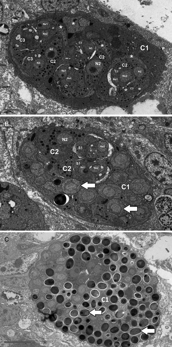
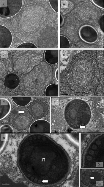
Uninfected M. cochillia primary cells show the characteristic development of secondary and tertiary cells with abundant ribosomes present as well as haplosporosomes and multivesicular bodies (Fig. 3A). Early stage infection with the microsporean results in merogonic proliferation within the primary cell with no involvement of the secondary cells (Fig. 3B). Subsequently, in late stage infections the primary cell of M. cochillia becomes replete with sporoblasts and mature spores (Fig. 3C). The earliest developmental stages of the microsporidian observed were thin-walled uninucleate meronts (Fig. 4A) with subsequent development into binucleate meronts (Fig. 4B). Maturation into sporonts and early sporoblasts is characterized by the formation of a thickened cell wall (Fig. 4C and D) and sporogony proceeds with the intracytoplasmic development of a membranous system and early polar vesicle formation (Fig. 4E). Further differentiation of the sporoblast can be seen in Fig. 4F where spore extrusion apparatus is under construction. Mature spores have dense osmiophilic cytoplasm, a single nucleus, four turns of the polar filament (Fig. 4G, Gi), Golgi apparatus and a polar vesicle (Fig. 4Gii).
Microsporidian hyperparasitism of Marteilia cochillia infecting cockles (Cerastoderma edule). (A). Uninfected M. cochillia. Primary cell (C1) contains three visible secondary cells (C2), each containing tertiary cells (C3). Key: *(cytoplasm of primary, secondary and tertiary cells), N1, N2, N3 (nucleus of primary, secondary and tertiary cells, respectively), Ns1, Ns2 (M. cochilla spore nuclei). Scale 1 µ m. (B) Microsporidian-infected M. cochilla. Microsporidian hyperparasites in this case are predominantly sporonts (arrows) and are confined to the cytoplasm of the paramyxean primary cell (C1). Two secondary cells (C2) of M. cochillia are also visible but not apparently parasitized. Key: as above but with S1 (M. cochillia spore). Scale 1 µ m. (C) Later stage infection of M. cochillia primary cell. The majority of microsporidian life stages are spores (arrows) but all appear to remain confined to the primary cell (C1) of M. cochillia. Scale = 2 µ m. All TEMs.

Microsporidian hyperparasite of Marteilia cochillia infecting cockles (Cerastoderma edule). (A) Uninucleate meronts. Scale 0·5 µ m; (B) Bi-nucleate (though not diplokaryotic) meronts. Scale 0·5 µ m; (C) Transition to sporont (thickening of cell wall). Scale 0·5 µ m; (D) Uninucleate sporont. Scale 0·5 µ m. (E) Maturation of uninucleate sporont to early sporoblast. Formation of membranous system and early polar vesicle within cytoplasm (arrow). Scale 0·5 µ m. (F) Sporoblast with precursors of spore extrusion apparatus (arrow). Scale 0·5 µ m. (G) Mature spore with single nucleus (n) and four turns of the polar filament (arrow). Scale 0·2 µ m. (Gi) Detail of polar filament. (Gii) Detail of Golgi apparatus (arrow) and polar vesicle. All TEMs.

Molecular characterization of microsporidian hyperparasites
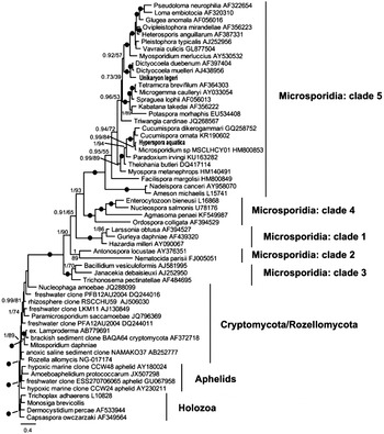
The primer pair V1f/1342AC amplified SSU sequences of the microsporidians parasitizing M. cochillia and M. minutus in cockles. The end fragment of the SSU, ITS and LSU partial region of the microsporidian hyperparasite from M. minutus was amplified using the primer pair HG4F/HG4R. Since these primers did not amplify the same region for the hyperparasite of M. cochillia, a new primer set (Micros_Mcochi_F and Micros_Mcochi_R) was designed. Via these analyses we obtained the following: (1) a sequence of 1911 bp for the microsporidian hyperparasite of M. minutus, and (2) a consensus sequence of 1599 bp for the microsporidian hyperparasite of M. cochillia. The SSU–ITS–LSU sequence of the microsporidian hyperparasite of M. cochillia was 99% similar (over 96% of the compared sequence) to that of an unassigned ‘Microsporidium sp.’ (HM800853), infecting the parasitic copepod Lepeophtheirus hospitalis on starry flounder Platichthys stellatus from British Columbia Canada. The hyperparasite from M. cochillia is also relatively closely related to numerous other microsporidian parasites described infecting marine and freshwater crustacean hosts and, is placed within clade 5 of the phylum according to the classification of Vossbrinck and Debrunner-Vossbrinck (Reference Vossbrinck and Debrunner-Vossbrinck2005). The SSU–ITS–LSU sequence of the microsporidian hyperparasite of M. minutus was 87% similar (over 99% of its sequence) to the crustacean amphipod pathogen Dictyocoela diporeiae (KF537632), similarly within clade 5 of the phylum. It is noteworthy that our results do not support a basal position of these hyperparasites within the phylum Microsporidia but rather, their close relationship to taxa described infecting aquatic crustacean and fish hosts (Fig. 5). We propose erection of a new genus and species to contain the hyperparasite infecting M. cochillia. The new taxon is named H. aquatica n. gen. n. sp.
Bayesian tree containing Hyperspora aquatica and Unikaryon legeri (this study) and 37 other microsporidian SSU sequences obtained from GenBank. Representative sequences from previously described Clades 1–5 are included. The tree is rooted to a subsection of Cryptomycota/Rozellomycota, Aphelid and Holozoan SSU sequences. Posterior probabilities and Bootstrap support appear next to each node.

ISH assay
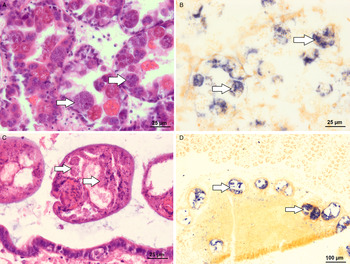
The MicrosMarteiliaISH probe hybridized strongly, without background hybridization, to the microsporidian hyperparasite infecting M. cochillia (cockles). Examination of adjacent tissue sections stained with H&E confirmed that hybridization was localized to hyperparasites within the host Marteilia primary cells. No hybridization signal was detected when the assay was made without probe (negative control, data not shown). The MicrosMarteiliaISH did not hybridize with the microsporidian hyperparasite of M. minutus in cockles. Instead, the MicrosMetacercariaISH probe hybridized strongly with M. minutus in cockles and cross-reactivity was not observed with the microsporidian hyperparasites infecting M. cochillia (Fig. 6).
ISH of Hyperspora aquatica n. gen., n. sp., and Unikaryon legeri. (A, B) Hyperspora aquatica n. gen., n. sp. infecting Marteilia cochillia in cockles. (A) H&E histology showing microsporidian infected paramyxeans in the digestive gland (arrows); (B) positive control ISH labelling of microsporidian hyperparasites (arrows) (unlabelled negative control images not shown). (C, D) Unikaryon legeri infecting Meiogymnophallus minutus in cockles; (C) H&E histology showing trematode parasites infected with U. legeri (arrows); (D) positive control ISH labelling of microsporidian parasites (arrows) (unlabelled negative control images not shown).

Taxonomic summary
Hyperparasite of Marteilia cochillia. Hyperspora aquatica n. gen., n. sp., Genus Hyperspora n. gen.
Definition. Parasite life stages infecting primary cells of M. cochillia (Cercozoa: Marteiliidae) within the digestive epithelium of cockles (C. edule). Spores spherical to ellipsoid, approximately 1·2 × 1·0 µ m with four polar filament coils in a single rank. Merogonic and sporogonic stages occur in direct contact with the cytoplasm of primary cells of M. cochillia. Observed life cycle is unikaryotic and progresses from uninucleate meronts to multi-nucleate meronts prior to initiation of sporogony. Sporogony is marked by thickening of the cell wall, budding of unincucleate sporonts and eventual development of the spore extrusion precursors in uninucleate sporoblasts. Sporoblasts mature to spores, which continue to occupy the cytoplasm of the primary cell of the host. Type species H. aquatica n. gen., n. sp.
-
Type Species: Hyperspora aquatica n. gn., n. sp.
-
Description: As for genus.
-
Diagnosis: Presence of a microsporidian parasite with descriptive features of the genus in the cytoplasm of primary cells of M. cochillia infecting the digestive gland of cockles (C. edule). Diagnosis of morphological features by histology/impression smear and TEM as described herein. Nucleic acid-based diagnosis via PCR amplification, analysis of the defined SSU–ITS–LSU gene sequence and comparison with GenBank Accession number (to be assigned).
-
Type host: Marteilia cochillia Carrasco et al. (Reference Carrasco, Hine, Durfort, Andree, Malchus, Lacuesta, González, Roque, Rodgers and Furones2013)
-
Type locality: Ría de Arousa, Galicia, NW Spain
-
Site of infection: Cytoplasm of primary cells of M. cochillia infecting the digestive gland epithelial cells of cockles (C. edule).
-
Etymology: The generic name refers to the hyperparasitic habit of the parasite in the paramyxean M. cochillia. The specific epithet refers to its presence in aquatic habitats and its phylogenetic similarity to microsporidian pathogens infecting hosts from different trophic positions within such habitats.
-
Type material: Voucher specimens of histological sections stained with H&E and TEM resin blocks have been deposited in the Registry of Aquatic Pathology (RAP) at the Cefas Weymouth Laboratory, UK and in the Registry of Pathology at the Centro de Investigacións Mariñas (CIMA), Spain. Hyperspora aquatica SSU–ITS–LSU gene sequences have been deposited in GenBank under accession numbers (to be assigned).
Hyperparasite of of M. minutus. Unikaryon legeri (Canning and Nicholas, Reference Canning and Nicholas1974).
The microsporidian hyperparasite infecting the digenean M. minutus in Spanish C. edule observed in the current study is presumed (via morphological similarity, data not shown) to be U. legeri Canning and Nicholas (Reference Canning and Nicholas1974). This hyperparasite was previously described infecting the same hosts on the south coast of England as far back as 1897 (Giard, 1897 in Canning and Nicholas, Reference Canning and Nicholas1974). Although a full taxonomic re-description of U. legeri is not required here, it is important to note that for the first time, molecular phylogenetic data have placed the parasite within clade 5 of the phylum Microsporidia (see Vossbrinck and Debrunner-Vossbrinck, Reference Vossbrinck and Debrunner-Vossbrinck2005) with relatively close relationship to parasites infecting crustacean and fish hosts [including numerous Dictyocoela spp. infecting amphipods and, Triwangia caridinae Wang et al. (Reference Wang, Nai, Wang, Solter, Hsu, Wang and Lo2013) infecting caridinid shrimps] (Fig. 5). This is first such information relating to the molecular phylogeny of microspordian hyperparasites of digenean hosts. Unikaryon legeri gene sequences have been deposited in GenBank under accession numbers (to be assigned).
DISCUSSION
In this paper, we describe the type genus and species of a novel microsporidian hyperparasite, H. aquatica n.gn. n.sp. infecting the paramyxid protist M. cochillia, in cockles. Furthermore, we provide clarity on the phylogenetic placement of the hyperparasitic microsporidian U. legeri Canning and Nicholas (Reference Canning and Nicholas1974) infecting the digenean M. minutus, also within cockles. Both Hyperspora and Unikaryon form distinct lineages within clade 5 (Marinosporidia) (Vossbrinck and Debrunner-Vossbrinck, Reference Vossbrinck and Debrunner-Vossbrinck2005), but both cluster phylogenetically with microsporidian pathogens previously described infecting aquatic arthropod and fish hosts (Stentiford et al. Reference Stentiford, Bateman, Feist, Stone and Dunn2013). Based upon SSU rRNA sequence data, H. aquatica is most closely related (99% sequence similarity) to an undesignated microsporidian pathogen (‘Microsporidium sp.’, GenBank accession HM800853) infecting the marine parasitic copepod L. hospitalis on starry flounder (P. stellatus) (Jones et al. Reference Jones, Prosperi-Porta and Kim2012). The SSU rRNA sequence HM800853 most closely resembles that derived from microsporidian pathogens infecting aquatic amphipod crustacean hosts. Jones et al. (Reference Jones, Prosperi-Porta and Kim2012) do not provide a detailed description of the nature of the association between the microsporidian and the host L. hospitalis (and thereby cannot discount the possibility that the host is simply a mechanical vector for the microsporidian from which sequence HM800853 was obtained). Observations relating to two other microsporidian pathogens infecting other Lepeophtheirus spp. within the same study (Facilispora margolisi and Desmozoon lepeophtherii) however, demonstrate susceptibility to microsporidian infection, and to disease, in this host genus (Freeman et al. Reference Freeman, Bell and Sommerville2003). Furthermore, Jones et al. (Reference Jones, Prosperi-Porta and Kim2012) demonstrate that detection of sequence relating to HM800853 was only found associated with the flounder ectoparasite L. hospitalis (suggesting that it was not a widespread contaminant in the environment). The current study, which reveals potential synonymy between H. aquatica, a microsporidian hyperparasite of paramyxid protists infecting bivalve molluscs, and one infecting ectoparasitic copepods on marine fish becomes the major focus for the remainder of this discussion. In particular, it is pertinent to consider whether H. aquatica from paramyxid hosts is also a pathogen of crustacean hosts and further, whether it utilizes a strategy of ‘hitch-hiking’ within the paramyxid in order to transmit between marine molluscs and susceptible crustacean hosts (Lefèvre et al. Reference Lefèvre, Lebarbenchon, Gauthier-Clerc, Missé, Poulin and Thomas2008; Thomas et al. Reference Thomas, Renaud and Poulin2008).
Does H. aquatica hitch-hike to crustacean hosts within Marteilia?
Within the molluscan digestive gland epithelium, H. aquatica infects the primary cell cytoplasm of M. cochillia. In addition, sporogonic developmental stages and eventually spores of Marteilia are also produced by endogeny within this primary cell, which are not infected. The unique cell-within-cell development of paramyxids therefore allows separation of spore development of H. aquatica and Marteilia, such that infection of the primary cell cytoplasm with H. aquatica does not appear to hinder sporogenesis of the paramyxid (since normal development of sporonts and mature spores are commonly observed) (see Fig. 3). The primary cell therefore contains the infective stages of two taxonomically distinct pathogens and conceivably, is a vehicle for co-transmission of paramyxid sporangia (containing sets of mature Marteilia spores), and of H. aquatica, via the digestive gland lumen, presumably for excretion via the gut, to the environment. There is an interesting parallel with the current case, of hyperparasitism with the microsporidian (Neoflabelliforma aurantiae) infecting another spore forming parasite, the myxozoan Aurantiactinomyxon-type actinosporean stage infecting the oligochaete Tubifex tubifex (Morris and Freeman, Reference Morris and Freeman2010). In this case the microsporidian infects both the actinosporean early stage bi-nucleate cells as well as oligochaete cells themselves. The infection progressively inhibits spore formation, unlike the case with H. aquatica where spore formation of the paramyxean host does not seems to be inhibited.
Numerous studies have proposed a role for copepods in the life cycle of sister parasite M. refringens. Audemard et al. (Reference Audemard, Le, Barnaud, Collins, Sautour, Sauria, de, Coustau, Combes and Berthe2002) and later Boyer et al. (Reference Boyer, Chollet, Bonnet and Arzul2013) demonstrated that the pathogen could be detected in the calanoid copepod Paracartia grani via PCR. Furthermore, the pathogen appeared to be localized to ovarian tissues using ISH. Carrasco et al. (Reference Carrasco, López-Flores, Alcaraz, Furones, Berthe and Arzul2007) extended the potential range of intermediate hosts to the orders Cyclopoida and Harpaticoida in a region where Marteilia is endemic within natural stocks of molluscs. Boyer et al. (Reference Boyer, Chollet, Bonnet and Arzul2013) demonstrated that infection prevalence peaks in copepods corresponded to peaks in prevalence in molluscs from the same locations and further, that up to 90% of the planktonic stages of copepods (including adults, nauplii and eggs) were retained during filter-feeding in mussels. Finally, Carrasco et al. (Reference Carrasco, Arzul, Berthe and Furones2008) showed using ISH that the presence of initial infective stages of M. refringens within the gill and mantle epithelia is strong evidence for acquisition of paramyxean parasite infection via filter feeding (see also Kleeman et al. Reference Kleeman, Adlard and Lester2002). The combined evidence from these studies implies that in addition to potential for direct transmission, Marteilia can infect intermediate hosts (such as copepods) and that the parasite may be released from, or contained within, intermediate host life stages (including eggs and larvae) prior to re-infection of susceptible molluscan hosts.
Given that H. aquatica is also likely to be liberated to the environment via the cockle gut, along with infective life stages of M. cochillia, it is conceivable that the microsporidian could be similarly transmitted to copepod (or other crustacean) hosts via the aforementioned route. Supporting evidence for the co-transmission theory arises from studies on the sister paramyxid Paramarteilia sp. in amphipod hosts. Here, although molluscan hosts have not yet been implicated in transmission cycles of Paramarteilia, high co-occurrence of microsporidian and paramyxean pathogens have been reported in Echinogammarus marinus (Short et al. Reference Short, Guler, Yang, Kille and Ford2012). In this case, unpublished work from our laboratory has shown that the microsporidian does not hyperparasitize the paramyxid but rather, the parasites form two separate infections at different locations within the same individual. In this instance, if both pathogens did enter the amphipod simultaneously (as proposed for the M. cochillia/H. aquatica model), the pathogens form separate infections thereafter. In other cases (e.g. Paramarteilia canceri infecting the European edible crab Cancer pagurus), although no consistent associations between paramyxid and microsporidian infection have been reported (Feist et al. Reference Feist, Hine, Bateman, Stentiford and Longshaw2009), several microsporidian genera are known to co-infect this crab host (Stentiford et al. Reference Stentiford, Bateman, Longshaw and Feist2007, Reference Stentiford2008, Reference Stentiford, Bateman, Dubuffett and Stone2011).
Based upon SSU rRNA sequence data, H. aquatica is most closely related (99% sequence similarity) to an undesignated microsporidian infecting a marine parasitic copepod L. hospitalis (Jones et al. Reference Jones, Prosperi-Porta and Kim2012). Interestingly, cross-taxa transmission has already been demonstrated for another microsporidian parasite (D. lepeophtherii (=Paranucleospora theridion) from its ectoparasitic copepod host (Lepeophtheirus salmonis) to fish (Nylund et al. Reference Nylund, Andersen, Saevareid, Plarre, Watanabe, Arnesen, Karlsbakk and Nylund2011). Although it is beyond the remit of the current study, apparent synonymy between H. aquatica and the ‘Microsporidium sp.’ infecting Lepeophtheirus sp. on flounder raises the potential for further cross-taxa transmission of microsporidians such as H. aquatica from protists (within bivalves) to crustaceans. Extending this concept to the study of three host transmission (protist, crustacean, fish) is worthy of additional consideration, particularly at sites where H. aquatica is prevalent and where co-habiting fauna (crustaceans, fish) can be collected during environmental surveys.
Hyperspora does not group with basal microsporidians
In recent years, a consensus molecular phylogenetic-based viewpoint has emerged which affiliates the phylum Microsporidia with the Fungi (Lee et al. Reference Lee, Corradi, Byrnes, Torres-Martinez, Dietrich, Keeling and Heitmand2008; Capella-Gutiérrez et al. Reference Capella-Gutiérrez, Marcet-Houben and Gabaldón2012). Whether they branch within (e.g. Tanabe et al. Reference Tanabe, Watanabe and Sugiyamaaff2002) or outside of the group (Jones et al. Reference Jones, Forn, Gadelha, Egan, Bass, Massana and Richards2011a , Reference Jones, Richards, Hawksworth and Bass b ; Capella-Gutierrez et al. Reference Capella-Gutiérrez, Marcet-Houben and Gabaldón2012), however, has been a topic of debate. More recent multi-gene evidence places Cryptomycota (=Rozellomycota) as sister to fungi (James et al. Reference James, Pelin, Bonen, Ahrendt, Sain, Corradi and Stajich2013), and rRNA gene phylogenies show a relationship between this clade, ‘core’ fungi, and aphelids (intracellular parasitoids of algae) although their relative branching orders are unresolved (Karpov et al. Reference Karpov, Mikhailov, Mirzaeva, Mirabdullaev, Mamkaeva, Titova and Aleoshin2013, Reference Karpov, Mamkaeva, Aleoshin, Nassonova, Lilje and Gleason2014; Corsaro et al. Reference Corsaro, Walochnik, Venditti, Steinmann, Müller and Michel2014). Several new lineages have been characterized branching within or between Cryptomycota and Microsporidia: Mitosporidium (Haag et al. Reference Haag, James, Pombert, Larsson, Schaer, Refardt and Ebert2014), Paramicrosporidium (Corsaro et al. Reference Corsaro, Walochnik, Venditti, Steinmann, Müller and Michel2014) and Nucleophaga (Corsaro et al. Reference Corsaro, Walochnik, Venditti, Steinmann, Müller and Michel2014). Mitosporidium and Paramicrosporidium are morphologically intermediate between Cryptomycota and Microsporidia, whereas Nucleophaga, although similar to Paramicrosporidium, has fewer microsporidian characters, and are more Rozella-like (Corsaro et al. Reference Corsaro, Walochnik, Venditti, Steinmann, Müller and Michel2014). Interestingly, our rDNA phylogenetic analyses (Fig. 5), the first to simultaneously analyse a broad representation of environmental cryptomycotans together with all available characterized lineages, show that the evolution of (albeit rudimentary) infection machinery (e.g. polar filament, anchoring disc) in taxa such as Mitosporidium occurred prior to the more extreme genome reduction events typical of many (sequenced) members of the phylum Microsporidia per se, and that there is no clear phylogenetic pattern to the development of these characters within the cryptomycotan radiation. Clearly more sampling and characterization of lineages within Cryptomycota as currently circumscribed and those that might branch between these and the longer-branch microsporidians will be very important to understand the evolution of highly specialized modes of parasitism seen in the latter.
An interesting group, the so-called ‘metchnikovellids’ are conspicuously absent from modern phylogenies of Microsporidia and their basal lineages. These gregarine-infecting parasites are morphologically similar to both the intranuclear parasite of amoeba, Paramicrosporidium (Rotari et al. Reference Rotari, Paskerova and Sokolova2015) and, to the gut-infecting parasite of Daphnia, Mitosporidium (see Haag et al. Reference Haag, James, Pombert, Larsson, Schaer, Refardt and Ebert2014). Their affiliation with Microsporidia, and their proposed ‘basal’ position was stated early by Vivier (Reference Vivier1965) and later, by Sprague (Reference Sprague, Bulla and Cheng1977), who suggested in fact that they may represent rudimentary forms (at the time, erecting the class Rudimicrosporea, consist of a single order, the Metchnikovellida). Whilst the literature tentatively reports SSU rDNA sequence data to support an affiliation of the metchnikovellids with the aforementioned basal groups (Simdianov et al. Reference Simdianov, Yudina and Aleoshin2009), primary data are absent. Nevertheless, morphological similarities between representatives (such as Metchnikovella incurvata infecting gregarines, Sokolova et al. Reference Sokolova, Paskerova, Rotari, Nassonova and Smirnov2013) and other members of this basal group infecting other single-celled eukaryotes (e.g. amoebae and algae), including Mitosporidium and Paramicrosporidium suggest that metchnikovellids branch within the cryptomycota radiation as defined by Karpov et al. (Reference Karpov, Mamkaeva, Aleoshin, Nassonova, Lilje and Gleason2014). These cases clearly contrast with the phylogenetic position and description of H. aquatica provided in this paper. In the latter, not only does meront, sporont and spore morphology fall within the standard variation observed across the phylum Microsporidia, but also, rRNA-based phylogenies place it well within a previously defined microsporidian group (Clade 5).
In summary, we demonstrate the presence of non-basal lineages of Microsporidia, morphologically and phylogenetically similar to taxa infecting aquatic crustaceans and fish, infecting paramyxid and digenean parasites of molluscs. Our results propose that these hyperparasites may have exploited the parasitic lifestyle of their hosts to ‘hitch hike’ between hosts at different trophic levels. Further, it is proposed that those cases of paramyxean and digenean hyperparasites reported herein represent more recent acquisitions than those infecting other hosts within the developing ‘basal’ clade of the Cryptomycota.
ACKNOWLEDGEMENTS
Beatriz Bustos, Elena Penas and Maribel Meléndez provided technical assistance. Sample processing and examination with transmission electron microscopy were performed in the Servicio de Microscopía Electrónica of CACTI (University of Vigo) with technical assistance by Inés Pazos.
FINANCIAL SUPPORT
The authors acknowledge support of the UK Department for Environment, Food and Rural Affairs (Defra) under project no. C6560 (to G. D. S. and D. B.), the European Commission under project no. C6928 (to G. D. S and K. B.) and the Consellería do Medio Rural e do Mar da Xunta de Galicia, through the project PGIDIT-CIMA 12/03 (to A. V.).