Introduction
The oogram methodology was initially described to evaluate slight qualitative alterations in the oviposition of Schistosoma mansoni. In the paper describing this method, Pellegrino et al. (Reference Pellegrino, Oliveira, Cunha and Siqueira1962) showed that when active drugs are given to mice infected with the parasite, a progressive change in the number of S. mansoni eggs and in the percentage of viable mature and immature eggs of distinct stages occurs in the intestine wall. This change is explained, according to these authors, by the interruption of oviposition due to the death of adult worms, mainly female, or momentary effects on them, and the consequent maturation of viable eggs already there. Thus, alterations observed in oograms can be irreversible or reversible.
In comparison with other methods for assessing the oviposition of the parasite, oogram methodology is easy to execute and provides results more quickly. Thus, it has been utilized since its establishment for the screening of new anti-schistosomal drugs, in the research and clinical control of schistosomiasis. In both cases its usefulness has already been thoroughly demonstrated (Pellegrino et al., Reference Pellegrino, Oliveira, Cunha and Siqueira1962; Cunha et al., Reference Cunha, Cançado, Pellegrino and Oliveira1963, Reference Cunha, Cançado and Rezende1987; Cançado et al., Reference Cançado, Cunha, Carvalho and Cambraia1965; Pellegrino & Faria, Reference Pellegrino and Faria1965; Katz et al., Reference Katz, Pellegrino and Memória1966; Katz & Pellegrino, Reference Katz and Pellegrino1974; Cunha, Reference Cunha1982; Coelho & Pereira, Reference Coelho and Pereira1991; Araújo et al., Reference Araújo, Kohn and Katz1999).
Furthermore, oogram methodology has been used to assess the biology of the parasite. Studies have investigated the oviposition pattern of S. mansoni in mice exposed to irradiated cercariae (Oliveira et al., Reference Oliveira, Katz and Pellegrino1971), the fecundity of only one trematode couple (Valadares et al., Reference Valadares, Coelho, Pellegrino and Sampaio1981), the effects of host dietary deficiency or overfeeding on the worms (Akpom, Reference Akpom1978; Neves, Reference Neves2006), vaccinal assays (Pacheco et al., Reference Pacheco, Zucconi, Mati, Garcia, Miyoshi, Oliveira, Melo and Azevedo2005, Reference Pacheco, Mati, Castro, Dorella, Oliveira, Miyoshi, Melo and Azevedo2008; Cardoso et al., Reference Cardoso, Macedo, Gava, Kitten, Mati, Melo, Caliari, Almeida, Venancio, Verjovski-Almeida and Oliveira2008; Garcia et al., Reference Garcia, Fonseca, Pacifico, Durães, Marinho, Penido, Caliari, Melo, Pinto, Barsante, Cunha-Neto and Oliveira2008) and have evaluated S. mansoni egg release, or not, into the intestinal lumen in mice treated with drugs that can change the immune response of the vertebrate host (Hermeto et al., Reference Hermeto, Bicalho, Silva, Melo and Pereira1994; Mati et al., Reference Mati, Freitas and Melo2010).
On the other hand, the ratio of second-stage S. mansoni eggs per g intestine/number of female parasites initially suggested by Pacheco et al. (Reference Pacheco, Zucconi, Mati, Garcia, Miyoshi, Oliveira, Melo and Azevedo2005) is a promising way to verify the individual fecundity of parasitic females and it has already been employed in other studies (Pacheco et al., Reference Pacheco, Mati, Castro, Dorella, Oliveira, Miyoshi, Melo and Azevedo2008; Mati et al., Reference Mati, Freitas and Melo2010). Nevertheless, complementary information, such as detailed statistical evaluation, is still necessary for a better understanding and utilization of this ratio in experimental schistosomiasis.
Hence, the higher number of dead eggs of the trematode observed by the oogram method during acute infection in the small intestine of Swiss mice that received pentoxifylline (PTX), a substituted methylxanthine with a wide range of anti-inflammatory effects, has not been sufficiently explained (Mati et al., Reference Mati, Freitas and Melo2010). In addition, no significant alteration in parasite fecundity was verified in rodents of this strain treated with this drug, which has been used as both a haemorheological agent and tumour necrosis factor-α (TNF-α) inhibitor. In fact, PTX improves red blood cell deformability and decreases the potential for platelet aggregation and blood viscosity. Its immunomodulatory properties are also now well established in several pathological processes (Ward & Clissold, Reference Ward and Clissold1987; Tilg et al., Reference Tilg, Eibl, Pichl, Gächter, Herold, Brankova, Huber and Niederwieser1993; Samlaska & Winfield, Reference Samlaska and Winfield1994; Bienvenu et al., Reference Bienvenu, Doche, Gutowski, Lenoble, Lepape and Perdrix1995; Heyderman et al., Reference Heyderman, Klein, Daramola and Levin1995; Louie et al., Reference Louie, Baltch, Franke, Ritz, Smith, Singh and Gordon1996; Navarro et al., Reference Navarro, Punzón, Pizarro, Fernández-Cruz, Fresno and Muñoz-Fernández1996; Muniz-Junqueira, Reference Muniz-Junqueira2007), including intestinal schistosomiasis (Reis et al., Reference Reis, Ventura, Souza, Arana-Pino, Pelajo-Machado, Pereira, Lenzi, Conceição and Takyia2001; Xiong et al., Reference Xiong, Zhu and Luo2003; El-Lakkany & Nosseir, Reference El-Lakkany and Nosseir2007; Mati et al., Reference Mati, Freitas and Melo2010; Rabia et al., Reference Rabia, Nagy, Aly, Mohamed, El-Assal and El-Amir2010).
The aim of the present study was to evaluate the individual fecundity of the female parasite and the number of eggs in the intestine of inbred mice of the AKR/J strain (Ben-Efraim & Cinader, Reference Ben-Efraim and Cinader1964; Wheat et al., Reference Wheat, Wetsel, Falus, Tack and Strunk1987; Curry et al., Reference Curry, Else, Jones, Bancroft, Grencis and Dunne1995; Guerassimov et al., Reference Guerassimov, Hoshino, Takubo, Turcotte, Yamamoto, Ghezzo, Triantafillopoulos, Whittaker, Hoidal and Cosio2004) subjected to short-term treatment with PTX. A further aim was to carry out a better appraisal, including the study of statistical parameters, of the promising utilization of the above-mentioned ratio in studies regarding the oviposition of S. mansoni and factors that change it. Without doubt, AKR/J mice exhibit immunological aspects that make them an interesting model for schistosomiasis, even for studies of immunomodulatory compounds. This issue is discussed, and additional considerations concerning aspects of experimental infection with the trematode by the peritoneal route in AKR/J mice and the current applications for the oogram method are also approached.
Materials and methods
Experimental infection and treatment of mice
Fifty 6- to 8-week-old male AKR/J mice, weighing 25 ± 2 g, were infected by the intraperitoneal route with 60 ± 5 S. mansoni cercariae (LE strain). Rodents were allocated to five experimental groups and another five groups were maintained without treatment as controls. Short-term treatments with PTX [1-(5-oxyohexyl)-3,7-dimethylxanthine, 1-(5-oxyohexyl) theobromine] (Trental®) at a dose of 200 mg/kg were performed daily by the subcutaneous route during five consecutive days (Mati et al., Reference Mati, Freitas and Melo2010), which started at 56, 63, 70, 77 and 84 days post-infection (DPI).
Recovery of adult worms from the portal system and peritoneal cavity
At 2 days after the end of each treatment (days 63, 70, 77, 84 and 91 post-infection), rodents were killed by cervical dislocation according to the local animal experimentation ethical committee [Comitê de Ética em Experimentação Animal (CETEA) of the Universidade Federal de Minas Gerais (UFMG)]. Worms were then recovered from the portal system through perfusion (Pellegrino & Siqueira, Reference Pellegrino and Siqueira1956). Also, in order to obtain parasitic forms from the peritoneal cavity, a methodology previously described was employed (Pereira et al., Reference Pereira, Pellegrino, Valadares, Mello and Coelho1974; Bicalho et al., Reference Bicalho, Melo and Pereira1993; Bicalho, Reference Bicalho2003). All worms and free granulomas were inspected, counted and fixed in 10% formalin.
Oogram procedures
Fragments of the small intestine (terminal ileum), of length 1 cm, were cut from all animals. Later on, these fragments were transferred to Petri dishes containing isotonic saline, opened lengthwise with scissors to remove the excess mucus, partially dried on absorbent paper, weighed and placed between a glass slide and a plastic cover. This preparation was pressed on a rubber surface padded with absorbent paper (Pellegrino et al., Reference Pellegrino, Oliveira, Cunha and Siqueira1962; Pellegrino & Faria, Reference Pellegrino and Faria1965). The terminal ileum was used because it is known that this segment of intestine is the preferred oviposition site of various strains of S. mansoni in mice (Valadares et al., Reference Valadares, Coelho, Pellegrino and Sampaio1981).
With the aid of a light microscope (magnification × 100), all eggs in each slide were counted and classified according to their developmental stage, following specific features of each stage described below. In case of doubt about the classification of an egg, higher (magnification × 400) was used in microscopic analysis.
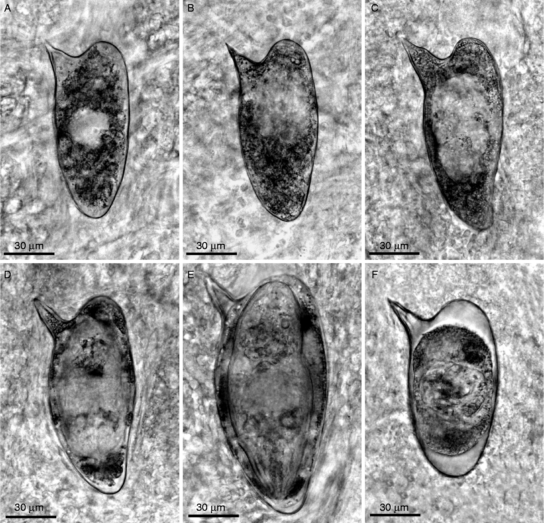
Qualitative oogram evaluation considered the shell, and dead and viable (mature and immature) eggs. Mature eggs contained an already developed miracidium and immature ones were classified into four different development stages. These were: first stage, when the embryo had one-third of the diameter of the egg; second stage, embryo with one-half of the length of the egg; third stage, embryo with two-thirds of the length of the egg; fourth stage, embryo almost entirely occupying the eggshell (Pellegrino et al., Reference Pellegrino, Oliveira, Cunha and Siqueira1962).
For the evaluation of quantitative oograms, the formula ‘number of eggs/g of tissue = number of eggs in intestinal fragment/weight of fragment in mg × 1000’ was employed (Cançado et al., Reference Cançado, Cunha, Carvalho and Cambraia1965; Katz et al., Reference Katz, Pellegrino and Memória1966; Katz & Pellegrino, Reference Katz and Pellegrino1974; Hermeto et al., Reference Hermeto, Bicalho, Silva, Melo and Pereira1994). Additionally, in order to verify the number of eggs per female parasite in a day, the ratio of second-stage S. mansoni eggs per g intestine/number of female parasites recovered from the portal system was utilized (Pacheco et al., Reference Pacheco, Zucconi, Mati, Garcia, Miyoshi, Oliveira, Melo and Azevedo2005, Reference Pacheco, Mati, Castro, Dorella, Oliveira, Miyoshi, Melo and Azevedo2008; Mati et al., Reference Mati, Freitas and Melo2010). Female worms from the peritoneal cavity were not used in the analysis of the rates their eggs, laid in the peritoneum, produce free granulomata in situ that do not normally reach the intestine.
Statistical analysis
Statistical analyses were carried out using computer software Statistica 6.0 for Windows (StatSoft Inc., Tulsa, Oklahoma, USA). Data obtained were submitted to Student's t-test or analysis of variance (ANOVA) followed by the Tukey test, as appropriate. For statistical assessment of which immature stage of S. mansoni egg is the most appropriate for evaluation of parasite fecundity, evidently performed before analysis on worm individual fecundity, ratios of each one of the four stages of immature eggs (first to fourth) per g intestine/number of female S. mansoni were compared. The coefficient of variation (CV) was utilized for this comparison of ratios among control and treated groups in the five different times of infection studied.
Results
Recovery of parasitic forms from the peritoneal cavity and portal system
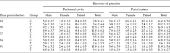
The mean number and standard deviation of male and female adult trematodes recovered from both the portal system and peritoneal cavity of rodents at different times of infection for control and treated groups are displayed in table 1. No significant change in worm burden was observed among AKR/J mice treated with PTX compared with their respective controls, despite macroscopic examination of abdominal viscera in AKR/J mice treated with the drug revealing a visible mesenteric vasodilatation.
Number of Schistosoma mansoni (means values±standard deviation) recovered from the peritoneal cavity and portal system of AKR/J mice treated with pentoxifylline.

C, Control group; T, treated group.
The total percentage of cercariae recovered as adult worms from the portal system and peritoneal cavity in the five periods studied ranged, respectively, between 36.7 and 48.3% and 30.8 and 46.0% in the control and treated groups. Treatment with PTX did not alter the sex ratio of parasites, and the average number of male schistosomes was always higher than female ones in all groups evaluated.
In the peritoneal cavity of both control and treated AKR/J mice, beyond the presence of S. mansoni couples, immature eggs and free granulomata around mature eggs of the parasite were also observed. In mice of this strain, treatment with PTX did not prevent the oviposition of female trematodes in situ. Eggs and granulomata were fixed and stored for more detailed study in the future.
Oogram analysis
The results provided by the oogram method were based on the examination of more than 50,000 S. mansoni eggs.
Treatment with the immunomodulatory drug PTX
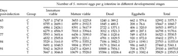
No change in the oviposition kinetics of worms was detected by oogram. Schistosoma mansoni eggs gradually passed through all four immature stages before they reached maturity. In addition, the development and distribution of eggs according to their stage presented minor intrinsic variations in treated and control groups (tables 2 and 3; figs 1 and 2).
Number of Schistosoma mansoni eggs (immature viable, mature viable, dead, eggshell and total; mean values ± standard deviations) observed through oogram methodology in the intestinal fragments of AKR/J mice treated with pentoxifylline at different times of infection.

C, Control group; T, treated group.
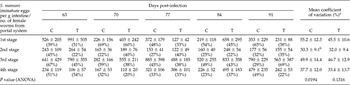
Immature eggs of Schistosoma mansoni observed by oogram in the small intestine of AKR/J mice treated with pentoxifylline.

For each stage of the parasite eggs the mean number, the standard deviation and the coefficient of variation (percentage) are presented.
C = control group, T = treated group.
a Mean of coefficient of variation was utilized to compare the variability by ANOVA among the stages of the S. mansoni immature eggs.
b Statistical difference by the post hoc Tukey test when compared with 1st stage (0.0232).
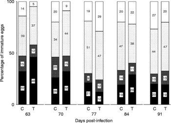
Percentage distribution of each stage (black area, first stage; grey shaded area, second stage; dotted area, third stage; white area, fourth stage) of Schistosoma mansoni immature eggs verified by oogram in intestinal fragments of AKR/J mice infected with the parasite and treated with pentoxifylline. C, Control group; T, treated group.

Schistosoma mansoni eggs from small intestine of AKR/J mice using oogram methodology: (A) first stage, (B) second stage, (C) third stage, (D) fourth stage, (E) mature, (F) dead – retracted embryo.

Although the mean numbers of dead and total eggs were slightly higher in all treated mice groups in comparison with the respective control groups, a significant difference was not observed (table 2). Similarly, there was no statistical difference between treated and control groups for mature and immature eggs.
Taking into consideration for each period studied the percentage proportions (not directly presented) between viable (immature and mature) and non-viable (dead) eggs, the number of non-viable eggs was proportionally higher in the groups of mice that had received PTX, and remained high as the time of infection progressed. Given all ten groups of animals in the five periods analysed, the only group that showed a mean number of dead eggs numerically superior to the average of immature and mature eggs was the group of AKR/J mice who received treatment with PTX and were killed in the last period analysed. Indeed, at 91 DPI the number of dead eggs virtually equalled the number of viable eggs in treated mice, in comparison with that observed in the control group (table 2).
Furthermore, as observed by evaluation of the ratio of second-stage S. mansoni eggs per g intestine/number of female parasites recovered from the portal system, treatment with PTX also did not alter the individual fecundity of S. mansoni females (table 3).
Ratio of eggs per g intestine/number of female worms
The average, standard deviation and CV of the ratios correlated with each evolutionary stage of immature eggs per g tissue/number of female parasites recovered from the portal system are shown in table 3.
In the five periods considered in the present study, the CV of second-stage eggs was the lowest in 40% of the cases (2/5 in both control and treated groups). In fact, the second and fourth stages of S. mansoni eggs presented, respectively, the smallest variabilities, with mean CVs of 30.3 ± 9.1% and 37.7 ± 12.0% in the control groups and 32.0 ± 9.4% and 33.4 ± 13.7% in the treated groups.
Comparison among the average CV of each one of the four stages of immature eggs by ANOVA was statistically significant (P= 0.0194), considering only the control groups, and statistical difference by the post hoc Tukey test was obtained between the first (higher CV) and second (lower CV) stages (P= 0.0232). On the other hand, no significant difference was observed if the five treated groups were independently analysed (P= 0.1316).
In addition, when the percentage of each stage of immature eggs in relation to the total number of immature eggs was considered, the second and fourth stages usually presented smaller values than the first and third stages (fig. 1). In the five periods of infection studied, percentages of second-stage eggs ranged from 9 to 20% in untreated and 11–15% in treated groups, whereas the corresponding percentages for fourth-stage eggs ranged from 14 to 27% and 5–29%. Moreover, second and third stages of S. mansoni eggs in the small intestine had the lowest variabilities among immature eggs.
Discussion
Although the major focus of the present study was the results provided by the oogram method in experimental schistosomiasis after treatment of AKR/J mice with PTX, other interesting aspects about the biology of S. mansoni in this model were verified.
In fact, up to now, this strain of rodents had not been utilized either in the evaluation of immunomodulatory drugs in the course of this parasitic infection, or in oogram analysis. AKR/J mice present deficiency in the complement system, a high incidence of leukaemia (Ben-Efraim & Cinader, Reference Ben-Efraim and Cinader1964; Wheat et al., Reference Wheat, Wetsel, Falus, Tack and Strunk1987), and in the peritoneal cavity of these rodents the complete development of the trematode occurs, reaching sexual maturity and laying eggs in situ (Bicalho et al., Reference Bicalho, Melo and Pereira1993; Bicalho, Reference Bicalho2003; Mati, Reference Mati2009). Therefore, AKR/J mice characterize a permissive and interesting model for research on S. mansoni biology. In the present study, similar findings in relation to the peritoneal cavity of these rodents were observed, mainly concerning the recovery of peritoneal granulomata. Furthermore, the oviposition kinetics evaluated by oogram methodology occurred typically, as evidenced by percentages of occurrence of the four stages of S. mansoni immature eggs, especially when considering the lowest percentage recorded for eggs of the second stage, which corroborated oogram results of mice infected with different strains of S. mansoni (Valadares et al., Reference Valadares, Coelho, Pellegrino and Sampaio1981).
AKR/J mice seem to have a tendency to present a polarized T-helper 1 cell (Th1) immune response (Curry et al., Reference Curry, Else, Jones, Bancroft, Grencis and Dunne1995; Guerassimov et al., Reference Guerassimov, Hoshino, Takubo, Turcotte, Yamamoto, Ghezzo, Triantafillopoulos, Whittaker, Hoidal and Cosio2004). It is possible that this feature explains, at least in part, the peculiarities in the immunomodulation observed in mice of this lineage infected with S. mansoni, since a reduction in the cellular infiltration in granulomata, which are large and with many eosinophils, at the beginning of their development and thus also in the hepatic pathology, is possibly associated with an increased production of Th1 cytokines in experimental schistosomiasis (Hoffmann et al., Reference Hoffmann, Cheever and Wynn2000, Sandor et al., Reference Sandor, Weinstock and Wynn2003; Pacheco et al., Reference Pacheco, Mati, Castro, Dorella, Oliveira, Miyoshi, Melo and Azevedo2008). In fact, liver granulomatous lesions are more pronounced and present a greater diameter in AKR/J mice about 2 months after infection, probably due to their uncommon process of modulation of the immune response. However, after this period, a significant regression in the size of hepatic granulomata in the AKR/J strain is verified and then hepatic lesions of these mice become more similar to those verified in Swiss mice, but with greater cellularity and lower fibre content. Thus, changes induced by drugs that alter the host immune response, such as PTX, might be easily perceived in mice of the AKR/J strain, an interesting model to study immunomodulatory drugs in this parasitic infection (Tristão, Reference Tristão2001; Mati, Reference Mati2009).
It is accepted that the passage of S. mansoni mature eggs from inside the mesenteric vessels into the intestinal lumen is due to the occurrence of an effective inflammatory reaction around these eggs and that the interruption of this response leads to them not being released (Doenhoff et al., Reference Doenhoff, Musallam, Bain and McGregor1978; Lenzi et al., Reference Lenzi, Lenzi and Sobral1987; Hermeto et al., Reference Hermeto, Bicalho, Silva, Melo and Pereira1994). In this regard, indirect evaluations of interference with the release or the retention of eggs in host tissues could be carried out, including studies concerning the changes arising from treatment with drugs that act on the immune system, such as PTX.
On the other hand, effects of treatment with this compound on aspects of the host–parasite relationship, such as adaptation of the parasite to the vertebrate host, the modulation of immunological response and the hepatic pathology in both acute and chronic infections are partially known (Reis et al., Reference Reis, Ventura, Souza, Arana-Pino, Pelajo-Machado, Pereira, Lenzi, Conceição and Takyia2001; Xiong et al., Reference Xiong, Zhu and Luo2003; El-Lakkany & Nosseir, Reference El-Lakkany and Nosseir2007; Mati, Reference Mati2009; Mati et al., Reference Mati, Freitas and Melo2010; Rabia et al., Reference Rabia, Nagy, Aly, Mohamed, El-Assal and El-Amir2010).
In the present study, no statistical difference was detected in the number of parasites recovered from the peritoneal cavity and portal system between the treated and control groups, indicating an absence of schistosomicidal effects of PTX, as already verified in C57 BL/6 (Rabia et al., Reference Rabia, Nagy, Aly, Mohamed, El-Assal and El-Amir2010) and Swiss mice (Mati et al., Reference Mati, Freitas and Melo2010). Besides, the macroscopical vasodilatation of the abdominal mesenteric vessels may be correlated with changes in the circulatory system due to treatment with PTX, as was suggested in our previous paper addressing the administration of the drug in this conventional strain of rodent. Furthermore, treatment of AKR/J mice with PTX at five different times of infection did not affect the occurrence of oviposition by peritoneal worms or the worm burden, as verified in Swiss mice. In addition, the treatment did not significantly change the average ratios of the second-stage S. mansoni eggs per g intestine/number of female worms from the portal system, despite the drug decreasing TNF-α levels (Ward & Clissold, Reference Ward and Clissold1987; Tilg et al., Reference Tilg, Eibl, Pichl, Gächter, Herold, Brankova, Huber and Niederwieser1993; Samlaska & Winfield, Reference Samlaska and Winfield1994; Bienvenu et al., Reference Bienvenu, Doche, Gutowski, Lenoble, Lepape and Perdrix1995; Heyderman et al., Reference Heyderman, Klein, Daramola and Levin1995; Louie et al., Reference Louie, Baltch, Franke, Ritz, Smith, Singh and Gordon1996; Navarro et al., Reference Navarro, Punzón, Pizarro, Fernández-Cruz, Fresno and Muñoz-Fernández1996; Muniz-Junqueira, Reference Muniz-Junqueira2007; El-Lakkany et al., Reference El-Lakkany, Seif-El-Din and Obeid2011). Previously, it was accepted that TNF-α was directly correlated with the fecundity of the schistosome (Amiri et al., Reference Amiri, Locksley, Parslow, Sadick, Rector, Ritter and McKerrow1992; Hagan et al., Reference Hagan, Garside and Kusel1993; Haseeb et al., Reference Haseeb, Solomon and Palma1996), but the administration of recombinant murine TNF-α in mice with severe combined immunodeficiency (SCID) did not alter granuloma formation in the liver or egg laying by S. mansoni and Schistosoma japonicum (Cheever et al., Reference Cheever, Poindexter and Wynn1999). Therefore, alterations in the individual fecundity of the parasite in rodents treated with PTX could not be expected, as verified.
In opposition to this finding, Reis et al. (Reference Reis, Ventura, Souza, Arana-Pino, Pelajo-Machado, Pereira, Lenzi, Conceição and Takyia2001) and El-Lakkany & Nosseir (Reference El-Lakkany and Nosseir2007) showed, by liver histopathology, a decrease in the number of hepatic eggs, and suggested a reduction in parasite oviposition in Swiss mice treated with PTX. However, they carried out distinct protocols of treatment with a lower dosage and longer time of drug administration than the protocol utilized in the present study. In addition, a significant increase was also observed, although slight (P < 0.05), in the number of dead eggs in the intestine of Swiss mice during acute infection when rodents that received short-term treatment with PTX were compared with untreated ones (Mati et al., Reference Mati, Freitas and Melo2010); nevertheless, no conclusive explanation has been made about this finding. A toxic effect of treatment with this drug on S. mansoni eggs is not very probable, and the non-existence of alterations in the number and distribution of immature eggs and in the ratio of second-stage S. mansoni eggs per g tissue/number of female worms in AKR/J mice treated with PTX confirm the absence of alterations in oviposition and egg viability, such as in Swiss mice. It is possible that the higher number of dead eggs previously observed in acute infection of Swiss mice and the non-significant increase in the mean number of dead eggs in all groups of AKR/J mice treated in the present study were correlated with changes in the intensity of the inflammatory process in the intestines, and maybe with the elimination of eggs into the intestinal lumen. Indeed, averages of dead and also total eggs were higher in animals treated with PTX, which might indicate only a subtle trend of retention of parasite eggs in host tissues, since these variations were not statistically significant in evaluated mice, and the mean number of mature eggs did not present the same profile of apparent increase in treated groups.
On the other hand, Hermeto et al. (Reference Hermeto, Bicalho, Silva, Melo and Pereira1994) observed, also utilizing the oogram method, higher number of eggs trapped in tissues of Swiss mice treated with dexamethasone. This reflects the effects of this drug, a potent glucocorticoid, on the passage of mature viable eggs of the parasite into the organ lumen of the host, probably characterizing a reduction in their elimination as a result of a reduction in the inflammatory infiltration around the eggs in the gut. However, in contrast to what was observed for dexamethasone, a significant blocking in the passage of mature eggs into the intestinal lumen of AKR/J mice treated with PTX was not verified.
Therefore, the effects of PTX in schistosomiasis seem not to be primarily and strongly immunosuppressant. Without doubt, major alterations arising out of PTX administration in rodents continue to be those observed previously in the liver (Reis et al., Reference Reis, Ventura, Souza, Arana-Pino, Pelajo-Machado, Pereira, Lenzi, Conceição and Takyia2001; Xiong et al., Reference Xiong, Zhu and Luo2003; El-Lakkany & Nosseir, Reference El-Lakkany and Nosseir2007; Mati et al., Reference Mati, Freitas and Melo2010; Rabia et al., Reference Rabia, Nagy, Aly, Mohamed, El-Assal and El-Amir2010). The liver is the organ where the immunological modulation process and its respective alterations on schistosomotic granulomata are more evident (Andrade & Warren, Reference Andrade and Warren1964; Carvalho et al., Reference Carvalho, Alvarenga and Melo1986; Silva et al., Reference Silva, Fernandes, Barbosa Junior, Oliveira and Andrade2000; Tristão et al., Reference Tristão, Melo, Vasconcelos and Grossi2000). Hence, data obtained are themselves more suggestive of immunomodulatory than immunosuppressant or just anti-inflammatory activity of PTX in experimental schistosomiasis.
Furthermore, Rabia et al. (Reference Rabia, Nagy, Aly, Mohamed, El-Assal and El-Amir2010) confirmed histopathological results showing that treatment with PTX of mice infected with S. mansoni induced a significant reduction in levels of cytokines of both Th1 and T-helper 2 cell (Th2) responses in serum, respectively, interferon-γ (IFN-γ) and interleukin-4 (IL-4). An increase in the value of the T-regulatory cell (Treg) cytokine, interleukin-10 (IL-10), was also observed. In addition, it is worth noting that this anti-inflammatory cytokine is involved with the delay in the genesis and size reduction of granulomatous lesions (Flores-Villanueva et al., Reference Flores-Villanueva, Zheng, Strom and Stadecker1996; Wynn et al., Reference Wynn, Cheever, Williams, Hieny, Caspar, Kühn, Müller and Sher1998; Sadler et al., Reference Sadler, Rutitzky, Stadecker and Wilson2003; Hesse et al., Reference Hesse, Piccirillo, Belkaid, Prufer, Mentink-Kane, Leusink, Cheever, Shevach and Wynn2004). As mentioned above, the absence of significant changes resulting from treatment with PTX on the passage of parasite eggs into the intestinal lumen of mice could be considered desirable and beneficial. Possibly this drug might be used in the future, mainly during chronic infection, as an antifibrotic compound in schistosomiasis (Xiong et al., Reference Xiong, Zhu and Luo2003; El-Lakkany & Nosseir, Reference El-Lakkany and Nosseir2007; Rabia et al., Reference Rabia, Nagy, Aly, Mohamed, El-Assal and El-Amir2010; El-Lakkany et al., Reference El-Lakkany, Seif-El-Din and Obeid2011), without which an excess in retention of S. mansoni eggs in host tissues and a consequent worsening of the pathological process is induced.
In statistical comparison to highlight which immature stage of S. mansoni eggs would be the most appropriate for the analyses of the individual fecundity of parasite females, the average values of the specific CV for the four ratios referring to each one of the immature stages of S. mansoni eggs per g intestine/number of female parasites were utilized. A similar pattern of variability among the egg stages has already been observed, as well as an occasional necessity of dividing the raw data obtained by the oogram analysis by the number of females recovered from the portal system, to reduce the possibility of errors (unpublished results). However, this division is not necessary in qualitative studies, as in those for screening of schistosomicidal drugs, or in cases where a reasonable homogeneity of data of the recovery of worms by perfusion of the portal system was obtained. The mean CVs of the second and fourth stages of S. mansoni eggs were smaller than those observed for the first and third stages, indicating lesser variability of these egg stages. That is, in the present study, the second and fourth stages of the parasite eggs each presented a more homogeneous distribution and are both probably the best ratios for the study of the individual fecundity of S. mansoni. Besides, as previously suggested by Pacheco et al. (Reference Pacheco, Zucconi, Mati, Garcia, Miyoshi, Oliveira, Melo and Azevedo2005), the ratio of second-stage S. mansoni eggs per g intestine/number of parasite females recovered from the portal system used to verify the number of eggs for each parasite female is really the most sensible parameter to analyse alterations in the fecundity of the female parasite, since it permits, more than fourth-stage immature eggs, the observation of newly laid eggs and presents fewer confounding factors. In fact, the morphology of the second stage (embryo with one-half of the egg) seems easier to identify and could result in oogram analyses being more secure and faster.
Oograms carried out at different times of experimentation, the suitable number of mice and groups studied and the statistical evaluation allow us to suggest that the utilization of the aforementioned ratio is promising and valuable in experimental schistosomiasis. Furthermore, the oogram method enables the passage of S. mansoni mature eggs into the intestinal lumen to be estimated indirectly, if greater or lesser, by observation of quantitative and qualitative changes in numbers of parasite eggs in host tissues. Thus, oograms can be utilized, beyond the schistosomicidal compound screening, in other studies on the host–parasite relationship, such as assessments of the individual fecundity of female S. mansoni and passage of mature parasite eggs into the intestinal lumen, including, for instance, assessments concerning the effects of treatment with immunomodulatory compounds or vaccine assays.
In conclusion, although PTX has previously been shown to have immunomodulatory effects, oogram analysis carried out in the present experiment indicates that treatment with this drug did not induce considerable egg retention in tissues of AKR/J mice or alterations in the fecundity of female worms. A statistical demonstration was also performed to set the ratio of second-stage S. mansoni eggs per g tissue/number of female parasites recovered from the portal system as a valid indicator of individual fecundity of parasitic females. Furthermore, it was highlighted that, half a century after the completion of the pioneering studies on the oogram, it remains a valuable methodology in experimental schistosomiasis.
Acknowledgements
We acknowledge the anonymous reviewers for helpful comments that greatly improved the manuscript.