INTRODUCTION
The enteric protozoan parasite Entamoeba histolytica is the aetiological agent of human amoebiasis, which is responsible for more than 50 million clinical cases and 50 000–100 000 deaths worldwide per year. The parasite can be found worldwide, but it is most common in tropical and subtropical areas of the world. (WHO/PAHO/UNESCO, 1997; Stanley, Reference Stanley2003; Pritt and Clark, Reference Pritt and Clark2008). Entamoeba histolytica trophozoites are highly motile and undergo complex, dynamic actin cytoskeletal rearrangements (Guillén, Reference Guillén1996). The complex properties of the actin cytoskeletal dynamics in E. histolytica are essential for cellular functions involved in pathogenicity, the interaction between the amoebic cell and its target cell (Singh et al. Reference Singh, Naik and Naik2004), phagocytosis of intestinal cells and human erythrocytes (Marion, Reference Marion, Laurent and Guillén2005), cytolysis of target cells (Leippe and Herbst, Reference Leippe and Herbst2004), cell migration (Blazquez et al. Reference Blazquez, Guigon, Weber, Syan, Sismero, Coppée, Labruyère and Guillén2008) and host immune system evasion through tissues, which are all processes that are regulated by actin-binding proteins (ABPs). ABPs are defined as proteins that are able to bind actin monomers, polymers or both and can control the dynamics of actin through distinct mechanisms. One important characteristic of some ABPs is the presence of a calponin-homology (CH) domain; tandem pairs of this domain ranging from 2 (e.g., a-actinin and filamin) to 4 copies (e.g., fimbrin) are required for its function. A number of cytoskeletal ABPs have been identified and characterized in E. histolytica (Binder et al. Reference Binder, Ortner, Erben, Scheiner, Wiedermann, Valenta and Duchêne1995; Vázquez et al. Reference Vázquez, Franco, Reyes and Meza1995; Vargas et al. Reference Vargas, Sansonetti and Guillén1996; Ebert et al. Reference Ebert, Guillén, Leippe and Tannich2000; Marion et al. Reference Marion, Tavares, Arhets and Guillén2004; Sahoo et al. Reference Sahoo, Labruyère, Bhattacharya, Sen, Guillén and Bhattacharya2004; Coudrier et al. Reference Coudrier, Amblard, Zimmer, Roux, Olivo-Marin and Guillén2005; Díaz-Valencia et al. Reference Díaz-Valencia, Almaraz-Barrera, Arias-Romero, Hernandez-Rivas, Rojo-Domínguez, Guillén and Vargas2005; Virel and Backman, Reference Virel and Backman2004, Reference Virel and Backman2006).
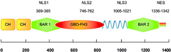
A phylogenic analysis of Entamoeba proteins that contain a CH domain suggested the existence of at least 5 phylogenetically distinct families of potential ABPs (Hon et al. Reference Hon, Nakada-Tsukui, Nozaki, Guillén, Graham Clark, Patricia and Adam2010). One of these proteins (Group D, NCBI Accession number: EHI_093850) is a nucleocytoplasmic ABP that was recently described by our group. EhNCABP166 consists of 1387 amino acids and contains 2 CH domains with high homology, 2 BAR domains and a GTPase-binding domain/formin 3 homology domain (GBD/FH3) (Fig. 1). In vitro, EhNCABP166 is able to bind diverse phosphoinositides, GTPases and filamentous actin (F-actin). In vivo, EhNCABP166 has the ability to bind F-actin (Campos-Parra et al. Reference Campos-Parra, Hernández-Cuevas, Hernandez-Rivas and Vargas2010). The presence of ABPs has recently been detected in nuclei of eukaryotic cells with specific functions (Uribe and Jay, Reference Uribe and Jay2009; Castano et al. Reference Castano, Philimonenko, Kahle, Fukalová, Kalendová, Yildirim, Dzijak, Dingová-Krásna and Hozák2010). In higher eukaryotes, it has been reported that diverse ABPs, including N-WASP, Flightless I and c-Abl, contain nuclear and export and localization signals (NES and NLS, respectively) and have been found in both the cytoplasm and nucleus (Van Etten et al. Reference Van Etten, Jackson and Baltimore1989; Taagepera et al. Reference Taagepera, McDonald, Loeb, Whitaker, McElroy, Wang and Hope1998; Lee et al. Reference Lee, Campbell and Stallcup2004; Wu et al. 2006). These NLSs and NESs mediate the transport of macromolecules between the nucleus and the cytoplasm. Proteins targeted to enter the nucleus due to containing a functional NLS could be carried in by importins (karyopherin) (Imamoto et al. Reference Imamoto, Shimamoto, Takao, Tachibana, Kose, Matsubae, Sekimoto, Shimonishi and Yoneda1995). There are 2 types of importins, α and β. Importin α proteins recognize the NLS on cargo proteins. Once the cargo is bound by importin α, the complex is recognized and bound by importin β, which subsequently binds to the fibrils of the nuclear pore complex (NPC) and is responsible for translocation of the cargo. Importin β is able to bind directly to some NLSs present in different proteins and transport them to the nucleus (Marfori et al. Reference Marfori, Mynott, Ellis, Mehdi, Saunders, Curmi, Forwood, Bodén and Kobe2011). Proteins that transfer cargo outside of the nucleus are called exportins (one of the most studied exportins is the protein CRM1). These proteins mediate the export of numerous proteins that carry an NES from the nucleus to the cytoplasm. To transport cargo to the cytoplasm, CRM1 must be associated with RanGTP (Boulo et al. Reference Boulo, Akarsu, Ruigrok and Baudin2007). In this regard, O'Reilly et al. (Reference O'Reilly, Dacks and Field2011) reported an in silico analysis of 36 predicted proteomes. They scanned the proteomes for candidate karyopherin-β (KAP-β) family members. KAP-βs are functionally classified as importins or exportins depending on the direction of transport they have been shown to mediate. Candidate members of each KAP-β subfamily have been found in all eukaryotic super groups (O'Reilly et al. Reference O'Reilly, Dacks and Field2011). Entamoeba histolytica was examined in this analysis, and the obtained data suggest the presence of KAP-βs in this parasite. Some KAP-βs mediate nuclear import through recognition of nuclear localization signals in cargo proteins (Chook and Süel, Reference Chook and Süel2011).
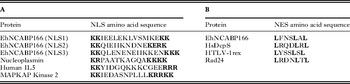
Schematic representation of the EhNCABP166 protein. EhNCABP166 contains an actin-binding domain comprised of 2 calponin homologue domains (CH) (ABD 1-233); 2 BAR domains, BAR1 (248-512) and BAR2 (1057-1314); a GTPase-binding domain (GBD/FH3 438-852); and an undefined alpha helix region (853-1056). In silico analysis showed that EhNCABP166 contains 3 bipartite nuclear localization signals (NLSs), which are located in the EhNCBAR1-166 domain (NLS1: 369-KKIEELEKLVSVMKEKKE-385), EhNCGBD/FH3-166 (NLS2: 746-KKQIEIIKNDNEKERKN-762) and the alpha helix region (NLS3: 1005-KKQLENENEIIKKENKK-1021). A nuclear export signal (NES 1336-LFNLSLAL-1342), which is located in the C-terminal region of the protein, is also described.

NLS motifs consist of clusters of positively charged amino acids (Arg and Lys) and can be mono- or bipartite (Xu et al. Reference Xu, Farmer and Chook2010). A wide variety of functional NES motifs have been identified, and their diversity is relatively high (Kutay and Güttinger, Reference Kutay and Güttinger2005). The presence of regularly spaced hydrophobic residues is a characteristic of these signals. There are few reports of the presence of NLSs or NESs in E. histolytica. However, the possible existence of an NLS and an NES in the protein Ehp53 has been reported. This protein is located in both the nucleus and cytoplasm (Mendoza et al. Reference Mendoza, Orozco, Rodríguez, García-Rivera, Sánchez, García and Gariglio2003). Furthermore, it has been reported that fusion of the SV40 large T antigen monopartite NLS to E. histolytica enolase results in increased levels of this protein in the nucleus (Tovy et al. Reference Tovy, Siman, Gaentzsch, Helm and Ankri2010), which suggests the existence of nuclear transport components in this parasite that allow the migration of proteins that contain NLSs to the nucleus. A preliminary in silico analysis showed that EhNCABP166 contains 3 NLSs and an NES. The location of each potential NLS within EhNCABP166 was determined via fusion of NLS1, 2 and 3 to a cytoplasmic domain of ehFLN, which is localized exclusively in the cytoplasm of E. histolytica trophozoites (Díaz-Valencia et al. Reference Díaz-Valencia, Almaraz-Barrera, Jay, Hernández-Cuevas, García, González-De la Rosa, Arias-Romero, Hernandez-Rivas, Rojo-Domínguez, Guillén and Vargas2007). Furthermore, due to the large size of EhNCABP166, we decided to evaluate the localization of the domains containing NLSs and the domains without predicted NLSs to evaluate the functionality of the NLSs in EhNCABP166. Similarly, we evaluated the effect of mutations in the first block of the NLSs. Furthermore, to determine the functionality of the NES, we fused this signal to a couple of constructs that localize in the nucleus. In this report, we provide evidence, for the first time, that EhNCABP166 contains functional NLSs and an NES that could allow transport between the nucleus and cytoplasm.
MATERIALS AND METHODS
Strain and culture conditions
Trophozoites of the pathogenic strain E. histolytica HM1:IMSS were axenically cultured in TYI-S-33 medium (Diamond et al. Reference Diamond, Harlow and Cunnick1978). The bacterial strain Escherichia coli DH5α was used in the recombinant DNA applications. This bacterial strain was cultured in Luria-Bertani medium at 37 °C in the presence of ampicillin (100 μg/ml).
Detection of EhNCABP166 and transfected constructs using transmission electron microscopy
To determine the specific location of EhNCABP166 and the 3 transfected constructs (NLS3Ac-d100-, EhNCR1C-166 and EhNCR1CK1005A/K1006A-166) in the nucleus of Entamoeba histolytica, trophozoites were fixed with 0·1% glutaraldehyde and 4% paraformaldehyde for 1 h at 25 °C, embedded in LR White, and then polymerized under UV at 4 °C overnight. Thin (60 nm) sections were obtained, mounted on Formvar-covered nickel grids and then they were incubated with an anti-EhNCABP166 polyclonal antibody (1:40 dilution) or an anti-HSV monoclonal antibody (1:100) (Novagen, Wisconsin, USA) for 1 h at 25 °C. Goat anti-mouse IgG conjugated to 20-nm gold particles (Ted Pella, Cardiff, UK) was used as the secondary antibody (1:40 dilution for the polyclonal antibody and 1:60 for the monoclonal antibody); samples were incubated for 1 h at 25 °C. Thin sections were observed with a Morgagni 268D Philips transmission electron microscope (FEI company, Eindhoven, The Netherlands).
DNA constructs and transfection assays
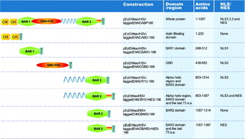
To determine the subcellular localization of the domains constituting the protein, the following 7 constructs were generated: EhNCABD-166 (1-233), EhNCBAR1-166 (248-512), EhNCGBD/FH3-166 (438–852), EhNCR1C-166 (853-1314), EhNCR1C + NES (853-1387), EhNCBAR2ΔNES-166 (1057-1314), and EhNCBAR2 + NES-166 (1057-1387) (Fig. 4). To generate fusions of the different regions of the EhNCABP166 gene to an HSV tag sequence, plasmid DNA containing the EhNCABP166 gene was amplified via PCR using specific primers for each construct that were modified at the 5′ and 3′ ends to incorporate KpnI and BamHI restriction sites (see Supplementary Table S1, online only).
To fuse the EhNCABP166 NLSs to Ac-d100- to obtain NLS1 Ac-d100--HSV, NLS2 Ac-d100--HSV and NLS3 Ac-d100--HSV, the construct pExEhNeo/HSV-tagged Ac-d100- was used as a template, which was previously generated by our research team (Díaz-Valencia et al. Reference Díaz-Valencia, Almaraz-Barrera, Jay, Hernández-Cuevas, García, González-De la Rosa, Arias-Romero, Hernandez-Rivas, Rojo-Domínguez, Guillén and Vargas2007). Plasmid DNA containing the Ac-d100- domain was amplified via PCR using a specific primer modified at the 5′ end to incorporate each NLS and a Kpn1 restriction site, together with a specific primer modified at the 3′ end to incorporate a BamHI restriction site (see Supplementary Table S1 online version only). In all cases, the reverse primer possessed a 3′ sequence that encoded an HSV (herpes simplex virus) tag (SQPELAPEDPED). The obtained PCR products were purified and digested with the KpnI and BamHI endonucleases and subsequently ligated into a pExEhNEO vector that had been cleaved at the same restriction sites. These plasmids were replicated in the E. coli DH5α strain. Plasmid DNA was purified using a Plasmid Maxi kit (Qiagen). Entamoeba histolytica trophozoites of the HM1:IMSS strain were transfected by electroporation with 200 μg DNA/107 cells (Hamann et al. Reference Hamann, Nickel and Tannich1995). Transfected cells grown for 48 h after electroporation were selected based on their resistance to 10 μg/ml G418 added to the culture. Positive clones were isolated and further subjected to higher concentrations of G418 (up to 30 μg/ml).
Directed mutagenesis of nuclear localization signals
To generate mutants of EhNCGBD/FH3-166 and EhNCR1C-166, both domains were cloned into the Pcr2.1 vector. Once they were cloned into this vector, mutants of EhNCGBD/FH3-166 (EhNCGBD/FH3K746A/K747A-166) and EhNCR1C-166 (EhNCR1CK1005A/K1006A-166) were generated via in vitro site-directed mutagenesis using the QuikChange© kit (Stratagene, Cedar Creek, Texas, USA) according to the manufacturer's suggested protocol and using specific primers (see Supplementary Table S1 online version only). All mutations were confirmed by automatic sequencing. Then, the mutated domains were cloned into the pExEhNeo vector. These plasmids were replicated in the E. coli DH5α strain. Plasmid DNA was purified using a Plasmid Maxi kit (Qiagen). Entamoeba histolytica trophozoites of the HM1:IMSS strain were transfected via electroporation with 200 μg DNA/107 cells (Hamann et al. Reference Hamann, Nickel and Tannich1995). Transfected cells that were grown for 48 h after electroporation were selected based on their resistance to 10 μg/ml G418 added to the culture. Positive clones were isolated and further subjected to higher concentrations of G418 (up to 30 μg/ml).
Confocal microscopy
The cellular localization of the constructs within transfected E. histolytica trophozoites was determined. Briefly, 2·4 × 105 trophozoites were pelleted and washed with 0·89% NaCl. Then, 1 ml of 4% paraformaldehyde, pre-warmed to 37 °C, was added, and the cells were incubated for 45 min at 37 °C. Following fixation, the cells were permeabilized with 0·1% Triton-PBS for 1 min. The fixed and permeabilized cells were washed with PBS 3 times, quenched using 50 mm NH4Cl in PBS for 30 min at room temperature, blocked with 1% BSA-PBS for 1 h at 37 °C and incubated with the anti-HSV Tag monoclonal antibody (Novagen, Wisconsin, USA) at a 1:150 dilution, in 1% BSA-PBS at 37 °C for 1 h. Then, the cells were washed 3 times with 1% BSA-PBS and incubated with a goat anti-mouse IgG Alexa Fluor® 488 secondary antibody (Molecular Probes® 1:900) at 37 °C for 1 h. The cells were subsequently washed again with PBS and mounted using 5 μl of Vectashield© with DAPI.
Also, cellular localization of PI(3,4) P2 within wild type trophozoites was determined. To determine cellular location of this lipid in the wild type, a purified anti-PI(3,4) P2 antibody mouse monoclonal (Echelon Biosciences®, Salt Lake City, USA) at a 1:50 dilution was used. As secondary antibody, a goat anti-mouse IgG Alexa Fluor® 488 (Molecular Probes® 1:800) was used. To determine the cellular location of PI(3,4) P2 and HSV tagged constructions within transfected cells (pExEhNeo/HSV tagged-EhNCBAR1-166 or pExEhNeo/HSV tagged- NLS1Ac-d100-), a purified anti-PI(3,4) P2 mouse monoclonal antibody (Echelon Biosciences®, Salt Lake City, USA) at a 1:50 dilution was used; a rabbit anti-HSV (GenScript® NJ, USA) at a 1:100 dilution was used. As secondary antibodies, a goat anti-mouse IgG TRITC (Zymed) and a goat anti-rabbit IgG FITC (Zymed) were used, both at a 1:800 dilution. For these samples the same microscopy protocol mentioned above was used.
The samples were analysed via confocal microscopy (FluoView 1000 Olympus and Leica SP5).
RESULTS
Nuclear location of EhNCABP166 under TEM
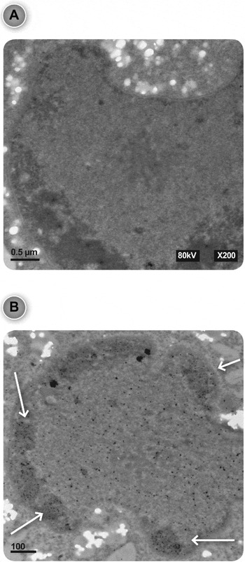
Through an immunostaining and confocal analysis of E. histolytica, we had previously demonstrated the presence of EhNCABP166 in both the nucleus and cytoplasm (Campos-Parra et al. Reference Campos-Parra, Hernández-Cuevas, Hernandez-Rivas and Vargas2010). To determine whether the association of EhNCABP166 with the nucleus corresponds to intranuclear localization, we decided to analyse this protein by transmission electronic microscopy (TEM), which is a very precise technique that allows determination of the position of the protein relative to the nuclear envelope. A polyclonal antibody that recognizes EhNCABP166 was used (Campos-Parra et al. Reference Campos-Parra, Hernández-Cuevas, Hernandez-Rivas and Vargas2010). As depicted in Fig. 2, the TEM micrograph showed that EhNCABP166 is located at the inner perinuclear zone and throughout the entire intranuclear space.
Transmission electron microscopy analysis of Entamoeba histolytica trophozoites. (A) Secondary antibody control. As depicted in the figure, the secondary antibody did not recognize any epitope. (B) Staining with the EhNCABP166 polyclonal antibody followed by the secondary antibody conjugated to gold particles. EhNCABP166 is localized in the nucleus in the perinuclear zone. Arrows indicate some areas of EhNCABP166 enrichment. Both images show the nuclei of wild-type trophozoites. Scale Bars = 0·5 μm (A) and 1 μm (B).

Subcellular location of NLS1-Ac-d100-, NLS2- Ac-d100- and NLS3 Ac-d100-
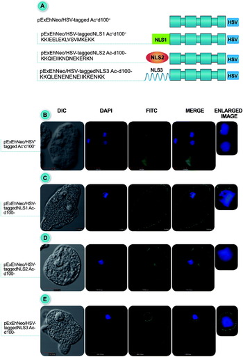
To determine whether NLS1, NLS2 and NLS3, which were found through an in silico analysis of EhNCABP166, were functional signals, each NLS was fused to the Ac-d100- domain (Fig. 3A), which is a cytoplasmic domain of ehFLN (Díaz-Valencia et al. Reference Díaz-Valencia, Almaraz-Barrera, Jay, Hernández-Cuevas, García, González-De la Rosa, Arias-Romero, Hernandez-Rivas, Rojo-Domínguez, Guillén and Vargas2007). The Ac-d100- domain has a molecular mass of 38·3 kDa and is comprised of 4 quasi-repetitive units, with repeats 1, 2 and 3 mainly forming β sheets and repeat 4 exhibiting a low content of β sheets. The Ac-d100- domain does not contain a predicted NLS and, as shown in Fig. 3B, it is located in the cytoplasm and at the cytoplasmic membrane. Fusion of each of the EhNCABP166 NLSs to Ac-d100- (Fig. 3C, D and E) altered the localization of Ac-d100-. The addition of these signals resulted in the migration of Ac-d100- toward the nucleus.
Cloning, expression and cellular analysis of the NLSs fused to Ac-d100- in transfected trophozoites. (A) Schematic diagram of NLS1, NLS2 and NLS3 fused to Ac-d100-, which was overexpressed in Entamoeba histolytica trophozoites. (B) The subcellular localization of Ac-d100- was analysed using confocal microscopy. Ac-d100- is distributed in the cytoplasm; this domain was used as a cytoplasmic control. Fusion of NLS1(C), NLS2 (D) and NLS3 (E) to Ac-d100- resulted in transport of this domain to the perinuclear zone. These results suggest that NLS1, 2 and 3 are functional signals. The localization of these constructs was determined using a monoclonal anti-HSV antibody and an FITC-labelled secondary antibody. Cells were stained with DAPI to visualize the nuclei. All sequences were fused to an HSV tag at their 3′ end (blue box). All transfected cells were grown under 30 μg ml−1 of G418. Scale Bar = 1000 μm.

These data suggest that NLS1 (369-KKIEELEKLVSVMKEKK-385), NLS2 (746-KKQIEIIKNDNEKERKN-762) and NLS3 (1005-KKQLENENEIIKKENKK-1021) are functional NLSs.
Subcellular localization of EhNCABP166 domains
To determine the subcellular localization of the diverse domains that make up EhNCABP166, every domain was cloned, fused with an HSV epitope tag and transfected into E. histolytica trophozoites. Seven constructs (Fig. 4) were produced, as described in the Materials and Methods section. The subcellular localization of these constructs was determined via confocal microscopy using an anti-HSV monoclonal antibody.
Schematic diagram of full-length EhNCABP166 and different EhNCABP166 constructs that were overexpressed in Entamoeba histolytica trophozoites. The diagram shows the name of the construction, the region and/or domain, and the amino acids that cover every construction. Also the diagram shows the presence of signals from every construction.

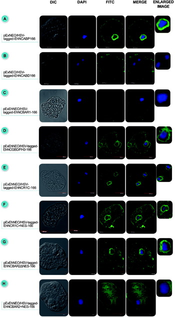
Analysis of the transfected cells using confocal microscopy showed that the intact EhNCABP166 protein localized to the inner perinuclear zone, cytoplasm and plasma membrane (Fig. 5A), as expected. However, the perinuclear staining appeared to be more intense. The actin-binding domain EhNCABD-166 is a protein fragment containing 2 CH domains but no NLS, and this fragment was clearly located on membrane extensions (Fig. 5B). Additionally, some trophozoites expressing EhNCABD-166 exhibited a different structure and shape, which suggested that EhNCABD-166 might be competing for F-actin with other ABPs present in E. histolytica. The EhNCBAR1-166 domain (31·8 kDa) was mainly localized to the cytoplasm (Fig. 5C). EhNCGBD/FH3-166 (50·7 kDa) and EhNCR1C-166 (55·7 kDa) were located in the inner perinuclear zone and in the cytoplasm, similar to intact EhNCABP166 (Fig. 5D and E). Additionally, EhNCR1C-166 was also found to be highly enriched in the inner perinuclear area. EhNCGBD/FH3-166 contains NLS2 (746-KKQIEIIKNDNEKERKN-762), and EhNCR1C-166 contains NLS3 (1005-KKQLENENEIIKKENKK-1021), which suggests that the NLSs present in these domains are responsible for the perinuclear location of EhNCABP166. Furthermore, EhNCR1C + NES-166 (64·4 kDa), which is a construct comprised of the alpha region and the EhNCBAR2 + NES-166 domain was analysed. This construct localized to the inner perinuclear area, cytoplasm and protrusions (Fig 5F). This domain contains NLS3 and the NES; therefore, as expected, this construct was found in both the nucleus and cytoplasm.
Cloning, expression and cellular analysis of EhNCABP166 and different constructs in Entamoeba histolytica trophozoites. (A–H) The distribution of all HSV-tagged domains and regions of EhNCABP166 in transfected trophozoites, analysed by confocal microscopy. (A) EhNCABP166 is located in the inner perinuclear area, cytoplasm and cytoplasmic membrane region; (B) EhNCABD-166 is located on membrane extensions; (C) EhNCBAR1-166 is located in the cytoplasm and the cytoplasmic membrane; (D) EhNCGBD-166 is located in the inner perinuclear area, cytoplasm and cytoplasmic membrane region; (E) EhNCR1C-166 is located in the inner perinuclear zone and cytoplasm; (F) EhNCR1C + NES-166 is located in the inner perinuclear zone, cytoplasm and membrane region; (G) EhNCBAR2ΔNES-166 is located in the inner perinuclear zone and cytoplasm; and (H) EhNCBAR2 + NES-166 is located in the cytoplasm and in the nucleus. Localization to these regions was determined using a monoclonal anti-HSV antibody and a FITC-labelled secondary antibody. Cells were stained with DAPI to visualize the nuclei. All sequences were fused to an HSV tag at their 3′ end (blue box). All of the transfected cells were grown under 30 μg/ml of G418. Scale Bar = 1000 μm.

EhNCBAR2ΔNES-166, which is a 31-kDa domain, localized to the inner perinuclear zone and cytoplasm (Fig. 5G). However, in silico analysis of this domain did not indicate the presence of an NLS. We added the last 73 amino acids of EhNCABP166, which includes an export signal (1336-LFNSLAL-1342), to this domain to obtain the construct EhNCBAR2 + NES-166. This construct was located mainly in the cytoplasm and, to a lesser extent compared to EhNCBAR2ΔNES-166, in the perinuclear zone (Fig. 5H). This result suggests that the decreased presence of EhNCBAR2 in the perinuclear area is due to the NES, which promotes export to the cytoplasm.
Mutation of EhNCABP166 NLSs
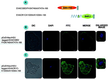
The classical bipartite NLS consists of 2 sets of 2–3 basic amino acids separated by 10–12 residues. The EhNCABP166 NLSs represent this type of signal. The first block in the 3 NLSs is conserved (Table 1). To ascertain whether the amino acid components of the first block of NLS2 and NLS3 are critical for the transport of EhNCABP166 to the perinuclear area, lysines 746 and 747 (NLS2) and lysines 1005 and 1006 (NLS3) were mutated to alanine (Fig. 6A). A mutated NLS1 was not generated because EhNCBAR1-166 did not localize to the inner perinuclear area as was observed for EhNCGBD/FH3-166 and EhNCRC1-166. The results show a diminished presence of the EhNCGBD/FH3K746A/K747A construct in the inner perinuclear area, mainly located close to the plasma membrane. Mutation of the lysines within the first block of NLS2 affected the shuttling of EhNCGBD/FH3-166 toward the perinuclear zone (Fig. 6B). EhNCR1CK1005A/K1006A-166 was located in the cytoplasm and in the cytoplasmic membrane, whereas its presence in the nucleus was diminished (Fig. 6C). These results suggest that the presence of lysines within the first block of the NLSs is critical to specify migration to the nucleus.
Cloning, expression and cellular analysis of mutated domains of EhNCABP166 in transfected trophozoites. (A) A schematic diagram of the mutated domains of EhNCABP166 that were overexpressed in Entamoeba histolytica trophozoites. Double mutations were generated in the EhNCABP166 NLSs involving an exchange of the lysines in the first block of the bipartite nuclear localization signals for alanines. (B) EhNCGBD/FH3K746A/K747A-166 shows a diminished presence in the perinuclear zone, while the mutated domain is located mainly at the cytoplasmic membrane. (C) EhNCR1CK1005A/K1006A-166 is located in the cytoplasm and at the cytoplasmic membrane and shows a diminished presence in the perinuclear zone. These results suggest that lysines in the first block of NLS2 and NLS3 are necessary for EhNCGBD/FH3-166 and EhNCR1C-166 to be transported to the perinuclear zone. All sequences were fused to a HSV tag at their 3′ end (blue box). All of the transfected cells were grown under 30 μl/ml of G418. Scale Bar = 1000 μm.

Bipartite NLS and NES of EhNCABP166

Nuclear location of the transfected constructs under TEM
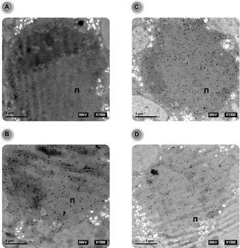
To confirm the results obtained using confocal microscopy concerning whether the location of the different constructs (NLS3Ac-d100-, EhNCR1C-166 and EhNCR1CK1005/1006) in the perinuclear zone corresponded to intranuclear localization, we sought to analyse the constructs via TEM. A monoclonal antibody that recognizes the HSV epitope was used. As depicted in Fig. 7B, C and D, TEM micrographs showed that all of the constructs were located in the nucleus in the inner perinuclear zone and throughout the entire intranuclear space. Mutation of lysines 1005 and 1006 resulted in a diminished presence of EhNCR1C-166 in the nucleus.
Transmission electron microscopy analysis of transfected Entamoeba histolytica trophozoites. (A) Secondary antibody control. As depicted in the figure, the secondary antibody does not recognize any epitope. (B) Staining with an anti-HSV monoclonal antibody was followed by a secondary antibody conjugated to gold particles. NLS3Ac-d100- is located in the nucleus. (C) Staining with an anti-HSV monoclonal antibody was followed by a secondary antibody conjugated to gold particles. EhNCR1C-166 was localized in the nucleus. (D) Staining with an anti-HSV monoclonal antibody was followed by a secondary antibody conjugated to gold particles. The degree of EhNCR1CK1005A/K1006A-166 localization to the nucleus is less than that of EhR1C-166. All images show the nuclei of transfected trophozoites. Scale Bar = 1 μm.

Location of PI(3,4)P2 within wild type and transfected cells
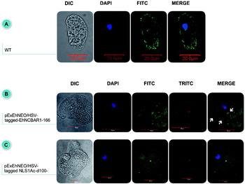
Transfected EhNCBAR1-166 domain was located outside the nucleus even when this domain contained the NLS1 (Fig. 5D), despite the fact that NLS1 is a functional signal that allows Ac-d100- to migrate to the nucleus upon fusion with this NLS (Fig. 3A). The EhNCBAR1-166 domain contains 2 consensus binding sites for phosphoinositide (3,4)P2 that overlaps with the NLS sequence. To determine the possible interaction of PI(3,4)P2 with the EhNCBAR1-166 domain within transfected trophozoites, the subcellular localization of both components was determined via confocal microscopy using an anti-PI(3,4)P2 monoclonal antibody and a rabbit anti-HSV antibody. First, the location of PI(3,4)P2 was determined within wild type cells. The result showed that the phosphoinositide localized to cytoplasm, plasma membrane and nucleus (Fig. 8A). In transfected cells with the EhNCBAR1-166 domain, PI(3,4)P2 and EhNCBAR1-166 domain were localized mainly to the cytoplasm. The result showed a co-localization of EhNCBAR1-166 domain with PI (3,4)P2 (Fig. 8B).The arrows show some orange dots, suggesting an interaction between these molecules within E. histolytica-transfected trophozoites. Also, we determined the location of PI(3,4)P2 within transfected cells with NLS1Ac-d100-; this construction only contains the NLS1 sequence attached to the Ac-d100- cytoplasmic domain. The results showed that PI(3,4)P2 and NLS1Ac-d100- were localized to cytoplasm and nucleus (Fig 8C). These results will be discussed below.
Cellular analysis of PI(3,4)P2 and EhNCBAR1-166 domain in wild type and transfected trophozoites. (A) PI(3,4)P2 was localized to cytoplasm (FITC), plasma membrane and nucleus within wild type trophozoites. (B) EhNCBAR1-166 domain was localized to the cytoplasm (FITC) and PI(3,4)P2 was localized mainly to the cytoplasm (TRITC) within transfected trophozoites with EhNCBAR1-166 construction. Arrows indicate co-localization of PI(3,4)P2 with EhNCBAR1-166 domain (C) NLS1Ac-d100- was localized to the cytoplasm and nucleus (FITC) and PI(3,4)P2 was localized to the cytoplasm and nucleus (TRITC) within transfected trophozoites with NLS1Ac-d100- construction. All of the transfected cells were grown under 30 μl/ml of G418. Scale Bars = 20 and 10 μm as indicated.

DISCUSSION
Here, we report the nuclear presence of EhNCABP166, a nucleocytoplasmic ABP in E. histolytica. In the nucleus, this protein is located both in the inner perinuclear area and throughout this organelle. Additionally, we report the existence of NLSs and an NES in EhNCABP166. To determine the functionality of the signals present in EhNCABP166, we applied different strategies. We demonstrated that the individual NLSs 1, 2 and 3 are functional signals that can specify the migration of Ac-d100- to the nucleus. The Ac-d100- domain alone is not shuttled to the nucleus, but fusion with the bipartite signals results in its being shuttled to this organelle. Additionally, cloning of the different domains and regions comprising EhNCABP166 allowed us to determine the functionality of the signals in each domain or construct. The results showed that EhNCABD-166 and EhNCBAR1-166 were located in the cytoplasm. Similar to other ABDs belonging to ABPs like filamin, EhNCABD-166 is located in the cortical region (Washington and Knecht, Reference Washington and Knecht2008), which is an area that is presumed to be rich in F-actin. EhNCABD-166 has a relatively small molecular mass of 27·7 kDa, which could allow it to be shuttled to the nucleus via diffusion, as has been observed for certain proteins (Fried and Kutay, Reference Fried and Kutay2003). The nuclear pore complex (NPC) permits the passive diffusion of small molecules, but this diffusion becomes inefficient when the molecular weight of the diffusive species approaches 20–40 kDa. The unique localization of EhNCABD-166 to areas where cell membrane ruffling occurs suggests the functionality of this domain, which is supported by the lack of an NLS and the absence of diffusion of this domain toward the nucleus. EhNCBAR1-166, which contains NLS1, was located mainly in the cytoplasm, despite the fact that NLS1 is a functional signal that allows Ac-d100- to migrate to the nucleus upon fusion with this NLS. These findings can be explained by the fact that the EhNCBAR1-166 domain contains 2 consensus binding sites for PI(3,4)P2 located at 359-ELRVQIKQKIKK-370 and 376-KLVSVMKEKK-385. The NLS1 located within 369-KKIEELEKLVSMKEKK-385 overlaps with the phosphoinositide-binding sites. A previous in vitro result showed that the EhNCBAR1 domain has an affinity for this phosphoinositide (Campos-Parra et al. Reference Campos-Parra, Hernández-Cuevas, Hernandez-Rivas and Vargas2010). To test the possible interaction of PI(3,4)P2 with EhNCBAR1-166 domain within transfected trophozoites, subcellular localization of the PI and the EhNCBAR1-166 domain was determined. Results showed that EhNCBAR1-166 co-localizes with PI(3,4)P2 in these cells. In vitro and in vivo results suggest the interaction between this PI and EhNCBAR1-166 domain. Furthermore, we determined the location of PI(3,4)P2 within transfected cells with NLS1Ac-d100- construction. The results showed that PI(3,4)P2 and NLS1Ac-d100- were localized in the cytoplasm and nucleus. The location of PI(3,4)P2 and NLS1Ac-d100- in these cells is similar to the location of this PI in wild type cells and similar to the location of this construction within transfected cells with NLS1Ac-d100-, so, the location of both components is not modified. Comparing the PI(3,4)P2 location among the images, a reduction in the presence of PI(3,4)P2 can be seen in the nucleus of transfected cells with EhNCBAR1-166. This suggests again the interaction between PI(3,4)P2 and EhNCBAR1-166 in the cytoplasm. The construct consisting of NLS1 fused to Ac-d100- only contained the NLS1 sequence and not the amino acids that surround the NLS, which are apparently important for the interaction with PI(3,4)P2. It has been proposed that the cellular location of some proteins is regulated by different phosphoinositides (Liu et al. Reference Liu, Wagner, Campbell, Nickerson, Schiffer and Ross2005; Saarikangas et al. Reference Saarikangas, Zhao and Lappalainen2010). These results suggest that the interaction of PI(3,4)P2 with consensus binding sites for PI(3,4)P2 could block the recognition site for importin and preclude the migration of EhNCBAR1-166 toward the nucleus.
EhNCGBD/FH3-166, which contains NLS2, was located at the nucleus, cytoplasm and plasma membrane. Fusion of NLS2 to Ac-d100- resulted in the localization of this domain in the nucleus, which depended on lysines 746 and 747, as mutation of these residues reduced the presence of EhNCGBD/FH3-166 in the inner perinuclear zone. Therefore, NLS2 is a functional signal that is partially responsible for the migration of EhNCABP166 to the nucleus. EhNCR1C-166, which contains NLS3, was located mainly in the inner perinuclear zone, similar to EhNCABP166, and to a lesser extent, in the cytoplasm and at the plasma membrane. Fusion of NLS3 to Ac-d100- resulted in this domain localizing to the nucleus, which depended on lysines 1005 and 1006 because mutation of these residues reduced the presence of EhNCR1C-166 in the nucleus. The results obtained using TEM corroborate the intranuclear location of these constructs. These results suggest that NLS3 is a functional signal that is responsible for the migration of EhNCABP166 to the nucleus. Addition of the nuclear export signal, which is within the last 73 amino acids of EhNCABP166, to EhNCR1C-166 (EhNCR1C + NES-166) resulted in a mainly cytoplasmic localization pattern, suggesting that the NES was responsible for the transport of EhNCR1C-166 towards the cytoplasm. EhNCBAR2ΔNES-166 was located at the inner perinuclear zone, cytoplasm and at the plasma membrane (Fig. 5g), but there was no indication of a predicted NLS in this domain; therefore, the nuclear location of EhNCBAR2ΔNES-166 was not expected. EhNCR1C-166 includes the EhNCBAR2ΔNES-166 domain and, as mentioned above, EhNCR1C-166 (containing NLS3) was located in the nucleus. Additionally, the EhNCR1CK1005A/1006A mutations within NLS3 led to a decrease in the nuclear localization of this sequence. However, EhNCBAR2ΔNES-166 was also located at the nucleus, which indicated that this domain contains a hypothetical NLS that is not functional (or is masked) in EhNCRIC-166. It is also possible that EhNCBAR2ΔNES-166 could interact with other proteins that allow its transportation toward the nucleus. For both EhNCR1C-166 and EhNCBAR2ΔNES-166, the addition of the last 73 amino acids of EhNCABP166 (containing the NES 1336-LFNSLAL-1342) shifted the localization of both constructs toward the cytoplasm. This type of export signal is present in proteins such as the human scavenger de-capping enzyme (DcpS) and the RAD24 protein (Shen et al. Reference Shen, Liu, Liu, Jiao and Kiledjan2008; Lopez-Girona et al. Reference Lopez-Girona, Furnari, Mondesert and Rusell1999).
These results suggest that the nuclear export signal present in EhNCABP166 is a functional signal and could be recognized by an importin β responsible for transporting proteins containing an NES to the cytoplasm (such as the CRM1 protein). Analysis in silico performed by O'Reilly et al. (Reference O'Reilly, Dacks and Field2011) showed the possible presence of CRM1 (also known as exportin 1) in E. histolytica. Exportins shuttle between the nucleus and cytoplasm, by binding cargo molecules under high RanGTP levels inside the nucleus, and release their cargo upon hydrolysis of Ran-bound GTP in the cytoplasm (Güttler et al. Reference Güttler, Madl, Neumann, Deichsel, Corsini, Monecke., Ficner, Sattler and Görlich2010). Additionally, in silico analysis revealed the presence of a Ran protein in E. histolytica. The putative Ran protein is designated by the access code EHI_148190, shares 73% identity and 85% homology with Homo sapiens Ran (data not shown) and is conserved in diverse organisms (Gorlich and Kutay, Reference Görlich and Kutay1999; Mosammaparast and Pemberton, Reference Mosammaparast and Pemberton2004). In light of these results, we suggest the existence of essential components belonging to a functional nucleocytoplasmic transport system, such as karyopherin α and β, exportins and Ran, in E. histolytica. Further studies will confirm the presence of all of the components of this system and its characteristics in this parasite. Moreover, some ABPs with specific functions have been reported in the nucleus (Uribe and Jay, Reference Uribe and Jay2009; Castano et al. Reference Castano, Philimonenko, Kahle, Fukalová, Kalendová, Yildirim, Dzijak, Dingová-Krásna and Hozák2010). ABPs are multifunctional proteins with different functions in specific compartments. Some ABPs contain an NLS that allows them be translocated to the nucleus, and similarly, some ABPs contain an NES. Certain proteins containing both signals have been reported to be components of signalling pathways, protein carriers or regulators of cellular processes (Malki et al. Reference Malki, Boizet-Bonhoure and Poulat2010; Rodríguez et al. Reference Rodríguez, Aburjania, Priedigkeit, DiFeo and Martignetti2010; Magico and Bell, Reference Magico and Bell2011). Following this idea, we can think about multi- functionality of several proteins from E. histolytica. The presence of NLSs or NES in proteins of E. histolytica suggests a role for these proteins in the nucleus besides having another function in the cytoplasm. We performes an in silico analysis to determine the existence of proteins containing NLSs or NESs in proteins from E. histolytica, and found that many proteins exist which possibly contain this kind of signal (Suppl. Table 2, online version only). The existence of these signals is logical in some proteins due their know function, but in other proteins the presence of these signals is not very obvious. So now it is important to study all the possible functions that a single protein with this kind of signal could have in this parasite. Therefore, our group is carrying out a variety of experiments to determine the function of the EhNCABP166 protein in the nucleus.
Taking all of our results together, we report for first time, the presence of functional NLSs and an NES in an ABP in E. histolytica, and we suggest that this protein could be shuttled between the nucleus and cytoplasm by a nucleocytoplasmic transport system in this parasite.
ACKNOWLEDGEMENTS
We thank biologist Anel Edith Lagunas Guillén for her kind technical assistance. Ricardo Uribe is a biomedical sciences Ph.D. Student from the Faculty of Medicine of the National Autonomous University of Mexico (UNAM), and we acknowledge his Doctoral Fellowship from the Mexican Science and Technology Council (CONACyT) (203209).
FINANCIAL SUPPORT
This research was supported by the Mexican Science and Technology Council (CONACyT) (130364) and by the French-Mexican collaborative program ANR-CONACyT PARACTIN (140364).