Uncontrolled bleeding has been determined to be a significant contributing factor to mortality in cases involving combat injuries, accidents and surgical procedures (Guo et al., Reference Guo, Wang, Liu, Liu, Wang and Li2023b). According to data from the World Health Organization, injuries and violence cause ~4.4 million deaths each year and account for nearly 8% of all deaths worldwide (Halvachizadeh et al., Reference Halvachizadeh, Mariani and Pfeifer2025). Uncontrolled bleeding can trigger a lethal triad of hypothermia, acidosis and coagulopathy, creating a vicious cycle (Thorsen et al., Reference Thorsen, Ringdal, Strand, Søreide, Hagemo and Søreide2011). Current clinical haemostatic materials generally suffer from limitations such as allowing high levels of blood loss, having long clotting times, being non-degradable, being difficult to fit onto complicated wounds and failing to meet patient expectations (Liu et al., Reference Liu, Hu, Yu, Xie, Lu and Lu2021). Consequently, the development of multifunctional haemostatic and wound-healing materials tailored for clinical application remains a major research priority (Guo et al., Reference Guo, Wang, Liu, Liu, Wang and Li2023b).
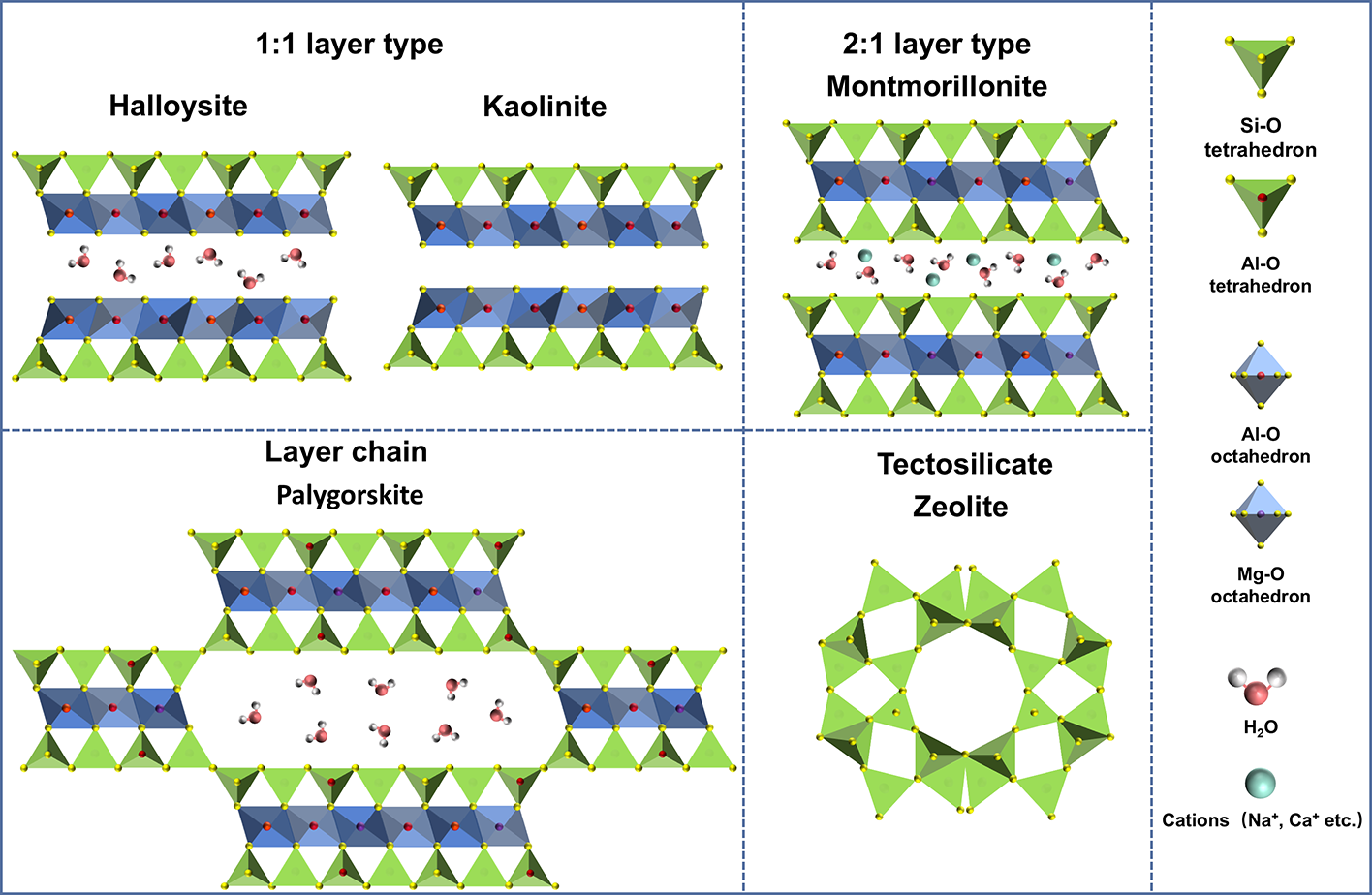
The application of clay mineral-based haemostatic agents in medicine has a long history, dating back thousands of years in China, Mesopotamia, Egypt, India, Greece and Rome, owing to their absorbency and tissue-adherence properties (Aziz et al., Reference Aziz, Gilani and Rindh2002). Clay minerals, especially nanoclays, can be classified into having one-dimensional, two-dimensional and three-dimensional structures based on their morphology and structure. One-dimensional clay minerals, represented by palygorskite (Pal), halloysite nanotubes (HNTs) and sepiolite, exhibit fibrous, rod or tube-like structures. Two-dimensional clay minerals, represented by kaolinite (Kaol) and montmorillonite (MMT), exhibit layered structures, while three-dimensional clay minerals, represented by zeolite (Ze), exhibit porous framework structures. Mixed-dimensional clay systems are natural coexisting aggregates of one-, two- or three-dimensional minerals, such as mixed-dimensional Pal clay (Wang et al., Reference Wang, Yang, Yang, Mu and Wang2024c) and mixed-dimensional Kaol. Most clay minerals, such as Kaol, Ze, HNTs, MMT and Pal, exhibit remarkable haemostatic capabilities due to their high water absorbency, large specific surface area, negative charge and ion-exchange properties (Katti et al., Reference Katti, Jasuja, Jaswandkar, Mohanty and Katti2022). Moreover, these clays are biocompatible, widely available and low cost (Katti et al., Reference Katti, Jasuja, Jaswandkar, Mohanty and Katti2022). A schematic diagram comparing the crystal structures of five typical silicate clay minerals used in haemostatic materials is provided in Fig. 1.
Schematic illustration of different structures of various clay minerals.

Figure 1 Long description
The diagram illustrates the structures of various clay minerals, divided into four sections. The top left section shows the 1:1-layer type minerals, Halloysite and Kaolinite, each with alternating layers of Si-O tetrahedrons and Al-O octahedrons. The top right section displays the 2:1-layer type mineral, Montmorillonite, with a similar arrangement but with an additional layer of Si-O tetrahedrons. The bottom left section features the layer-chain type mineral, Palygorskite, characterized by a chain-like structure with Si-O tetrahedrons and Al-O octahedrons. The bottom right section presents the tectosilicate type mineral, Zeolite, with a porous framework structure formed by interconnected Si-O tetrahedrons. A legend on the right side identifies the symbols used: Si-O tetrahedron, Al-O octahedron, Mg-O octahedron, H2O and cations such as Na plus, K plus, Ca superscript 2 plus and Mg superscript 2 plus.
Commercial products with clay minerals as the core haemostatic component have been developed. For instance, Kaol has been shown to activate coagulation factor XII through a specific negative-charge interaction, thereby initiating the intrinsic coagulation pathway (Shuo et al., Reference Shuo, Haoting, Yuqing, Liuyun and Xiang2024). Kaol has also been used as a principal component of QuikClot Combat Gauze®, a combat trauma dressing widely used by the US military and manufactured by Z-Medica (Basadonna, Reference Basadonna2012). Ze can release Ca2+ into the blood, thus accelerating the intrinsic pathway of blood coagulation and shortening the clot-formation time (Li et al., Reference Li, Cao, Lv, Jiang, Li and Li2013). However, this Ze-based haemostat also produces heat whilst stopping bleeding, and this can damage a tissue’s nerves and induce wound burns. HNTs – a porous material consisting of a tubular clay – can effectively concentrate blood components and activate the coagulation pathway through their nanotubular structure (Feng et al., Reference Feng, Luo, Wu, Liu, Liang, He and Liu2022). Basic research on HNT-based haemostats, such as sponges, hydrogels, fibres and gauzes, indicates that these have good application potential. MMT and Pal can promote blood coagulation through their layered/rod-shaped structures and ion-exchange properties (Li et al., Reference Li, Quan, Liang, Li, Yuan and Tao2016; Yang et al., Reference Yang, Wang, Li, Mu, Yang, Wang and Liu2024c). Although certain clay mineral-based materials (e.g. first-generation Ze-based QuikClot® and MMT-based WoundStat®) have demonstrated risks of heat generation and embolism during their application (Katti et al., Reference Katti, Jasuja, Jaswandkar, Mohanty and Katti2022), much recent research work on reducing heat generation and particle shedding has been conducted to increase their safety in comparison to inorganic-based haemostatic gauzes (Wang et al., Reference Wang, Shi, Huang, Wei, Shi, Xiao and Fan2024a).
Establishing adequate haemostasis is the cornerstone of wound healing (Liang et al., Reference Liang, He and Guo2021). In recent years, researchers have focused on developing safe and multifunctional integrated haemostatic materials with minerals as their core components. The article reviews the latest research progress on Kaol, Ze, HNTs, MMT, Pal and other clay minerals in the field of haemostasis, aiming to provide new perspectives for designing the next generation of high-performance and functional haemostatic materials.
Research progress on various clay minerals in rapid haemostasis
Kaolinite
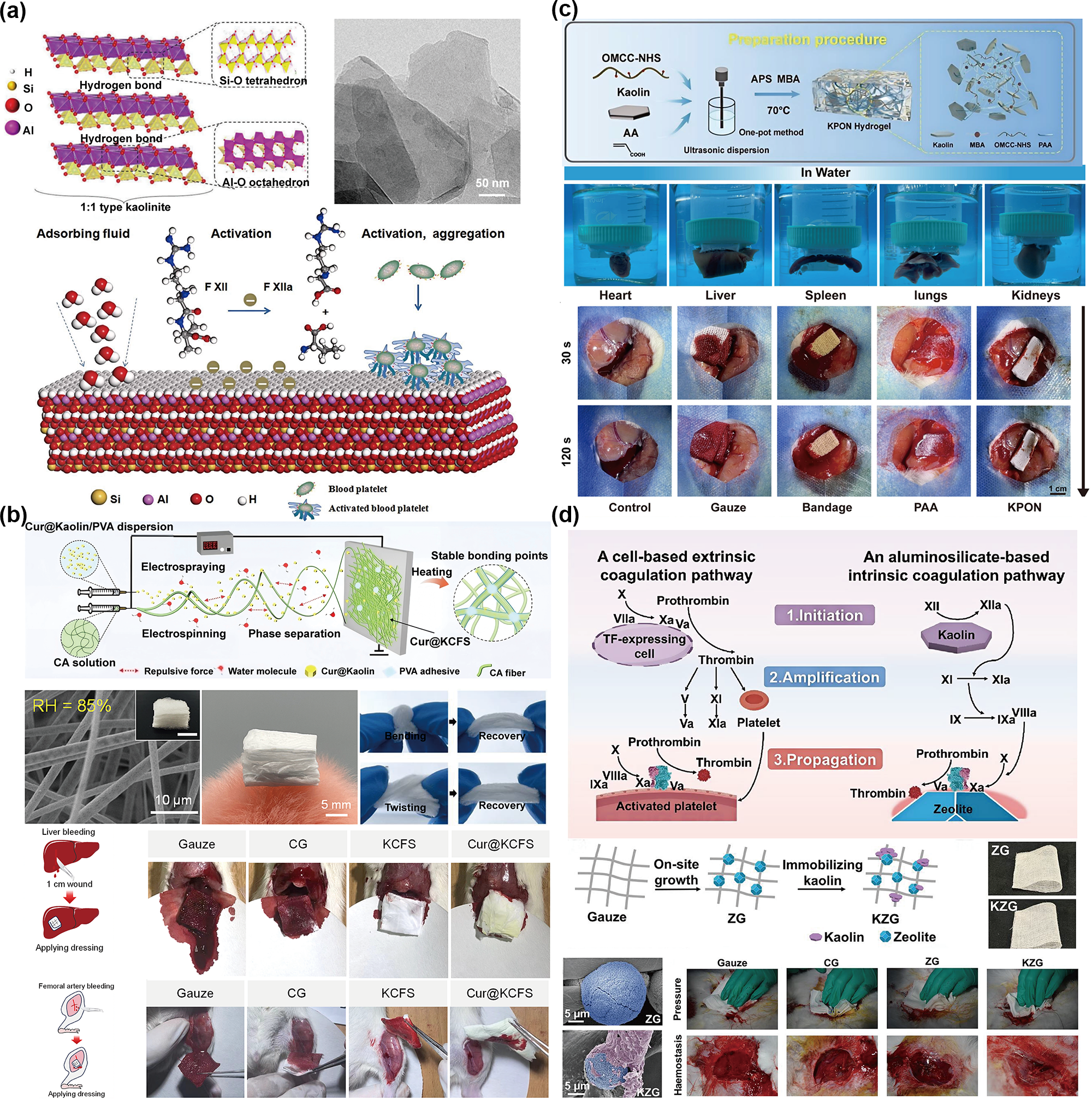
Kaol, with the structural formula Al2Si2O5(OH)4, is a 1:1 layered silicate with a negatively charged surface (Long et al., Reference Long, Zhang, Huang, Chang, Hu and Yang2018). The layered structure of Kaol consists of tetrahedral units and octahedral units (Fig. 2a; Wu et al., Reference Wu, Liao and Yang2023). The surface negative charge of Kaol specifically activates coagulation factor XII, thereby initiating the intrinsic coagulation pathway and ultimately promoting fibrin formation (Fig. 2a; Long et al., Reference Long, Zhang, Peng, Liao, Zhang and Wang2019; Shuo et al., Reference Shuo, Haoting, Yuqing, Liuyun and Xiang2024). Since 2008, Kaol has been replacing Ze products (e.g. the first-generation QuikClot®, QuikClot™, ACS™ and ACS+™) in battlefield medical gauzes, with the QuikClot Combat Gauze® now occupying a dominant market position (Gan et al., Reference Gan, Hu, Meng, Zhu, Gu and Wu2019). Additionally, an analysis revealed that Kaol from various regions conforms to fundamental biomedical standards (Tan et al., Reference Tan, Zheng and Yang2025). In this section, the research progress of various forms of Kaol-based haemostatic materials will be reviewed.
Kaol-based products used for haemostasis. (a) Crystal structure of Kaol, adapted under the terms of the Creative Commons CC BY licence (Wu et al., Reference Wu, Liao and Yang2023); transmission electron microscopy image and haemostatic mechanism of Kaol, adapted with permission from the published work (Long et al., Reference Long, Zhang, Peng, Liao, Zhang and Wang2019). Copyright 2019 Elsevier B.V. (b) Bird nest-inspired fluffy Kaol sponges for haemostasis and wound healing, adapted with permission from the published work (Dong et al., Reference Dong, Chen, Gao, Li, Yu and Ding2025). Copyright 2025 Elsevier B.V. CA = cellulose acetate; CG = Combat Gauze; Cur = curcumin; KCFS = kaolin/cellulose acetate fibrous sponge; PVA = poly(vinyl alcohol); RH = relative humidity. (c) Multi-crosslinking Kaol/oxidized cellulose hydrogel with robust properties for emergency haemostasis, adapted with permission from the published work (Zhao et al., Reference Zhao, Ren, Qiao, Tang, Chi, Jiang and Liu2025). Copyright 2024 Elsevier B.V. AA = acrylic acid; APS = ammonium persulfate; KPON = kaolin/OMCC-NHS/PAA hydrogel; MBA = N,N’-methylenebisacrylamide; OMCC-NHS = N-hydroxysuccinimide-grafted oxidized microcrystalline cellulose; PAA = poly(acrylic acid). (d) Kaol–Ze composite: a synergistic haemostat for effective haemorrhage control, adapted with permission from the published work (Wang et al., Reference Wang, Shi, Huang, Wei, Shi, Xiao and Fan2024a). Copyright 2024 American Chemical Society. CG = Combat Gauze; KZG = kaolin-modified zeolite gauze; TF = tissue factor; ZG = zeolite gauze.

Figure 2 Long description
The infographic presents various aspects of Kaol-based haemostatic products. (a) Illustrates the crystal structure of Kaol, showing its 1:1 layered silicate structure with tetrahedral and octahedral units. The diagram explains the activation of coagulation factor XII, leading to fibrin formation. (b) Describes bird nest-inspired fluffy Kaol sponges for haemostasis and wound healing. It includes images of the sponges and their application in wound healing, with labels such as CA (cellulose acetate), CG (Combat Gauze), Cur (curcumin), KCFS (kaolin/cellulose acetate fibrous sponge) and PVA (poly(vinyl alcohol)). (c) Shows a multi-crosslinking Kaol/oxidized cellulose hydrogel with robust properties for emergency haemostasis. The process involves OMCC-NHS, PAA and other components, with images of the hydrogel in water and its application on various organs like heart, liver, spleen, lungs and kidneys. (d) Depicts a Kaol–Ze composite as a synergistic haemostat for effective haemorrhage control. It includes a diagram of the coagulation pathway, showing the roles of tissue factor, prothrombin and thrombin, with images of gauze applications on wounds. Labels include CG (Combat Gauze), KZG (kaolin-modified zeolite gauze), TF (tissue factor) and ZG (zeolite gauze).
Haemostatic materials in various forms based on Kaol are prepared through simple mixing, electrospinning, freeze-drying and freeze–thaw methods. For instance, Kaol/iron oxide powder can efficiently absorb blood, activate platelets and shorten bleeding times (Long et al., Reference Long, Zhang, Huang, Chang, Hu and Yang2018). Compared to pure polyvinyl alcohol non-woven fabric, the polyvinyl alcohol/Kaol/γ-chitosan non-woven fabric reduces blood clotting times by ∼2.5-fold (Lee et al., Reference Lee, Kim, Myung, Jung, Han, Kim and Lee2024). Chitosan/poly(ethylene oxide)/Kaol nanofibre membranes exhibited optimal haemostatic performance when Kaol was added at a concentration of 10% (Liu et al., Reference Liu, Zhang, Liu, Dong, Tian, Li and Meng2022). To overcome Ze’s heat generation and improve haemostasis, Kaol-modified Ze gauze reduces particle shedding and heat production, resulting in superior haemostasis (75% less blood loss, 33% shorter haemostasis time). This mechanism involves Kaol activating coagulation factor XII, followed by Ze adsorbing activated factor X/activated factor V to form prothrombinase, thereby accelerating coagulation (Fig. 2d; Wang et al., Reference Wang, Shi, Huang, Wei, Shi, Xiao and Fan2024a). Additionally, three composite sponges prepared by the freeze-drying process, namely carboxymethyl chitosan/sodium alginate/Kaol composite sponges (Song et al., Reference Song, Li, Chen, Han, Duns and Dessie2023), carboxylated sisal fibre/chitosan/Kaol composite sponges (Shuo et al., Reference Shuo, Haoting, Yuqing, Liuyun and Xiang2024) and gelatin/Kaol composite sponges (Zhou et al., Reference Zhou, Li, Chang, Chen, Wang, Guo and Wang2024a), exhibited significantly shorter haemostasis times than a commercially available gelatin sponge.
Several biomimetic strategies have been employed to develop high-performance haemostatic materials based on Kaol. Inspired by the fluffy structure of bird nests, a curcumin@Kaol/polyvinyl alcohol fibre sponge prepared via electrospinning/electrostatic spraying co-processing exhibited both ultra-high water absorption (3384%) and wound-healing promotion effects (Fig. 2b; Dong et al., Reference Dong, Chen, Gao, Li, Yu and Ding2025). Inspired by marjoram essential oil and Kaol, a polyvinyl alcohol/Kaol/marjoram composite sponge prepared via freeze–thaw cycling exhibited haemostatic, antibacterial, antioxidant and wound-healing properties (Tamer et al., Reference Tamer, Alsehli, Omer, Afifi, Sabet and Mohy-Eldin2021). Similarly, a Kaol/cedarwood essential oil hydrogel exhibited haemostatic, antibacterial and antioxidant properties, leveraging the inherent antimicrobial and antioxidant characteristics of cedarwood essential oil (Tamer et al., Reference Tamer, Sabet, Alhalili, Ismail, Mohy-Eldin and Hassan2022). Additionally, a hydrogel powder composed of gelatin, Kaol, oxidized glucan and strontium-doped hydroxyapatite shortened haemostasis times and promoted early osteogenic differentiation (Jiang & Liang, Reference Jiang and Liang2026).
For moist surfaces such as internal organs, achieving both strong wet adhesion and highly efficient haemostasis presents a challenge. A wet-adhesive hydrogel composed of Kaol, polyacrylic acid and oxidized microcrystalline cellulose demonstrated a blood loss rate of only 32% compared to medical gauze (Fig. 2c; Zhao et al., Reference Zhao, Ren, Qiao, Tang, Chi, Jiang and Liu2025). Inspired by mussels and brown algae, polyacrylamide- tannic acid/Kaol hydrogels exhibit strong and durable adhesive properties (Fan et al., Reference Fan, Wang, Fang, Li, Zhou and Wang2020). A biomimetic adhesive composed of dopamine, polyethylene glycol diacrylate, tannic acid, gelatin and Kaol achieved complete haemostasis within 30 s in a porcine femoral artery injury model (Zhang et al., Reference Zhang, Mao, Yang, Shen, Zeng and Tang2025). Furthermore, adhesive-enhanced gallic acid-grafted chitosan/Kaol microneedle patches achieved haemostasis within 35 s (Tang et al., Reference Tang, Liu, Zhang, Cheng, Wang and Zhou2025). Beyond wet adhesion, another critical design consideration is post-operative tissue adhesion. To address this, a Janus dressing with a hydrophobic polylactic acid/carboxymethyl chitosan layer (anti-adhesion) and a hydrophilic polyvinyl alcohol/carboxymethyl chitosan/Ca2+/Kaol layer (platelet activation) demonstrated both easy removal and rapid haemostasis (Yu et al., Reference Yu, Wang, Deng, Su, Zhao and Zhao2024).
In summary, Kaol maintains an irreplaceable position in the field of haemostasis due to its specificity and high efficiency in activating coagulation factor XII through contact. However, Kaol haemostatic materials are highly dependent on an intact endogenous coagulation pathway, limiting their effectiveness in patients with coagulation disorders. In the future, multifunctional Kaol haemostatic materials targeting a broader patient population will better meet clinical needs.
Zeolite
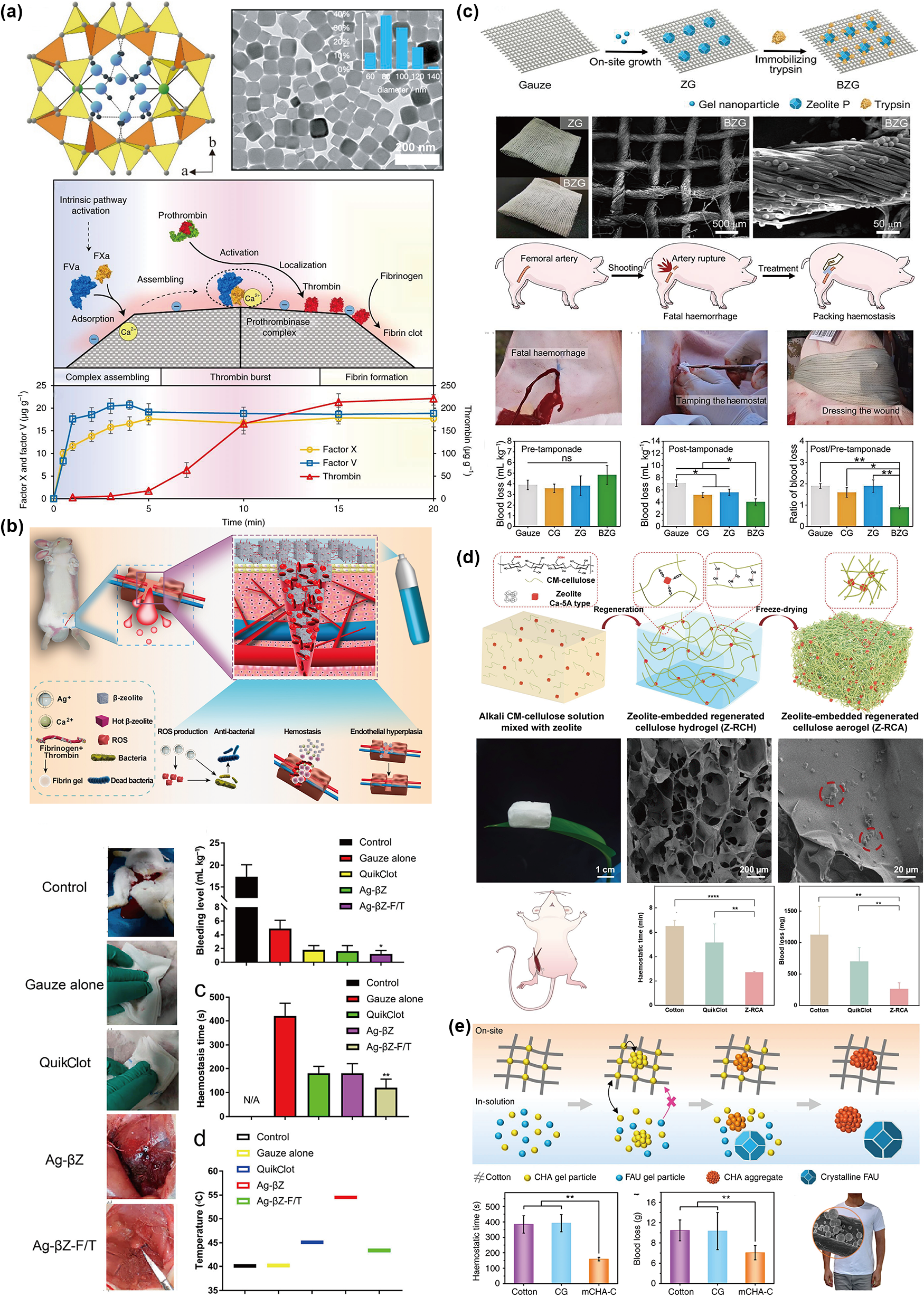
Ze, with the structural formula A(x/q)((AlO2)x(SiO2)y)·n(H2O), is a crystalline microporous aluminosilicate (natural or synthetic) featuring a regular microporous crystal structure (Fig. 3a; Tong et al., Reference Tong, Zhu, Tian, Cui, Si and Chen2025), along with an exceptional ion-exchange capacity and high specific surface area (Fig. 3a; Shang et al., Reference Shang, Chen, Castagnola, Liu, Boselli and Petseva2021). The surfaces of Ze, mediated by calcium ions, can enrich and assemble coagulation factors (e.g. activated factor X; Fig. 3a; Shang et al., Reference Shang, Chen, Castagnola, Liu, Boselli and Petseva2021), forming a highly efficient catalytic interface similar to the natural ‘prothrombinase complex’ (activated factor X/activated factor V) on platelet surfaces. As a result, the coagulation cascade is markedly accelerated, leading to rapid haemostasis (Shi et al., Reference Shi, He, Chen, Wei, Xiao and Shang2025).
Ze-based products used for haemostasis. (a) Crystal structure of Ze, adapted with permission from the published work (Tong et al., Reference Tong, Zhu, Tian, Cui, Si and Chen2025). Copyright 2025 Elsevier B.V. Transmission electron microscopy image and haemostatic mechanism of Ze, adapted with permission from the published work (Shang et al., Reference Shang, Chen, Castagnola, Liu, Boselli and Petseva2021). Copyright 2021 Springer Nature. (b) Composite haemostat spray staunches post-surgical bleeding and ameliorates bacteria-induced inflammation to expedite wound healing, adapted with permission from the published work (Xu et al., Reference Xu, Hu, Fang, Luo, Liu and Zhang2023b). Copyright 2023 American Chemical Society. Ag-bZ-F/T = silver-loaded beta-zeolite combined with fibrinogen and thrombin; ROS = reactive oxygen species. (c) A bio-inorganic hybrid haemostatic gauze for haemorrhage control within the ‘platinum ten minutes’, adapted with permission from the published work (Yu et al., Reference Yu, Zhang, Xiao, Fan and Li2022). Copyright 2022 American Chemical Society. BZG = bio-zeolite gauze; CG = Combat Gauze; ns = non-significant; ZG = zeolite gauze; * and ** denote statistical significance at p < 0.05 and p < 0.01, respectively. (d) Ze-anchored regenerated cellulose aerogel for efficient and biosafe haemostasis, adapted with permission from the published work (Jin et al., Reference Jin, Chen, Liu, Xia, Li, Wang and Zhang2025). Copyright 2025 Elsevier B.V. CM = first 10 min immediately after severe traumatic hemorrhage. (e) A flexible mesoporous Ze–cotton hybrid haemostat, adapted under the terms of the Creative Commons CC BY licence (Yu et al., Reference Yu, Shang, Chen, Xiao, Zhu and Fan2019). CG = Combat Gauze; CHA = chabazite-type; FAU = faujasite; mCHA-C = mesoporous chabazite-type/zeolite/cotton hybrid.

Figure 3 Long description
The infographic consists of five panels labeled a–e, read left-to-right, top-to-bottom, comparing zeolite-based haemostatic materials. (a) Displays the crystal structure of Ze and its haemostatic mechanism, with a graph showing accelerated coagulation over time. (b) Illustrates a composite haemostat spray for post-surgical bleeding. Images compare control, gauze alone, QuikClot, Ag-bZ and Ag-bZ-F/T. Bar graphs indicate effectiveness, with Ag-bZ-F/T showing superior performance. (c) Describes a bio-inorganic hybrid haemostatic gauze. Images show gauze, 3D cell nanoscaffold, zeolite and tropism. Graphs demonstrate haemostatic performance in animal models, highlighting rapid haemostasis. (d) Features a Ze-anchored regenerated cellulose aerogel. Diagrams and SEM images depict its structure. Bar graphs compare haemostatic efficiency, with the aerogel performing effectively. (e) Depicts a flexible mesoporous Ze-cotton hybrid haemostat. Illustrations show its structure and graphs display haemostatic performance, emphasizing its flexibility and efficiency. The infographic highlights the superior performance of zeolite-based haemostats in various applications.
For instance, under severe high-altitude exposure conditions, a bio-Ze gauze demonstrated stable haemostatic performance, whereas a conventional gauze and QuikClot Combat Gauze® showed significantly reduced efficacy (Jia et al., Reference Jia, Du, Zong, Jiang, Zhong and Ye2023). Ion-exchanged P-type calcium–copper Ze (Cu: 0.64–6.30 mg g–1) showed strong haemostatic and antibacterial effect (Wang et al., Reference Wang, Shi, Huang, Xiao and Fan2025), and its gauze counterpart is similarly effective (Wang et al., Reference Wang, Zhang, Wang, Xiao, Yu and Fan2024b). Y-type Ze combined with trypsin-modified gauze catalyses coagulation, outperforming a commercially available gauze (Fig. 3c; Yu et al., Reference Yu, Zhang, Xiao, Fan and Li2022). Ammonium Ze/pectin/calcium chloride/cotton gauze promotes coagulation and adhesion (Edwards et al., Reference Edwards, Prevost, Cintron, Edwards, Prevost and Cintron2023a), with further incorporation of silver/ascorbic acid improving antimicrobial properties (Edwards et al., Reference Edwards, Prevost, Hinchliffe, Nam, Chang and Hron2023b). To reduce Ze heat release, a haemostatic spray composed of silver/Ze/fibrinogen/thrombin can be used for post-operative haemostasis and to achieve antibacterial effects (Fig. 3b; Xu et al., Reference Xu, Hu, Fang, Luo, Liu and Zhang2023b). Mesoporous Ze crystals can be firmly attached to cotton fibres via template-free in situ growth, with a leaching rate below 1% after 10 min of sonication (Fig. 3e; Yu et al., Reference Yu, Shang, Chen, Xiao, Zhu and Fan2019).
To further address the limitations of single-component Ze (poor fixation, high heat generation and poor wound-healing properties), researchers have developed a Ze composite sponge/aerogel. For example, a Bletilla polysaccharide/Ze composite sponge addressed the aforementioned shortcomings (Zhu et al., Reference Zhu, Cheng, Zhao, Hu, Zhou and Zhou2023). A cellulose nanofibre/calcium ion/Ze aerogel (2.89% Ze loading), possessing an ultra-high specific surface area, significantly improved the conversion efficiency of prothrombin to thrombin and avoided the temperature rise associated with Ze addition (Chen et al., Reference Chen, Jin, Kamiya, Liu, Tian and Xia2025b). Similarly, Ze-loaded bacterial cellulose nanofibre aerogels also demonstrated highly effective haemostatic properties, reducing clotting times by 71% (Wang et al., Reference Wang, Li, Liao, Kong, Wang and Wei2023c). In a regenerated cellulose nanofibre/Ze aerogel composite, 3% of the Ze was firmly anchored via hydrogen bonds, resulting in minimal exothermic reactions and highly efficient, safe haemostasis (Fig. 3d; Jin et al., Reference Jin, Chen, Liu, Xia, Li, Wang and Zhang2025). Compared to commercially available sponges, a silver nanoparticle@Ze/chitin/bamboo powder composite sponge exhibited less bleeding and shorter haemostasis times. The bamboo quinones in bamboo powder provide antibacterial properties, while long, flexible microfibres improve this sponge’s resilience (Zheng et al., Reference Zheng, Li, Xu, Xu, Zeng and Tam2023).
In summary, Ze enables rapid haemostasis via its highly efficient water absorption capacity and calcium ion-mediated action. Ze composites, prepared through techniques such as ion exchange and in situ growth, incorporate antimicrobial components and biopolymer components, producing haemostasis, antimicrobial, anti-inflammatory and wound-healing activities while reducing heat release and particle shedding. In China, Professor Jie Fan’s group at Zhejiang University has commercialized a Ze-based haemostat and has reported that the heat release and shedding of particles can be controlled. Future research will focus on producing safe, multifunctional Ze haemostatic materials.
Halloysite nanotubes
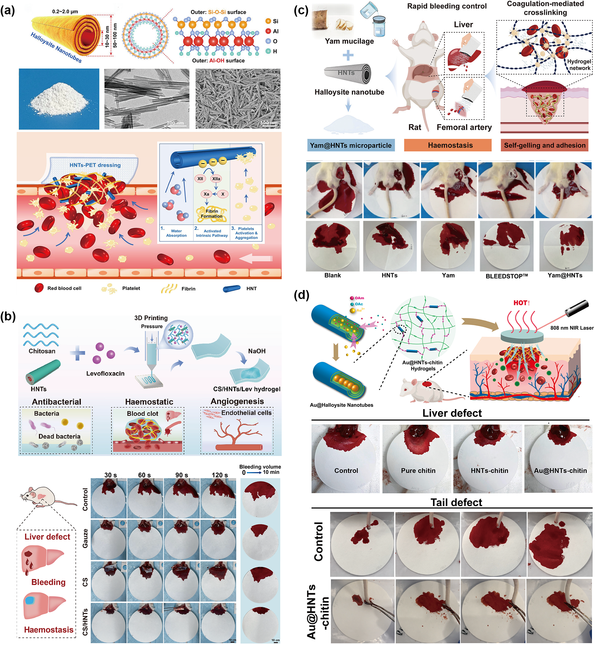
HNTs, with the structural formula Al2Si2O5(OH)4·nH2O, are a naturally occurring silica–aluminate clay. HNTs exhibit a high aspect ratio and a unique hollow tubular structure (Fig. 4a; Li et al., Reference Li, Li, Li, Lin, Chen and Chen2021), characterized by lengths of 200–1000 nm, inner diameters of 10–30 nm and outer diameters of 50–70 nm (Fig. 4a; Berahman et al., Reference Berahman, Raiati, Mehrabi Mazidi and Paran2016). The outer surface of HNTs consists of negatively charged siloxane (Si–O) groups, while the inner surface comprises positively charged aluminol (Al–OH) groups (Fig. 4a; Feng et al., Reference Feng, Luo, Wu, Liu, Liang, He and Liu2022). As a naturally occurring aluminosilicate mineral material with reported biocompatibility, HNTs display coagulation-promoting capabilities intermediate between Ze and MMT and ∼2.3 times that of Kaol (Feng et al., Reference Feng, Luo, Wu, Liu, Liang, He and Liu2022). The haemostatic mechanism of HNTs involves the hollow tubular structure rapidly concentrating blood, the negative charges triggering the intrinsic coagulation cascade and platelets being activated and aggregated to accelerate thrombus formation (Fig. 4a; Feng et al., Reference Feng, Luo, Wu, Liu, Liang, He and Liu2022). In this section, we review recent research progress on HNTs as haemostatic materials.
HNT-based products used for haemostasis. (a) Morphological characterization (photograph, transmission electron microscopy and scanning electron microscopy images), adapted with permission from the published work (Feng et al., Reference Feng, He, Lin, Xie, Liu and Lvov2023). Copyright 2023 John Wiley and Sons. Crystal structure, adapted with permission from the published work (Feng et al., Reference Feng, Chen, He, Liu, Lvov and Liu2024), Copyright 2024 American Chemical Society. Haemostatic mechanism of HNTs, adapted with permission from the published work (Feng et al., Reference Feng, Luo, Wu, Liu, Liang, He and Liu2022), Copyright 2022 Elsevier B.V. PET = poly(ethylene terephthalate). (b) 3D-printed HNT/chitosan hydrogel for haemostasis and infected wound healing, adapted under the terms of the Creative Commons CC BY licence (Chen et al., Reference Chen, Zhou, Zhou, Zhang, Zeng, Zhou and Liu2025d). CS = chitosan; Lev = levofloxacin. (c) HNT-triggered self-gelling yam microparticles for wet tissue adhesion and instant haemostasis, adapted with permission from the published work (Xu et al., Reference Xu, He, Hu, Hu, Liu and Rao2025b). Copyright 2025 American Chemical Society. (d) Au@HNT/chitin composite hydrogel with antibacterial and haemostatic activities for wound healing, adapted under the terms of the Creative Commons CC BY licence (Zhao et al., Reference Zhao, Guo, Wang, Zhou, Huang and Dong2023a). NIR = near-infrared; OAc = oleic acid; OAm = oleylamine.

Figure 4 Long description
The infographic presents HNT-based products for haemostasis across four sections. (a) Shows morphological characterization of halloysite nanotubes (HNTs) with images from transmission electron microscopy and scanning electron microscopy. It illustrates the haemostatic mechanism involving HNTs concentrating blood, activating platelets and accelerating thrombus formation. (b) Describes a 3D-printed HNT/chitosan hydrogel for haemostasis and wound healing. It highlights antibacterial and angiogenesis properties, with images showing liver defect, bleeding and haemostasis in a rat model. (c) Depicts HNT-triggered self-gelling yam microparticles for wet tissue adhesion and instant haemostasis. It includes images of rapid bleeding control in a rat femoral artery model, showing haemostasis and self-gelling adhesion. (d) Features Au@HNT/chitin composite hydrogel with antibacterial and haemostatic activities for wound healing. It includes images of liver and tail defects treated with control, pure chitin, HNTs-chitin and Au@HNTs-chitin, demonstrating haemostasis and healing under near-infrared laser treatment.
A series of fibre membranes based on electrospinning have been prepared. Electrospun amino-HNT/polylactic acid nanofibre mats exhibited rapid haemostatic capabilities (Delyanee et al., Reference Delyanee, Solouk, Akbari and Daliri Joupari2021). Electrospun astilbin/HNT/polyvinyl alcohol nanofibre membranes integrated haemostatic, anti-inflammatory, antibacterial and wound-healing properties (Tang et al., Reference Tang, Wang, Liu, Ding, Yang and Wu2024). Electrospun polyvinyl pyrrolidone/HNT haemostatic membranes promoted blood coagulation through multiple pathways, with haemostatic efficacy comparable to that of commercial products (Cui et al., Reference Cui, Huang, Lei, Li, Jiang and Zeng2021). Electrospun HNTs/polylactic acid membranes displayed haemostatic properties comparable to QuikClot Combat Gauze® and promoted angiogenesis and collagen deposition (Delyanee et al., Reference Delyanee, Solouk, Akbari and Daliri2022). Electrospun cellulose/HNTs membranes increased plasma coagulation speeds 2.4-fold and whole-blood coagulation by 1.3-fold vs QuikClot Combat Gauze®, with no cytotoxicity (Udangawa et al., Reference Udangawa, Mikael, Mancinelli, Chapman, Willard, Simmons and Linhardt2019). Unlike electrospinning, solution blow spinning uses high-speed airflow instead of high-voltage electric fields (Padovani et al., Reference Padovani, Sanches, Moura Aouada, Malmonge and de Paula2024). HNT/chitosan/poly(oxyethylene) fibre membranes prepared via solution blow spinning exhibited excellent hydrophilicity, breathability, flexibility and antibacterial properties, with an in vitro haemostasis time of 217 s (Chen et al., Reference Chen, Mo, Zhang, Zhang, Cui, Wang and Li2024).
Beyond fibre membranes, other HNT-based haemostatic dressings and porous scaffolds have also been developed. A HNT-coated polyester fibre dressing was developed using a simple impregnation method with 21% HNT loading (Feng et al., Reference Feng, Luo, Wu, Liu, Liang, He and Liu2022). To resolve HNT detachment, a HNT/alginate/cotton composite dressing prepared via alginate mediation and Ca2+ crosslinking demonstrated excellent haemostatic performance, with over 80% of the coagulation components remaining intact after ultrasonication (Feng et al., Reference Feng, He, Lin, Xie, Liu and Lvov2023). Similarly, HNT microspheres connected to an electrospun polylactic acid nanofibre membrane reduced the risk of inflammation and distal thrombosis caused by HNT shedding, with an in vitro clotting time of 171 s (Huang et al., Reference Huang, Qian, Tian, Shen, Liu, Yang and Zhang2023). Compared to chitosan sponges, chitosan/HNT sponges prepared by freeze-drying showed an 89% increase in haemostatic capacity (Liu et al., Reference Liu, Shen, Ao, Dai, Liu and Zhou2014). HNT/chitosan/collagen non-symmetrical haemostatic sponges prepared via directional freezing reduced clotting times by 40% and exhibited antimicrobial properties (Lin et al., Reference Lin, Feng, He, Ding and Liu2023). A freeze-dried HNT/oxidized cellulose aerogel, with a hierarchical porous structure, was shown to be capable of rapidly absorbing blood (Long et al., Reference Long, Yang, Shi and Yang2024). A carboxymethyl chitosan/sericin/silver/HNT haemostatic dressing demonstrated increased antibacterial properties, upregulated vascular endothelial growth factor and promoted wound healing (Li et al., Reference Li, Jiang, Zhang, Xie, Li and Ming2023). A biodegradable modified starch@HNT composite sponge precisely sealed bleeding sites, reducing haemostasis times by 53% (Yang et al., Reference Yang, Chen, Wang, Si, Hao and Shi2024a).
In addition to fibre membranes and dressings, various HNT-based hydrogels have also been developed for haemostasis. A HNT/oxidized sodium alginate/gelatin hybrid hydrogel reduced clotting times from 255 to 207 s (Lin et al., Reference Lin, Zhang, Chen and Liu2024). A HNT/silk fibroin/chitosan hydrogel synergistically promoted blood coagulation, hair follicle regeneration, angiogenesis and scar reduction (Xu et al., Reference Xu, Gao, Li, Dai, Zhu and Meng2021). A HNT/chitosan/oxidized dextran adhesive haemostatic hydrogel was shown to be specifically adaptable to various wound shapes (Li et al., Reference Li, Li, Li, Lin, Chen and Chen2021). Tetracycline hydrochloride salt encapsulated within HNTs formed a hydrogel upon mixing with silk nanofibres, whose clotting time was 59% shorter than that of a tetracycline hydrochloride/silk nanofibre hydrogel (Xiao et al., Reference Xiao, Liu, Chen, Zhao, Sun and Yang2025a). Compared to chitosan hydrogels, chitosan/HNT hydrogels prepared via 3D printing technology demonstrated a 42% reduction in clotting times (Fig. 4b; Chen et al., Reference Chen, Zhou, Zhou, Zhang, Zeng, Zhou and Liu2025d). A hierarchically orientated foam for haemorrhage control and wound healing has also been developed. Specifically, the outer layer of this multilayer foam consists of polyvinyl alcohol and a phase-change material (to prevent thermal stress), the middle layer consists of a silk fibroin membrane (to block blood flow) and the inner layer consists of polyvinyl alcohol and HNTs (to improve haemostatic effects; Jia et al., Reference Jia, Luo, Yang, Wu, Diao, Ojo and Lyu2025).
Furthermore, functionalization of HNTs based on their unique hollow structure has become a current focus of research. FeS2@HNT/polyvinyl alcohol/alginate composite hydrogels simultaneously exhibit haemostatic, antibacterial, anti-inflammatory, photothermal and wound-healing properties. The FeS2 modification on HNTs endows HNTs with peroxidase activity and significantly enhances antibacterial efficacy under near-infrared irradiation (Zeng et al., Reference Zeng, Chen, Zhou, Zhang, Luo, Zhou and Liu2026). Similarly, HNTs loaded with linalool and a photothermal agent can release linalool upon infrared activation, facilitating rapid haemostasis, sustained antibiotic delivery and the healing of infected wounds (Zhou et al., Reference Zhou, Li, Hu and Fu2024b). Au@HNT/chitin hydrogels were prepared by filling 5–10 nm gold nanoparticles into the lumens of HNTs and crosslinking with epichlorohydrin. As a result, the hydrogel exhibited haemostatic, photothermal, antibacterial and wound-healing properties (Fig. 4d; Zhao et al., Reference Zhao, Feng, Zhou, Tan and Liu2023b). Peptide RG5 was loaded into the lumens of HNTs to form a haemostatic powder, which was then encapsulated within an injectable gelatin/alginate hydrogel. The results showed that the clotting times of peptide RG5/HNT powders were shorter than those of HNTs alone and comparable to Yunnan Baiyao; the haemostatic efficacy of peptide RG5/HNT/gelatin/alginate was greater than that of the haemostatic agent Celox® (Zhang et al., Reference Zhang, Zhao, Lv, Liu, Wang and Li2021), a commercial haemostatic material based on chitosan that is frequently utilized in complex emergency scenarios.
To overcome the insufficient adhesion of conventional hydrogels in wet environments, a highly crosslinked methacrylated carboxymethyl chitosan/oxidized dextran/tannic acid/HNT hydrogel was developed. The HNTs improved the mechanical properties and haemostatic performance of the hydrogel, while the tannic acid improved the adhesion of the material (Zhao et al., Reference Zhao, Guo, Wang, Zhou, Huang and Dong2023a). Inspired by traditional Chinese medicine, a yam mucilage/HNT self-gelling microparticle system was developed. Synergistic interaction between yam mucilage and HNTs enabled robust adhesion to wet tissues and rapid haemostasis (Fig. 4c; Xu et al., Reference Xu, Wu, Liu, Geng, Li and Liao2025a). Polydopamine@HNT/gellan gum/sericin hydrogels achieved rapid haemostasis and photothermal antibacterial effects while downregulating inflammatory factors to promote wound healing (Yuan et al., Reference Yuan, Jiang, Jiang, Guo, Liu and Ming2023).
In summary, HNTs have emerged as a core component for multifunctional haemostatic materials due to their hollow tubular structure, high specific surface area and surface negative charge. Through techniques such as electrospinning and freeze-drying, HNT-based materials load antimicrobial and adhesive components within the nanotubes, achieving haemostatic, adhesion, anti-inflammatory and wound-healing functions. However, the risk of embolism caused by HNT detachment remains a potential core issue limiting the clinical translation of these materials. Therefore, commercializing high-performance HNT-based haemostats adaptable to the wound microenvironment is crucial.
Montmorillonite
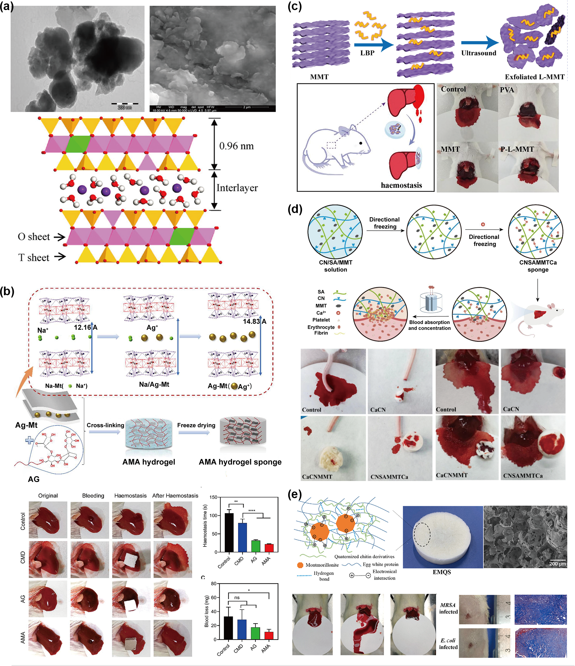
MMT, with the structural formula (Na,Ca)0.33(Al,Mg)2(Si4O10)(OH)2·nH2O, is a naturally occurring 2:1-type layered silicate (Yang et al., Reference Yang, Wang, Yang, Mu and Wang2023). Its unit structure consists of two layers of silicate tetrahedra sandwiching one layer of alumina octahedra (Fig. 5a; Paluszkiewicz et al., Reference Paluszkiewicz, Stodolak, Hasik and Blazewicz2011; Chen et al., Reference Chen, Zheng, Wang and Hu2025c). As a historically established haemostatic material, MMT exhibits high water absorbency and possesses a large specific surface area and abundant negative charge (Guo et al., Reference Guo, Dong, Liang and Li2021). The core haemostatic mechanism of MMT stems from its surface negative charges activating the coagulation system together with its high specific surface area enabling the physical adsorption and concentration of blood (Li et al., Reference Li, Quan, Liang, Li, Yuan and Tao2016). However, early commercial MMT powders such as WoundStat® carried risks of inducing vascular embolism, limiting their direct application (Katti et al., Reference Katti, Jasuja, Jaswandkar, Mohanty and Katti2022). In recent years, developing safe, multifunctional, integrated haemostatic materials with MMT as the core component has become a research focus.
MMT-based products used for haemostasis. (a) Transmission electron microscopy image and scanning electron microscopy image, adapted with permission from the published work (Paluszkiewicz et al., Reference Paluszkiewicz, Stodolak, Hasik and Blazewicz2011). Copyright 2011 Elsevier B.V. Crystal structure of MMT (Chen et al., Reference Chen, Zheng, Wang and Hu2025c), adapted under the terms of the Creative Commons CC BY licence. (b) MMT/agarose 3D network gelatin sponge for wound healing with haemostatic and durable antibacterial properties, adapted with permission from the published work (Zhang et al., Reference Zhang, Pan, Dong, Yang, Huang, Yan and Gao2023). Copyright 2023 American Chemical Society. AG = agarose gel sponge; AMA = Ag-Mt/agarose composite gel sponge; CMD = commercial medical dressing; Mt = montmorillonite; ns = non-significant; *, ** and **** denote statistical significance at p < 0.05, p < 0.01 and p < 0.0001, respectively. (c) Hydrogel loaded with exfoliated MMT using anti-inflammatory Lycium barbarum polysaccharides for advanced wound dressing, adapted with permission from the published work (Shi et al., Reference Shi, Lan, Feng, Ni, Zhu, Wang and Sun2022). Copyright 2022 Elsevier B.V. A-MMT = polysaccharide (LBP)-functionalized ultrathin MMT nanosheets; LBP = polysaccharides extracted from Lycium barbarum L.; P-L-MMT = PVA hydrogel loaded with L-MMT; PVA = poly(vinyl alcohol). (d) Water-triggered shape memory cellulose/alginate/MMT composite sponge for rapid haemostasis, adapted with permission from the published work (Liu et al., Reference Liu, Yin, Hu, Kang, Hu and Wei2024c). Copyright 2024 Elsevier B.V. CN = carboxylated cellulose; SA = sodium alginate. (e) Quaternized chitin/egg white protein/MMT composite sponge with antibacterial and haemostatic effects for wound healing, adapted with permission from the published work (Yang et al., Reference Yang, Zhang, Zeng, Jia, Zhang, Yu and Duan2022). Copyright 2022 Elsevier B.V. EMQS = egg white protein/quaternized chitin derivative/MMT composite sponge; MRSA = methicillin-resistant Staphylococcus aureus.

Figure 5 Long description
The image contains multiple panels showing MMT-based haemostatic products. (a) Displays transmission and scanning electron microscopy images, along with a diagram of the crystal structure of MMT, indicating an interlayer distance of 0.96 nm. (b) Illustrates the process of creating an AMA hydrogel sponge, including steps like cross-linking and freeze-drying. It shows test results of haemostasis and antibacterial properties, with a bar graph comparing different samples. (c) Depicts the exfoliation of MMT using LBP and its application in haemostasis, with images of test results on mice. (d) Shows the creation of a CN/SA/MMT composite sponge, with diagrams of directional freezing and images of haemostasis tests. (e) Illustrates the development of a quaternized chitin/egg white protein/MMT composite sponge, with diagrams and images of antibacterial and haemostatic tests, including a bar graph of results.
To prevent MMT detachment, electrospun polycaprolactone/chitosan oligosaccharide/MMT composite membranes, with fibre diameters of 60–80 nm, have been prepared, and these have demonstrated clotting times comparable to commercial gauzes and reduced bleeding volumes by more than half (Tran et al., Reference Tran, Vu, Le, Le, Nguyen and Vu2025). The MMT-ferric oxide/polyvinylpyrrolidone electrospun magnetic film, prepared via electrospinning, as well as inhibiting liver cancer recurrence, also demonstrated superior haemostatic capability compared to a commercial gauze group (Xiang et al., Reference Xiang, Hu, Yan, Yang, Ji and Xu2025). Silver ions were loaded onto MMT via ion exchange followed by one-step electrodeposition with chitosan to produce a transparent hydrogel membrane. This membrane exhibited excellent antibacterial, wound-healing and haemostatic properties (Xu et al., Reference Xu, He, Hu, Hu, Liu and Rao2025b). Notably, a quaternized cellulose ammonium/polyethylene glycol/tannic acid/MMT haemostatic powder absorbed interfacial fluids, hydrated into a stable hydrogel and adhered to tissue surfaces, making it suitable for irregularly shaped wounds (Liu et al., Reference Liu, Hu, Hu, Bai, Kang, Wei and Huang2024b).
Sponges represent a common form of haemostatic material. Carboxylated nano-cellulose/MMT composite sponges prepared via directional freezing technology rapidly absorbed water and concentrated blood, exhibiting an activated partial thromboplastin time comparable to that of pure MMT (Yin et al., Reference Yin, Hu, Kang, Hu, Wu and Liu2023). A cellulose/alginate/MMT composite sponge prepared via directional freezing technology exhibited an anisotropic three-dimensional porous structure, enabling the rapid coagulation of blood (Fig. 5d; Liu et al., Reference Liu, Yin, Hu, Kang, Hu and Wei2024c). A silk fibroin/MMT sponge prepared via electrospinning, with a porous structure, showed an absorption capacity as high as 1688% (Fang et al., Reference Fang, Wang, Yang, Tang and Huang2024a). A graphene/MMT sponge achieved rapid haemostasis in ∼85 s, with angiography confirming no thrombus formation. Within the composite, MMT was stably embedded within crosslinked graphene layers (Li et al., Reference Li, Quan, Liang, Li, Yuan and Tao2016). A MMT/silver/agarose composite sponge, with sustained release of silver ions between layers, achieved long-lasting antibacterial effects (Fig. 5b; Zhang et al., Reference Zhang, Pan, Dong, Yang, Huang, Yan and Gao2023). Ciprofloxacin was incorporated into silk fibroin/chitosan/MMT sponges to achieve sustained release for 1 week (Ngo et al., Reference Ngo, Nguyen, Nguyen, Tran, Nguyen and Luu2024). Developed to overcome the resistance issues posed by traditional antibiotics, a polydopamine/cerium dioxide/MMT composite material exhibited antibacterial and haemostatic properties while accelerating wound healing (Liu et al., Reference Liu, She, Li, Li, Yang and Li2024a).
Furthermore, introducing adhesive components is a key strategy for treating moist and dynamic wounds. With exceptional tissue adhesion (∼47 kPa), a polyaldehyde dextran/MMT composite sponge rapidly sealed wounds, reduced clotting times from 480 to 3–10 s in vitro and reduced bleeding volumes by nearly 95% in vivo (Liu et al., Reference Liu, Liu, Yu, Wang, Yao and Liu2020). Similarly, an oxidized bacterial cellulose/dopamine@MMT/silver composite sponge exhibited strong adhesion and haemostatic capabilities (Cao et al., Reference Cao, Yang, Zhang, Liu and Chen2021). Inspired by pastry-making techniques, a fully natural material-based sponge constructed from egg white protein, quaternized chitin derivatives and MMT exhibited antibacterial, haemostatic and wound-healing acceleration functions (Fig. 5e; Yang et al., Reference Yang, Zhang, Zeng, Jia, Zhang, Yu and Duan2022). Additionally, for anticoagulation patients who are at high risk of post-extraction bleeding and impaired bone regeneration, a silk fibroin/MMT sponge prepared via ethanol-enhanced cryopreservation exhibited shape memory properties. Upon contact with water or blood, the sponge expanded to achieve compression haemostasis while promoting new bone formation in the extraction socket (Xiao et al., Reference Xiao, Sun, Fu, Yang, Yao and Shao2025b).
Additionally, researchers have incorporated MMT as the core component of hydrogels. A goji berry polysaccharide@MMT/polyvinyl alcohol hydrogel exhibited excellent haemostatic effects and promoted wound healing. Notably, the ultra-thin nanolayers of goji berry polysaccharide@MMT, with a thickness of 1 nm, were prepared via liquid-phase exfoliation (Fig. 5c; Shi et al., Reference Shi, Lan, Feng, Ni, Zhu, Wang and Sun2022). A γ-polyglutamic acid/polyethyleneimine/MMT hydrogel absorbed wound exudate to maintain wound cleanliness and accelerate blood coagulation. The incorporation of γ-polyglutamic acid/polyethyleneimine endowed the hydrogel with anti-inflammatory properties and promoted wound healing (Li et al., Reference Li, Han, Zhang, Tan, Cui, Wang and Shi2024a). A polydopamine/MMT/polyacrylamide hydrogel exhibited outstanding haemostatic and antibacterial properties. In this system, polydopamine was intercalated into MMT, thereby inhibiting dopamine oxidation while preserving the anti-inflammatory and antibacterial catechol groups (Huang et al., Reference Huang, Zhao, Wu, Han, Li and Xing2024).
In summary, MMT activates the coagulation cascade via its surface negative charge and concurrently absorbs blood through its high specific surface area. MMT-based haemostatic materials, prepared through intercalation, electrospinning and freeze-drying, incorporate adhesive and antimicrobial components to achieve multiple functions, including haemostasis, strong adhesion, antibacterial activity and wound healing. The risk of embolism when using MMT alone has been noted. This underscores the critical research focus on developing safe, multifunctional MMT haemostatic materials and establishing methods to securely anchor MMT within composite structures.
Palygorskite
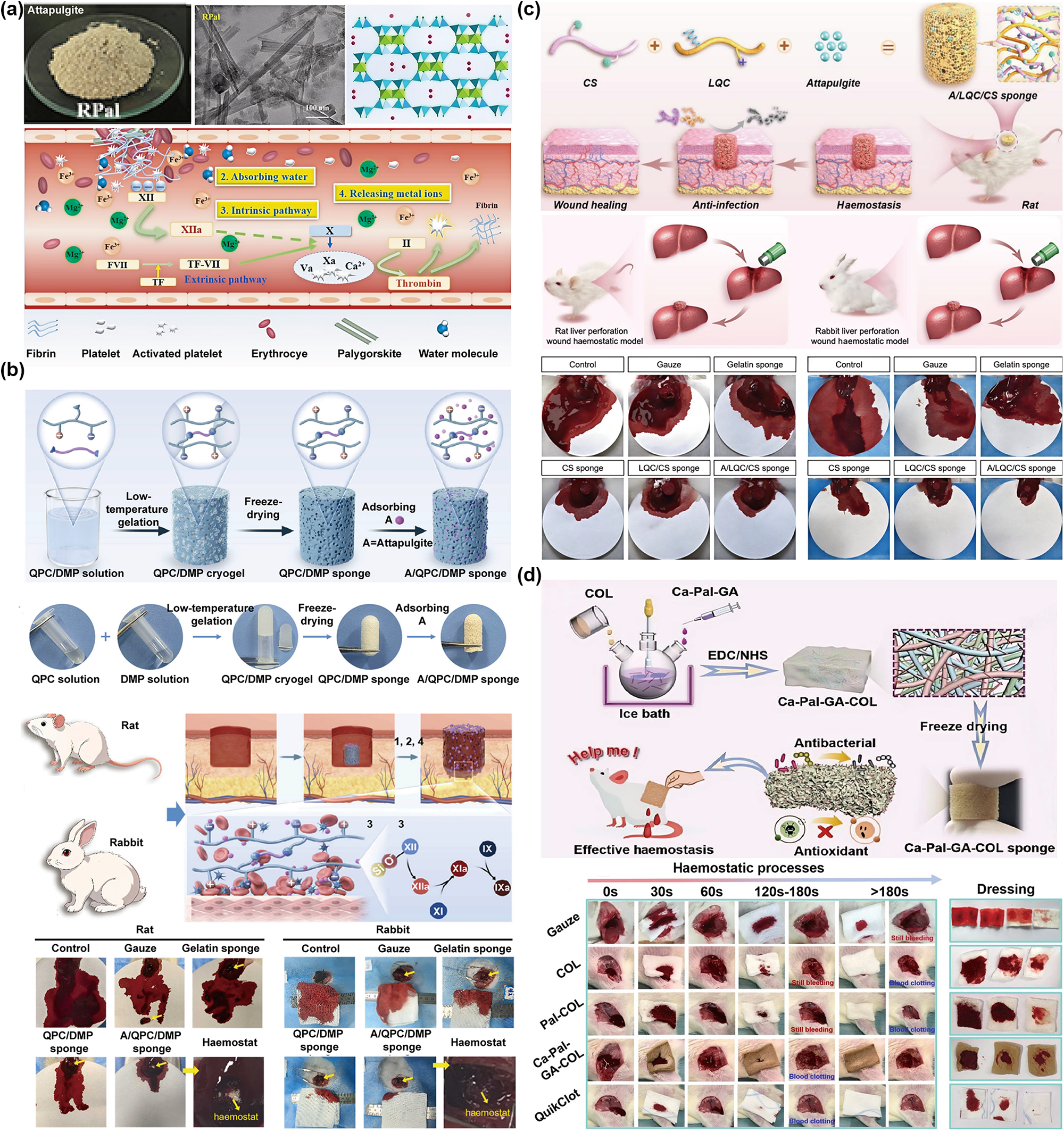
Pal, with the structural formula (Mg,Al)5((OH)2(Si,Al)8O20)·nH2O, is a naturally occurring layer-chain silicate clay. Pal exhibits a nanorod morphology and pores of 0.64 × 0.37 nm, demonstrating excellent haemostatic potential (Fig. 6a; Yang et al., Reference Yang, Wang, Li, Mu, Yang, Wang and Liu2024c). The haemostatic mechanism of Pal involves its nanorod morphology providing physical adsorption sites, while its ultra-high hydrophilicity promotes platelet aggregation. Upon contact with blood, Pal absorbs Fe3+ and Mg2+ while releasing Ca2+, effectively accelerating fibrin formation (Fig. 6a; Wang et al., Reference Wang, Yang, Yang, Mu and Wang2024c; Yang et al., Reference Yang, Wang, Li, Mu, Yang, Wang and Liu2024c). In this section, we review research progress on Pal haemostatic materials in various material forms.
Pal-based products used for haemostasis. (a) Photograph, transmission electron microscopy image, crystal structure and haemostatic mechanism of Pal, adapted with permission from the published work (Yang et al., Reference Yang, Wang, Li, Mu, Yang, Wang and Liu2024c). Copyright 2024 IOP Publishing Ltd. R-Pal = raw Pal. (b) Anti-infective and pro-coagulant chitosan composite sponge with shape memory for haemostasis in multi-tissue defects, adapted with permission from the published work (Du et al., Reference Du, Li, Liu, Hou, Sun and Wang2025a). Copyright 2025 Elsevier B.V. DMP = disuccinimide carbonate-modified polyethylene glycol; QPC = quaternized and phosphated chitosan. (c) Lauric acid-modified quaternized chitosan and Pal-reinforced self-elastic chitosan sponge for non-compressible haemorrhage and wound healing, adapted with permission from the published work (Du et al., Reference Du, Zhao, Liu, Chen, Li, Zhang and Wang2025b). Copyright 2025 Elsevier B.V. CS = chitosan; LQC = lauric acid-modified quaternized chitosan. (d) Antibacterial and antioxidant adherent sponge for arterial bleeding control via gallic acid-mediated fibrous clay assembly in collagen, adapted with permission from the published work (Guo et al., Reference Guo, Zeng, He, Zhang, Shi, Ran and Li2025). Copyright 2025 American Chemical Society. Ca-Pal-GA = Ca2+-loaded Pal assembled with gallic acid; COL = collagen; EDC/NHS = 1-ethyl-3-(3-dimethylaminopropy)carbodimide/N-hydroxysuccinimide.

Figure 6 Long description
This infographic presents Pal-based haemostatic materials designed to enhance haemostasis, antibacterial properties and wound healing. It consists of four panels labeled (a) to (d). Panel (a) shows R-Pal, including a photograph, electron microscopy image and crystal structure, illustrating its haemostatic mechanism. It highlights the absorption of Fe superscript 3 plus and Mg superscript 2 plus and the release of Ca superscript 2 plus, promoting fibrin formation. Panel (b) features a chitosan composite sponge for haemostasis in multi-tissue defects, with images of QPC solution, QPC/DMP solution and QPC/DMP sponge. Experimental results on rats and rabbits demonstrate the sponge′s effectiveness in reducing bleeding. Panel (c) depicts a lauric acid-modified chitosan sponge for non-compressible haemorrhage and wound healing, with images of CS, LQC and ALQC/PEG sponge. Experiments on rats show significant haemostasis and wound closure. Panel (d) illustrates an antibacterial and antioxidant sponge for arterial bleeding control, featuring a gallic acid-mediated fibrous clay assembly in collagen. It includes images of haemostatic processes at time intervals (0s, 30s, 60s, 120-180s and dressing), showing the effectiveness of various sponges like Pr-Pal and QPC/CS. The infographic highlights the superior performance of these materials in achieving rapid haemostasis and effective wound healing.
A systematic study was conducted on the effects of oxalic acid gradient leaching on the haemostatic properties of Pal. The data indicated that Pal leached with oxalic acid for 1 h exhibited the optimal procoagulant effect (Wang et al., Reference Wang, Yang, Yang, Mu and Wang2024c). When the Pal concentration was 20%, a Pal/chitosan/polyvinylpyrrolidone film exhibited the strongest tensile properties and haemostatic performance (Wang et al., Reference Wang, Mu, Zhang, Du, Yang and Wang2023b). Based on these findings, an improved Pal/chitosan/polyvinylpyrrolidone film incorporating dencichine was developed, endowed with additional antibacterial properties (Yang et al., Reference Yang, Wang, Li, Yang, Liu and Wang2024b). Additionally, ZnO/Pal nanocomposites prepared via a hydrothermal method mediated by lemon peel extract also demonstrated haemostatic, antibacterial and biocompatible properties (Li et al., Reference Li, Yang, Yang, Mu, Liu and Wang2024b). Moreover, red peony extract-mediated ferric oxide nanoparticles were uniformly distributed on the Pal surface and exhibited significantly stronger adhesion to platelets and red blood cells than isolated ferric oxide nanoparticles or Pal. This can be attributed to the interaction between Pal’s negative charge and Fe3+, which enhances platelet/red blood cell adhesion and aggregation while accelerating coagulation factor activation (Wang et al., Reference Wang, Li, Yang, Yang, Mu and Wang2026).
A Pal/alkylated chitosan composite sponge achieved a 100% survival rate in a rabbit carotid artery injury model, with total blood losses of 8.4 ± 4.0 g, outperforming Celox® (Chen et al., Reference Chen, Yan, Chen, Yang and Xu2023). Pal-enhanced chitosan sponges demonstrated superior haemostatic efficacy compared to a commercial gauze and gel sponges in a non-compressible liver perforation model (Fig. 6c; Du et al., Reference Du, Zhao, Liu, Chen, Li, Zhang and Wang2025b). Physically incorporating Pal into quaternary ammonium/phosphorylated chitosan sponges improved their mechanical strength and hydrophilicity, imparting shape memory functionality. After absorbing water or blood, the sponge rapidly regained its original shape, effectively sealing wounds and promoting haemostasis (Fig. 6b; Du et al., Reference Du, Li, Liu, Hou, Sun and Wang2025a). Furthermore, to reduce the risk of thrombosis caused by clay particle shedding, Pal loaded with Ca2+ was modified with gallic acid and assembled with collagen into a composite sponge. QuikClot Combat Gauze® became noticeably turbid after 2 min of ultrasonic cleaning, whereas the composite sponge remained clear after 10 min of ultrasonic cleaning, with 83% mass retention and tissue adhesion. Its haemostatic performance approached that of QuikClot Combat Gauze® (Fig. 6d; Guo et al., Reference Guo, Zeng, He, Zhang, Shi, Ran and Li2025).
In summary, Pal, with its unique morphology and high specific surface area, emerges as an ideal component for synergistic haemostasis. By employing strategies that combine Pal with other materials, its inherent drawbacks (e.g. powder shedding, difficult cleanup and potential thrombogenicity) can be effectively mitigated. In the future, research on Pal-based haemostatic materials will focus on regulating the wound microenvironment following haemostasis (i.e. infection prevention, exudate management and the modulation of early tissue repair), thereby expanding their functionality from rapid haemostasis to effective wound healing.
Other clay minerals
Laponite
Laponite (Lap), with the chemical formula Na0.7((Si8Mg5.5Li0.3)O20(OH)4)–0.7, is a synthetic layered silicate clay. Unlike naturally occurring clays, Lap exhibits high batch consistency, high purity and a low swelling rate (Zheng et al., Reference Zheng, Zhang, Wang, Liu, Pan and Li2021). Lap has a unique disc-shaped structure with a diameter of 20–30 nm and a thickness of ∼1–2 nm. Its outer surface exhibits a negative charge, and it possesses excellent haemostatic capability and biocompatibility. The haemostatic mechanism of Lap is based on its nanodisc structure and negative charge, which effectively activate coagulation factor XII and aggregate red blood cells, thereby achieving rapid haemostasis (Fang et al., Reference Fang, Lin, Wang, Weng, Chen and Liu2024b).
Constructing materials in a sponge form is a common strategy in Lap composite preparation. Haemostatic powders prepared from quaternized chitosan, catechol-modified alginate and Lap exhibited adhesive, haemostatic and antibacterial capabilities (Fang et al., Reference Fang, Lin, Wang, Weng, Chen and Liu2024b). A supramolecular sponge composed of gelatin methacrylate, Lap and branched supramolecular polymers had a porous structure dependent on Lap concentration and effectively promoted blood coagulation, collagen deposition and granulation (i.e. tissue formation; Zhang et al., Reference Zhang, Peng, Wei, Sun, Xie, Yu and Chen2024a). Based on carboxymethyl cellulose and hydroxyethyl cellulose, a composite sponge with high water absorption, strong adhesion and haemostatic properties was successfully prepared through citric acid crosslinking and the incorporation of Lap and dopamine (Mahmoodzadeh et al., Reference Mahmoodzadeh, Valizadeh, Edalati, Khordadmehr, Zakeri, Salehi and Jarolmasjed2024). A shape-memory silk fibroin/glycerol/Lap composite scaffold exhibited excellent haemostatic performance and promoted osteogenic regeneration (Lyu et al., Reference Lyu, Chen, Luo, Lin, Yang, Zhou and Tao2024).
To treat active moist wounds, various adhesive hydrogels have been developed. A gelatin methacrylate/Lap/dopamine@gelatin pore-adhesive hydrogel exhibited a clotting time of less than 1 min and effectively sealed wounds (Xie et al., Reference Xie, Zheng, Pu, Jia, Wang and Chen2022). A Lap/polyacrylic acid/oxidized carboxymethyl cellulose/tannic acid hydrogel effectively stopped bleeding (Xu et al., Reference Xu, Jiao, Huang, Ren, Liang, Wei and Zhang2024). The in vitro clotting index of a gelatin/tannic acid/Lap haemostatic hydrogel was 16% (Xu et al., Reference Xu, Xu, Gao, Gao, Zhuang, Zhang and Dong2023a). β-Glucan/Lap hybrid hydrogels demonstrated positive outcomes in diabetic wound models, including reduced haemostasis time, normal vascularization and hair follicle regeneration (Zhang et al., Reference Zhang, Ning, Chen and Dong2024b). Lap/chitosan/alginate/lignin/acrylamide hydrogels exhibited improved mechanical properties and haemostatic performance with the incorporation of Lap (Zhao et al., Reference Zhao, Huang, Tian, Luo, Wang and Wang2022). A Lap/polyvinyl alcohol/alginate hydrogel, at a Lap concentration of 0.5%, significantly promoted fibroblast proliferation and improved haemostatic capacity, which indirectly demonstrated Lap’s biocompatibility (Golafshan et al., Reference Golafshan, Rezahasani, Tarkesh Esfahani, Kharaziha and Khorasani2017). A dual-layer hydrogel strip composed of a thermosensitive layer and an inert layer has been prepared previously. Lap/polydopamine nanoparticles were coated onto the outer surface of the hydrogel strip. Here, polydopamine provided adhesion functionality, while Lap activated coagulation factors and exerted haemostatic effects (Davis Jr et al., Reference Davis, Biswas, Aragon, Dana, Sureka and Ramanuja2026).
Furthermore, haemostatic hydrogels based on Lap have been designed to address distinct clinical challenges. For bone defect bleeding, gelatin methacrylate/Lap hydrogels prepared via ice-templating polymerization demonstrated excellent shape recovery, enabling non-compressive haemostasis and bone regeneration (Chen et al., Reference Chen, Yang, Tang, Wang, Zhang and Sun2025a). For persistent bleeding and alveolar bone resorption after tooth extraction, a sandwich sponge composed of alginate, chitosan, Lap and soluble eggshell membrane was developed. This sponge demonstrated superior haemostatic performance, supported stem cell adhesion and proliferation and improved osteogenic differentiation (Farsinezhad et al., Reference Farsinezhad, Niknafs, Samieifard, Rezaie, Sadeghzadeh and Vahdat2025). Ulcers formed after endoscopic submucosal dissection remain a significant clinical challenge due to the risk of uncontrollable bleeding. To address this issue, an injectable shear-thinning hydrogel composed of carboxymethyl chitosan/Lap/polyvinyl alcohol reduced bleeding by 83% in both rat liver incision and gastric perforation models (Guo et al., Reference Guo, Shen, Ma, Wang, Yao and Zhang2023a). For incompressible wounds such as visceral injuries, a Lap/gelatin haemostatic porous hydrogel with shape-memory functionality was developed using an ice-crystal template method (Teng et al., Reference Teng, Xia, Qian, Hu, Hong and Liao2022).
For use in limited oral surgical spaces, a Lap/gelatin methacrylate/tannic acid hydrogel exhibited haemostatic, adhesive, low-swelling and antibacterial properties, promoting the healing of full-thickness oral mucosal defects (Zhu et al., Reference Zhu, Li, Xie, Yang, Li and Wang2022). For use in non-compression haemorrhage and skin regeneration, a dual-layer biomimetic scaffold was constructed, consisting of a gelatin nanofibre membrane simulating the dermis layer and a gelatin methacrylate/Lap hydrogel loaded with epidermal growth factor simulating the epidermis layer. This scaffold achieved rapid wound sealing, complete re-epithelialization, ordered collagen deposition, hair follicle regeneration and scarless healing (Zandi et al., Reference Zandi, Dolatyar, Lotfi, Shallageh, Shokrgozar and Tamjid2021). To address bleeding and poor healing in diabetic wounds, a hydrogel was developed using arginine-functionalized chitosan and gallic acid-functionalized gelatin/methacrylic anhydride as the main components, incorporating zinc-loaded Lap. This hydrogel exhibited antibacterial, antioxidant and anti-inflammatory properties. It intelligently released nitric oxide (NO) and Zn2+: NO inhibited bacterial growth, while Zn2+ directly killed bacteria (Wei et al., Reference Wei, Zheng, Zhong, Ding, Huang and Liu2025).
Diatomite
Diatomite, with the chemical formula SiO2·nH2O, is a biogenic siliceous sedimentary rock. Diatomite exhibits high porosity, a high specific surface area and excellent haemostatic properties (Youn et al., Reference Youn, Ki, Min, Abdelhamid and Pack2024). Diatomite exerts its haemostatic effect by physically adsorbing blood and concentrating platelets (Youn et al., Reference Youn, Ki, Min, Abdelhamid and Pack2024). Diatomite serves as a drug carrier for antibiotics such as vancomycin, gentamicin sulfate and doxycycline hydrochloride. Data indicate that diatomite exhibits over 90% adsorption efficiency for doxycycline. Compared to plain gelatin sponges, antibiotic-loaded diatomite/gel sponges demonstrated superior haemostatic efficacy and sustained antimicrobial capability (Youn et al., Reference Youn, Ki, Min, Abdelhamid and Pack2024). Diatomite particles prepared using silica sol as a binder demonstrated rapid haemostatic efficacy without residual toxicity or side effects (Su et al., Reference Su, Jiang, Sun, Cao, Mu and Cong2022). Chitosan/diatomite haemostatic agents were shown to induce red blood cell adsorption and aggregation (Feng et al., Reference Feng, Li, Wu, Mu, Kong and Jiang2016). The incorporation of chitosan overcame diatomite’s haemolytic and cytotoxic properties. To address potential dry sockets and bone resorption following tooth extraction, alginate/diatomite/quaternized chitosan composite sponges were moulded into root-shaped structures. These sponges featured interconnected hierarchical porous structures, and they had haemostatic, antibacterial and bone regeneration-promoting properties (Wang et al., Reference Wang, Sun, Zhang, Kong, Wang and Lu2023a).
Sepiolite
Sepiolite, with the chemical formula Mg8Si12O30(OH)4·12H2O, is a naturally occurring layered chain silicate clay. Its structure consists of alternating layers of silicate tetrahedra and magnesium oxide octahedra. Sepiolite exhibits fibrous or acicular nanostructures with diameters of ∼10–30 nm, and it has a high adsorption capacity and ion-exchange capability, a surface negative charge, abundant pore channels, a high specific surface area, excellent biocompatibility and haemostatic properties (Jiang et al., Reference Jiang, Wang, Qi, Yin, Liao, Luo and Ding2024). Its haemostatic mechanism involves physically constructing a thrombus by excessively adsorbing water and proteins from blood (Jiang et al., Reference Jiang, Wang, Qi, Yin, Liao, Luo and Ding2024). A sepiolite@silver nanoparticle composite material stopped bleeding within 103 ± 8 s without exothermic reactions and inhibited bacterial growth (Jiang et al., Reference Jiang, Yang, Peng, Li, Peng and Zhang2023). CuxO-anchored sepiolite incorporated into polyvinyl alcohol hydrogels demonstrated significantly improved toughness and haemostatic performance (Jiang et al., Reference Jiang, Wang, Qi, Yin, Liao, Luo and Ding2024).
Haemostatic mechanisms and clinical applications
Wound healing is a complex process comprising four distinct phases: haemostasis, inflammation, proliferation and remodelling (Liang et al., Reference Liang, He and Guo2021). An ideal wound dressing should promote effective coagulation during the haemostasis phase, inhibit infection and facilitate debridement during the inflammation phase, serve as a cellular scaffold supporting tissue regeneration during the proliferation phase and, during the remodelling phase, emphasize material biodegradability and scar formation inhibition (Cao et al., Reference Cao, Deng, Xu, Wang, Tang and Han2024). Haemostasis and wound healing are two closely interrelated processes. Effective haemostasis is the first step in wound healing and lays the foundation for subsequent healing processes.
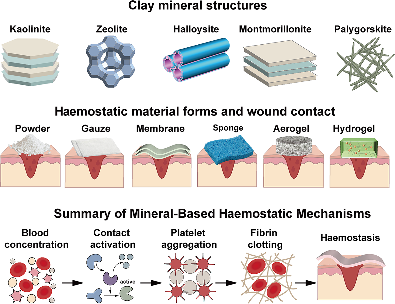
The haemostatic action of clay minerals is a multidirectional synergistic process, which can be summarized as involving four key factors: presence of a surface negative charge, water absorption, ion exchange and activation of the coagulation pathway (Katti et al., Reference Katti, Jasuja, Jaswandkar, Mohanty and Katti2022). For example, Kaol specifically activates coagulation factor XII to initiate the intrinsic coagulation pathway (Shuo et al., Reference Shuo, Haoting, Yuqing, Liuyun and Xiang2024). Ze releases Ca2+ through ion exchange and constructs a porous topological interface to recruit and assemble the activated factor X/activated factor V prothrombinase complex (Shang et al., Reference Shang, Chen, Castagnola, Liu, Boselli and Petseva2021). HNT-mediated haemostasis relies on structural advantages and multi-effect synergism: the unique hollow tubular structure of HNTs rapidly concentrates blood, abundant negative charges trigger the intrinsic coagulation cascade and topology-induced platelet aggregation accelerates thrombus formation (Feng et al., Reference Feng, Luo, Wu, Liu, Liang, He and Liu2022). With its ultra-high specific surface area, MMT physically adsorbs blood, while its surface negative charges further activate the coagulation system (Li et al., Reference Li, Quan, Liang, Li, Yuan and Tao2016). Nanobars of Pal provide physical adsorption sites that promote platelet aggregation. Upon blood contact, Pal absorbs Fe3+ and Mg2+ while releasing Ca2+, further catalysing fibrin generation (Wang et al., Reference Wang, Yang, Yang, Mu and Wang2024c). Additionally, Lap utilizes its nanodisc structure and electronegativity to initiate the intrinsic coagulation pathway (Fang et al., Reference Fang, Lin, Wang, Weng, Chen and Liu2024b). Sepiolite achieves physical thrombus construction by adsorbing blood components (Jiang et al., Reference Jiang, Wang, Qi, Yin, Liao, Luo and Ding2024). The structures, forms and haemostatic mechanisms of mineral-based haemostatic materials are summarized in Fig. 7.
Structures, forms and haemostatic mechanisms of mineral-based haemostatic materials.

Figure 7 Long description
The infographic is divided into three sections. The first section, ′Clay mineral structures,′ includes images of Kaolinite, Zeolite, Halloysite, Montmorillonite and Palygorskite. The second section, ′Haemostatic material forms and wound contact,′ shows different forms like Powder, Gauze, Membrane, Sponge, Aerogel and Hydrogel, each depicted in contact with a wound. The third section, ′Summary of Mineral-Based Haemostatic Mechanisms,′ illustrates the processes of Blood concentration, Contact activation, Platelet aggregation, Fibrin clotting and Haemostasis, with corresponding images for each step.
The haemostatic capabilities of clay minerals have been repeatedly demonstrated. However, clinical translation and ultimate haemostatic efficacy are significantly constrained by the materials’ delivery form. Powders and granules possess a large specific surface area, enabling rapid and thorough contact with blood. However, clay powders carry an embolism risk and are prone to being washed away (Katti et al., Reference Katti, Jasuja, Jaswandkar, Mohanty and Katti2022). Therefore, powder particles are commonly used for superficial wounds, irregular wounds and ‘sprinkling-style’ haemostasis in emergency care, but they are not suitable for large-vessel injuries or visceral surgery haemostasis.
Nanofibre membranes possess a high specific surface area and haemostatic efficiency, being capable of guiding cell adhesion and migration. They are suitable for conventional surgical haemostasis and localized bleeding in precision surgeries. However, fibre membranes often suffer from insufficient mechanical properties, and these materials carry high costs associated with their large-scale production.
Gauzes achieve haemostasis through physical compression and capillary absorption, making them suitable for open wounds requiring external pressure, such as battlefield haemorrhages, limb trauma and surgical field bleeding control. However, gauzes lack active procoagulant properties, tend to adhere to wounds and are limited in their applicability to non-compressible areas such as internal organs and body cavities.
Sponge and aerogel materials, due to their high fluid absorption capacity and space-filling properties, can effectively seal deep penetrating injuries (e.g. gunshot wounds or sharp-object punctures) and non-compressible bleeding in internal organs (e.g. liver, spleen, kidneys, groin, pelvis, chest cavity). However, excessive expansion of these materials may compress surrounding tissues, posing a risk of secondary injury.
Hydrogels exhibit excellent biocompatibility and maintain a moist environment, but their limited mechanical strength and insufficient wet adhesion restrict their haemostatic efficacy. Hydrogels are particularly suitable for precise haemostasis in minimally invasive procedures and diffuse organ bleeding. However, compared to traditional carriers, hydrogels can more efficiently encapsulate and release target drugs in a sustained manner, enabling integrated haemostasis, antibacterial effects, repair and regeneration. In recent years, in situ-formed hydrogels have been shown to be adaptable to various wound types.
In summary, clay minerals achieve haemostasis through multiple mechanisms, including surface negative charge, water absorption and ion exchange. Beyond the inherent haemostatic activity of the clay itself, the materials formed may either enhance or limit the final haemostatic efficacy. Clearly, no single form of material can perfectly suit all haemostatic scenarios. In the future, haemostatic materials tailored to different application scenarios using advanced processing techniques such as 3D or 4D printing methods should be developed.
Challenges and development trends
Ideal haemostatic materials should be multifunctional (i.e. haemostatic, antibacterial, anti-inflammatory, drug delivery, promotion of healing and regeneration properties), highly biosafe, reliably anti-embolic, clinically applicable across diverse scenarios and either degradable in situ or easily removable.
To date, Kaol-based QuikClot Combat Gauze® remains the preferred US military combat dressing due to its efficacy and biocompatibility (Basadonna, Reference Basadonna2012). In contrast, the Ze-based QuikClot® powder was discontinued due to the exothermic reactions it causes; in addition, the MMT-based WoundStat® haemostatic agent, although superior to QuikClot Combat Gauze® in terms of haemostatic efficacy, was discontinued for use in combat due to the risk of embolism posed by the shedding of free particles (Katti et al., Reference Katti, Jasuja, Jaswandkar, Mohanty and Katti2022). In fact, QuikClot Combat Gauze® does not pose an embolism risk due to its physical loading technology, which firmly anchors Kaol to the gauze fibres, thereby preventing its potential escape. Such incidents reveal the importance of maintaining a balance between haemostatic efficacy and safety.
Based on this, a series of crucial considerations should be prioritized when developing multifunctional haemostatic materials. Specifically, while ensuring that the materials achieve multifunctionality (haemostasis, antibacterial properties, repair and regeneration), clay particles must be immobilized within the polymer networks (e.g. hydrogels, sponges or gauzes). Furthermore, long-term in vivo experiments must conclusively rule out the risk of circulatory system embolism. Additionally, any clay particles shed in the wound site should either be excreted from the body or be safely retained as biocompatible substances.
Summary
Uncontrolled bleeding and acute haemorrhage remain critical factors contributing to mortality. Conventional clinical haemostatic materials have multiple limitations and fail to meet clinical requirements. This review focused on recent advances in clay minerals in the haemostat field. Clay minerals such as Kaol, Ze, HNTs, MMT and Pal have demonstrated significant advantages in haemostasis due to their high water absorption, surface negative charges and ion-exchange capacities.
The widespread clinical application of clay-based haemostatic materials still faces significant challenges. To further improve the haemostatic efficacy of clay minerals, clay-based haemostatic materials should be meticulously designed in various forms (e.g. powders, gauzes, sponges, hydrogels, membranes) and tailored to various application scenarios. A thorough elucidation of the biocompatibility of mineral materials holds promise for expanding the market applications of clay materials. Furthermore, developing multifunctional composite materials based on clay-derived haemostatic agents, integrating haemostatic, antimicrobial, tissue repair and regeneration properties rather than relying solely on haemostatic functionality, holds promise for better meeting clinical demands so as to achieve versatile haemostatic solutions.
Author contributions
Shuiqing Zhou, Xiangyu Chen and Mingxian Liu developed the concept for the review. Shuiqing Zhou wrote the initial drafts of the manuscript. Xiangyu Chen assisted in the manuscript writing. Mingxian Liu revised the manuscript. Shuiqing Zhou and Mingxian Liu wrote the final version of the manuscript. All authors have approved the final version of the manuscript.
Financial support
We acknowledge the financial support received from the National Natural Science Foundation of China (52573297), Natural Science Foundation of Guangdong Province (2025A1515011860) and ‘Master Mentor Plan’ of Jinan University (YDXS2507).
Competing interests
The authors declare none.