Introduction
The diagnosis of cholesteatoma is based on clinical symptoms and otoscopic findings. Computed tomography (CT) can demonstrate a middle-ear mass and show the extent of any bone erosion, but cholesteatoma has no diagnostic CT features. Following combined approach tympanoplasty for cholesteatoma, ‘second-look’ surgery approximately a year later is the ‘gold standard’ to exclude residual or recurrent disease.
A number of investigators have assessed the use of imaging to identify residual cholesteatoma following combined approach tympanoplasty.Reference Lemmerling, De Foer, Vandevyver, Vercruysse and Verstraete1
Delayed contrast (gadolinium) enhanced, T1-weighted magnetic resonance imaging (MRI) had promised a more accurate method of scanning, including the detection of cholesteatomas as small as 3 mm, but accuracy is often variable. Agreement between radiological and surgical findings has varied from almost 100 per cent to 50 per cent.Reference Vanden, Coen, Parizel and Van de Heyning2, Reference Ayache, Williams, Lejeune and Corre3
Diffusion-weighted imaging has also been extensively evaluated, as it is better at differentiating cholesteatoma from granulation tissue.Reference Lemmerling, De Foer, Vandevyver, Vercruysse and Verstraete1, Reference Venail, Bonafe, Poirrier, Mondain and Uziel4 This technique was initially carried out using echo planar imaging, but detection of small cholesteatomas was hampered by susceptibility artefacts in the temporal bone. More recently, a new type of diffusion-weighted imaging has been developed, based on non echo planar imaging techniques, which are less affected by susceptibility artefact. Published reports have shown that non echo planar imaging has superseded conventional echo planar imaging in the detection of post-operative residual or recurrent cholesteatoma. De Foer and colleaguesReference De Foer, Vercruysse, Bernaerts, Deckers, Pouillon and Somers5, Reference De Foer, Vercruysse, Bernaerts, Maes, Deckers and Michiels6 have demonstrated that non echo planar imaging incorporating the single-shot turbo spin-echo diffusion-weighted technique can detect cholesteatomas as small as 2 mm. The Belgian group have claimed that the use of non echo planar imaging will eliminate the need for second-look surgery. They have attributed the single-shot turbo spin-echo sequence's improved results to better image resolution and less susceptibility to artefact, compared with other forms of diffusion-weighted imaging.
Recently, a new non echo planar sequence, not previously evaluated for the detection of cholesteatoma, has been shown to be capable of detecting and helping identify the nature of soft tissue lesions 3 mm and larger.Reference Lehmann, Saliou, Brochart, Page, Deschepper and Vallee7 Lehmann et al. obtained exceptional results from a 3T MRI machine (GE Healthcare, Fairfield, Connecticut, USA) using the periodically rotated overlapping parallel lines with enhanced reconstruction (PROPELLER) sequence.
The radiological appearance of cholesteatoma varies from sequence to sequence, and requires considerable interpretative skill. T1-weighted MRI with gadolinium shows enhancement in scar tissue but not in cholesteatoma. A 45 minute delay is necessary after the introduction of the gadolinium, in order to allow the scar tissue to enhance. However, diffusion-weighted imaging shows cholesteatoma as a hyperintense lesion; this is thought to be due to a T2 shine-through effect rather than restricted diffusion.Reference Lemmerling, De Foer, Vandevyver, Vercruysse and Verstraete1
In an effort to reproduce the success of Lehmann et al., we investigated the use of PROPELLER with enhanced reconstruction diffusion-weighted MRI in the detection of residual or recurrent cholesteatoma. This sequence is in widespread use in UK hospitals, but not currently in the field of otology. It is primarily used for the reduction of motion artefacts in uncooperative patients.Reference Forbes, Pipe, Karis and Heiserman8
We compared a diffusion-weighted, multishot fast spin-echo, periodically rotated overlapping parallel lines with enhanced reconstruction sequence at 1.5 T with a diffusion-weighted, echo planar, array spatial sensitivity encoding technique MRI (ASSET) sequence, in the detection of cholesteatoma in patients awaiting primary or stage one combined approach tympanoplasty, canal wall up surgery, or second-look or second-stage combined approach tympanoplasty surgery. If the PROPELLER sequence was found to be specific and sensitive enough, this would add to the growing body of evidence from different centres, and would enable otolaryngology departments to benefit from readily available technology and to prevent unnecessary surgery.
Materials and methods
Between October 2008 and July 2009, we prospectively imaged a total of 19 patients. Sixteen patients were awaiting second-look combined approach tympanoplasty following discovery of cholesteatoma at the initial operation, and three patients were awaiting first-stage combined approach tympanoplasty. Patients were aged between 15 and 67 years (mean, 44 years).
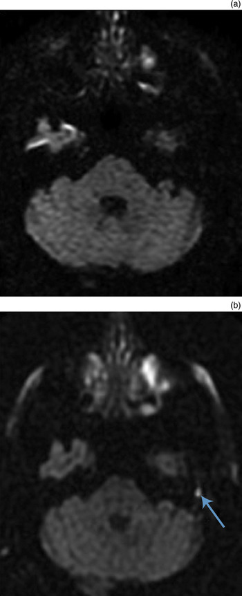
All patients underwent MRI (Signa, 1.5 T; GE Healthcare, Milwaukee, Wisconsin, USA) using both the PROPELLER sequence and the ASSET sequence (see Table I for imaging parameters). A standard head coil was used with eight elements (8 HR Brain; GE Healthcare). We used 3 mm sections with no gaps for both sequence types. Apparent diffusion coefficient maps and exponential diffusion-weighted imaging were not used. All scans were independently reported by two neuroradiologists who were blinded to patients' clinical histories. Neuroradiologists one and two had five and 10 years' experience in neuroradiology, respectively. Images produced by the two sequence types were reviewed independently of each other. For both sequence types, the criterion for the diagnosis of cholesteatoma was an area of hyperintense signal (as compared with brain cortex) (see Figure 1). The neuroradiologists classified the images as showing either positive or negative findings for cholesteatoma.
Example of an ASSET (1a) and PROPELLER (1b) sequence image of a 1 cm cholesteatoma in the left attic region (arrowed 1B). The signal created by the cholesteatoma is hyperintense in comparison to brain cortex on PROPELLER imaging and was thus reported as positive for cholesteatoma. Based on ASSET imaging alone cholesteatoma was not identified. ASSET, Array Spatial Sensitivity Encoding Technique; EPI, echo-planar imaging; PROPELLER, Periodically Rotated Overlapping Parallel Lines with Enhanced Reconstruction; non-EPI, non-echo-planar imaging.

Mri sequence parameters

MRI = magnetic resonance imaging; TR = repetition time; TE = echo time; FOV = field of view; ASSET DWI = array spatial sensitivity encoding technique sequence with diffusion-weighted imaging; PROPELLER = periodically rotated overlapping parallel lines with enhanced reconstruction sequence
Surgery took place an average of six weeks after MRI scanning. The surgical findings were correlated with those of the two imaging sequences. We then calculated the sensitivity, specificity, and positive and negative predictive values of the two imaging sequences in establishing a diagnosis of cholesteatoma. Inter-observer correlation was also calculated using the κ coefficient.
Results and analysis
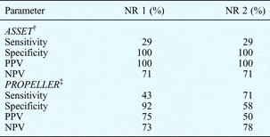
Cholesteatomas were found in all three patients undergoing first-stage combined approach tympanoplasty (100 per cent), and in four of the 16 undergoing second-stage combined approach tympanoplasty (25 per cent). The cholesteatomas ranged in size from 2 to 10 mm. Both of the assessed imaging sequences identified only cholesteatomas of 4 mm or larger. Table II displays results for the individual neuroradiologists.
Neuroradiologist reporting accuracy*

* Compared with surgical findings. †Echo planar imaging; ‡non echo planar imaging. NR = neuroradiologist; ASSET = array spatial sensitivity encoding technique sequence; PPV = positive predictive value; NPV = negative predictive value; PROPELLER = periodically rotated overlapping parallel lines with enhanced reconstruction sequence
When using the array spatial sensitivity encoding technique sequence alone, both neuroradiologists correctly identified only two out of seven patients with cholesteatoma (29 per cent); they also had false positive results. Use of the PROPELLER with enhanced reconstruction sequence produced mixed results (see Table II). In particular, the second neuroradiologist made a false positive report of cholesteatoma using this sequence; this was later surgically identified as cerumen in the external auditory canal.
Analysis of inter-observer agreement, using the κ coefficient, revealed perfect agreement between the neuroradiologists (κ = 1.00) when reporting the ASSET sequence scans, but only fair agreement (κ = 0.39) when reporting the PROPELLER sequence scans. (Agreement criteria were: poor <0.20, fair 0.21–0.40, moderate 0.41–0.60, good 0.61–0.80 and very good 0.81–1.00.)
Discussion
In this study, both the ASSET sequence and the PROPELLER sequence produced results that could not be relied upon to preclude second-look surgery.
The array spatial sensitivity encoding technique sequence had a 100 per cent positive predictive value, due to its lack of false positives. However, it missed five other cholesteatomas: two of 5 mm and one of 10 mm (all three were first-stage combined approach tympanoplasty); this resulted in very poor sensitivity.
The PROPELLER sequence detected more cholesteatomas but had more false positives, resulting in very variable sensitivity and specificity. Both neuroradiologists reported at least one of these scans as positive for cholesteatoma, in patients for whom surgical exploration revealed only granulation tissue.
The inter-observer agreement was perfect for the ASSET sequence, but only fair for the PROPELLER sequence. This perhaps reflected difficulties in interpreting the non echo planar imaging sequence. The disparity between the observers related to differences of opinion regarding whether a focus of signal change in the temporal bone was truly hyperintense compared with brain cortex.
Overall, the study results clearly demonstrate that the decision of whether or not to undertake cholesteatoma surgery cannot be made based purely on the results of ASSET sequence or PROPELLER sequence MRI scanning.
Forbes et al. Reference Forbes, Pipe, Karis and Heiserman8 demonstrated that the PROPELLER diffusion-weighted imaging sequence offered better image quality and detection of acute cerebral infarction, compared with diffusion-weighted echo planar imaging. The former sequence required only a short increase in scanning time.Reference Forbes, Pipe, Karis and Heiserman8
Our results for middle-ear imaging do not match the recently reported success of non echo planar imagingReference Lemmerling, De Foer, Vandevyver, Vercruysse and Verstraete1 and PROPELLER sequence imaging.Reference Lehmann, Saliou, Brochart, Page, Deschepper and Vallee7 Lehmann et al. Reference Lehmann, Saliou, Brochart, Page, Deschepper and Vallee7 have shown that it is possible to achieve remarkable results using 3 mm slices. They achieved sensitivity and specificity, and positive and negative predictive values of 100 per cent, for both senior neuroradiologists who reported the scans. They were able to detect cholesteatoma sizes down to 3 mm, compared with our 4 mm limit. The major difference in these authors' scanning method was the use of a 3 T MRI machine, in contrast to our 1.5 T MRI scanner. A stronger magnet allows a better signal-to-noise ratio to be achieved when imaging, but has the disadvantage of more pronounced susceptibility artefacts.Reference Schmitz, Aschoff, Hoffmann and Gron9 Another reason for our poorer results might be that the PROPELLER sequence, even though it is a form of non echo planar imaging, is not designed to detect cholesteatoma. This is partly due to the image acquisition process.Reference Pipe, Farthing and Forbes10
De Foer et al. Reference De Foer, Vercruysse, Bernaerts, Deckers, Pouillon and Somers5, Reference De Foer, Vercruysse, Bernaerts, Maes, Deckers and Michiels6 also obtained impressive results for non echo planar imaging, using the single-shot turbo spin-echo diffusion-weighted sequence rather than the PROPELLER sequence. They obtained sensitivity, specificity, and positive and negative predictive value results of 90, 100, 100 and 96 per cent, respectively. The single-shot turbo spin-echo diffusion-weighted sequence they used is a vendor-specific sequence available on Siemens MRI equipment (Siemens Medical Solutions, Erlangen, Germany) but not on our GE Healthcare MRI equipment; this prevented us from comparing this sequence to the PROPELLER sequence. There were also differences in the scanning protocol. The single-shot turbo spin-echo diffusion-weighted sequence images used 2 mm slices, compared to our 3 mm slices. This alone could account for the 4 mm size threshold below which the PROPELLER sequence was unable to detect cholesteatoma. Scanning at 3 mm in effect captures all the individual signals within the 3 mm thickness of the temporal bone being examined, and produces an averaged signal intensity on the image produced. If a 1 mm cholesteatoma was present within a 3 mm slice, its signal intensity would be reduced by partial volume artefact and it would not appear as hyperintense, compared with brain cortex, on diffusion-weighted imaging. Overlapping the slices would not circumvent this problem.
There was one false positive case in the PROPELLER sequence neuroradiology reports, which was partly due to difficulty in anatomical localisation. The PROPELLER sequence available to us was only able to obtain axial images, whereas the single-shot turbo spin-echo diffusion-weighted imaging sequence used by De Foer et al. Reference De Foer, Vercruysse, Bernaerts, Deckers, Pouillon and Somers5, Reference De Foer, Vercruysse, Bernaerts, Maes, Deckers and Michiels6 was able to produce both axial and coronal images. Localisation of a lesion on axial images alone is more difficult, compared with using both axial and coronal images. De Foer et al. Reference De Foer, Vercruysse, Bernaerts, Deckers, Pouillon and Somers5 also reported more artefact with axial compared with coronal imaging. In our false positive case, better anatomical localisation would have pinpointed the lesion as being located in the external auditory canal rather than the middle ear. It is known that cerumen appears to be as hyperintense as cholesteatoma on non echo planar imaging sequences.Reference Casselman and Kasbekar11 Therefore, cerumen debris within the ear canal or mastoid bowl could give the impression of a cholesteatoma and thus lead to a false positive report, if using only axial images obtained with the PROPELLER sequence.
• Non echo planar imaging is a relatively new technique for identifying middle-ear cholesteatoma, and can detect lesions as small as 2 mm
• The periodically rotated overlapping parallel lines with enhanced reconstruction magnetic resonance imaging (MRI) sequence has recently been shown capable of detecting cholesteatoma down to 3 mm in size, using a 3 T MRI machine
• This study used this sequence and a 1.5 T MRI machine, and could detect cholesteatoma only down to 4 mm
• This study's poor positive and negative predictive values were due in part to the technological limitations of the MRI sequence used, and to difficulties in radiological interpretation
• These problems will be encountered by other departments looking to incorporate non echo planar MRI imaging into their clinical practice
The use of any new technology involves an operator learning curve, and this certainly applies to the introduction of the PROPELLER sequence to detect middle-ear cholesteatoma. The neuroradiologist must interpret a combination of sequences to decide whether a cholesteatoma is present or not. We used a b-factor of 1000 when undertaking both types of MRI sequence, which has been shown to be effective in the detection of middle-ear cholesteatoma.Reference De Foer, Vercruysse, Bernaerts, Deckers, Pouillon and Somers5 Cholesteatoma appears as a hyperintense signal on diffusion-weighted imaging, whereas inflammation does not. Cerumen resembles cholesteatoma and appears as hyperintense on the PROPELLER sequence, but hypointense when using apparent diffusion coefficient mapping. The periodically rotated overlapping parallel lines with enhanced reconstruction sequence produces apparent diffusion coefficient maps. We did not use apparent diffusion coefficient mapping in the interpretation of pre-operative imaging. A recent publication has reported that high apparent diffusion coefficient values correlate with pure cholesteatoma but not with infection, which has low apparent diffusion coefficient values.Reference Thiriat, Riehm, Kremer, Martin and Veillon12 Cholesteatoma present together with infection gives an intermediate apparent diffusion coefficient value. Abscesses also resemble cholesteatoma on diffusion-weighted imaging scanning, and should be differentiated clinically. Finally, cholesterol granules are isointense on both diffusion-weighted imaging scanning and apparent diffusion coefficient mapping.Reference Casselman and Kasbekar11
There were limitations within our study. Two of the seven cholesteatomas found were 10 mm or larger. They were thus relatively easy for MRI scanning to detect. In addition, the large size would perhaps not challenge the neuroradiologist when determining whether the lesion was a cholesteatoma or not. Poor image resolution of the scan would also be less problematic. For our study purposes, the cholesteatoma lesions being investigated would ideally have been 1 to 6 mm in size, so that all factors involved in determining whether a scan was positive or negative for cholesteatoma would be challenged. This is understandably a difficult variable to control in clinical practice. A larger number of patients might have provided more statistical power; however, we feel that, given the results, it is clear that much work is needed in order to improve the sensitivity and specificity of the PROPELLER sequence in our hands. Given the above-mentioned inherent difficulties of using this sequence, such as its lack of accurate anatomical localisation and 3 mm minimum slice thickness, we believe it unlikely that this sequence could identify cholesteatomas smaller than 3 mm.
Conclusion
Our findings suggest that the PROPELLER sequence is not an ideal form of non echo planar imaging for the detection of middle-ear cholesteatoma. Casselman and De Foer et al. continue to report on their success with their particular non echo planar imaging sequences. Further studies should assess the reproducibility of results, using Casselman and De Foer and colleagues' protocols and MRI machines. Future research should also examine the diagnostic performance of a newly released version of the PROPELLER sequence, version 2.0, which has the capacity to produce coronal as well as axial images.
Acknowledgements
The work of Anand Kasbekar has been partly funded by grants from the Cambridge Hearing Trust and the Royal Society of Medicine. The work of Daniel Scoffings is funded by the National Institute for Health Research Cambridge Biomedical Research Centre.