Introduction
Distinguishing dementia subtypes facilitates optimal management and enables the provision of appropriate information to patients and carers about likely symptomatology and prognosis. Clinical trials also rely on accurate diagnosis of dementia subtypes. Current guidelines recommend blood flow (hexamethylpropyleneamine oxime, HMPAO), SPECT, or functional brain imaging using glucose (fludeoxyglucose, FDG) PET to facilitate differential diagnosis of the cause of the dementia syndrome when diagnosis is in doubt (NICE/SCIE, 2006; Hort et al., Reference Hort2010; Gauthier et al., 2012). There is growing evidence that PET is more accurate as a diagnostic tool (Ishiiand Minoshima, Reference Ishii and Minoshima2005; Herholz, Reference Herholz2011; O’Brien et al., Reference O’Brien2014). Decisions about the use of PET or SPECT may also be influenced by availability, financial cost, and perceived acceptability to patients. SPECT has been more readily available and significantly cheaper than PET (Colloby and O’Brien, Reference Colloby and O’Brien2004). However, PET is now more widely available and the price of FDG, used for PET, has reduced significantly over the last ten years (O’Brien et al., Reference O’Brien2014). Anecdotally PET is perceived as more burdensome for patients than SPECT (NICE/SCIE, 2006) but little is known about the patient experience of undergoing imaging or patient preferences (Davison and O’Brien, Reference Davison and O’Brien2014). Although PET and SPECT are both nuclear medicine scans, the procedures are more involved for PET than for SPECT. For example, patients have to fast and have their glucose checked prior to PET and while carers are able to stay with patients throughout SPECT scans, they are usually not allowed in the room during PET scans due to concerns about their exposure to the higher level of radiation associated with PET. Patient views on the acceptability of SPECT and PET could help inform future decisions regarding the most appropriate imaging procedure (Davison and O’Brien, Reference Davison and O’Brien2014).
As part of a study which investigated the diagnostic utility of PET with SPECT in distinguishing between people with neurodegenerative dementia (Alzheimer's disease (AD) and dementia with Lewy bodies (DLB)) and normal controls (O’Brien et al., Reference O’Brien2014), we collected data on participants’ views of SPECT and PET brain imaging. Our aim was to develop a detailed understanding of participant views and experiences of the two scans and to explore whether the anecdotal view that PET is more burdensome and less patient friendly than SPECT was supported by empirical data.
Methods
Study population
Patients aged over 60 years with mild to moderate dementia (MMSE >12) (Folstein et al., Reference Folstein, Folstein and McHugh1975) who met the criteria for probable AD (McKhann et al., Reference McKhann, Drachman, Folstein, Katzman, Price and Stadlan1984) or probable DLB (McKeith et al., Reference McKeith2005) were recruited prospectively from clinical services in north-east England. We also recruited age-matched controls with no signs or symptoms of dementia who had expressed a willingness to participate in research studies. All participants underwent detailed neuropsychiatric investigation prior to imaging (O’Brien et al., Reference O’Brien2014). SPECT and PET scans were completed on separate occasions within one month. The order of the scans was alternated between consecutive participants in each group. The views of carers accompanying patients to the scans were also sought.
SPECT head scans were undertaken using a Siemens Symbia dual-detector gamma camera. Participants were comfortably seated in a quiet room, with eyes open for a bolus intravenous injection of 500 MBq of Tc-99m HMPAO (Ceretec (exametazime)). They then either returned to the nuclear medicine waiting room for up to 30 minutes or proceeded directly to the camera room. Imaging time was 25 minutes. This process reflected the standard practice in a busy clinical department.
PET-CT scans were undertaken using a Siemens Biograph TruePoint PET-CT. Imaging took place approximately 30 minutes after intravenous injection of 250 MBq 18F-FDG. Participants were asked to fast for four hours pre-injection and blood glucose was tested. During the injection, participants were comfortably seated in a quiet room, with eyes open. They then remained alone in an individual cubicle until their scan. Imaging took approximately 10 minutes. Although the equipment and procedures used were similar to those used in clinical settings, the PET scans took place in a university clinical research facility, while the SPECT scans were done in a UK NHS hospital department. All scans were performed by NHS staff.
Data collection
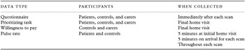
An overview of all data collection procedures is provided in Table 1. The development, content, and administration of each measure are described below.
Overview of study procedures

Questionnaires
While patients have made direct comparisons of different diagnostic procedures in previous studies (Liang et al., Reference Liang2003; Sparrow et al., Reference Sparrow, Plein, Jones, Thorley, Hale and Sivananthan2004), people with dementia were unlikely to be able to recall details of the imaging procedures after any length of time. To capture participants’ experiences and views on the two scans, the questionnaires were therefore administered immediately after each scan.
Parallel questionnaires were produced for patients, controls, and carers accompanying patients to the scan. The content of the questionnaires was informed by
-
1. existing literature on patient views of diagnostic procedures (Sparrow et al., Reference Sparrow, Plein, Jones, Thorley, Hale and Sivananthan2004; Wollman et al., Reference Wollman, Beeri, Weinberger, Cheng, Silverman and Prohovnik2004),
-
2. observation of routine practice in PET and SPECT suites, and
-
3. semi-structured interviews with five people with dementia referred for SPECT imaging and accompanying carers (these participants were not part of the main study).
Questionnaires for patients and controls were interviewer administered and took around 10 minutes to complete. The carer questionnaire was usually self-completed. Three open questions were included on each questionnaire to elicit views on the best and worst things about the scan and to identify any suggested improvements. Three further questions were added after use with the first five participants and carers recruited to the main study.
Prioritizing task
We used a simplified version of Q methodology (Baker et al., Reference Baker, Thompson and Mannion2006) to explore the relative priority given to attributes of the scan identified as potentially important in the development work:
-
1. being able to get comfortable in the scanner
-
2. length of time for the injection and scan
-
3. dose of radiation given
-
4. accuracy of the scan
-
5. how far you have to travel for the scan
-
6. how confined you feel in the scanner
-
7. whether my relative can stay in the room
-
8. helpful staff
-
9. noise of the scanner
A further item – having to fast before the scan – was added after the first five participants and carers recruited to the main study. Each item was printed on a card and respondents were asked to sort the cards according to importance. Additional blank cards were available so that respondents could add other factors if needed. Since the prioritizing task did not require detailed recall of the scans it was completed during a final HV after completion of both scans.
Willingness to pay questionnaire
The WTP questions were completed during the final HV. We used an “incremental approach” (Donaldson and Shackley, Reference Donaldson and Shackley1997) whereby participants were asked which scan they preferred (PET or SPECT) and the maximum amount they would be willing to pay for their preferred option. We presented cards with amounts ranging from £1 to £5,000 in random order and asked respondents to sort the cards into three piles: definitely willing to pay that amount; definitely not willing to pay; or unsure. Any cards in the unsure pile were then reviewed to identify the maximum amount the respondent was willing to pay for the preferred scan. Due to concerns over the ability of people with dementia to remember and compare both scans, ethical concerns over potential confusion over whether they would be required to pay for the procedures and desire to minimize respondent burden, the WTP questions were not administered to patients.
Physiological measurement
Information on average heart rate was collected as an indicator of stress (Sparrow et al., Reference Sparrow, Plein, Jones, Thorley, Hale and Sivananthan2004) using a fingertip pulse oximeter (Pulmolink CMS 50E-W). Normal heart rate was ascertained by measurements taken for 5 minutes during the initial HV. Further pulse data were collected for 5 minutes on arrival at the department and throughout each scan.
Study approvals
The study was sponsored by Northumberland, Tyne, and Wear NHS Foundation Trust. Ethical approval was given by Newcastle and North Tyneside 1 Research Ethics Committee (reference 09/H090688).
Statistical analysis
All questionnaire items were coded so that higher scores indicate more positive views. To test for differences in views of PET and SPECT scans, questions were compared within respondent type (patients, controls, carers) using the Wilcoxon signed-rank test for questions with scaled response categories and the McNemar test for those with dichotomous responses. Views of patients and controls on each type of scan were compared using a Mann–Whitney test for two independent samples. Responses to the open questions were recoded as a series of binary variables and compared across all three groups of respondents (Kruskal–Wallis test for independent samples).
The priority given to each attribute of the scan ranked in the prioritizing task was recoded as important (ranked most, second most, or third most important), unimportant (ranked as least, second least, or third least important), or as neither important nor unimportant (all items not ranked at either extreme). The priorities of patients, controls, and carers were compared using the Kruskal–Wallis test for independent samples. The WTP data were subject to simple descriptive analyses and a Mann–Whitney U test to compare the amounts respondents were willing to pay for their preferred scan.
Since the PET scans were considerably shorter than SPECT scans (approximately 10 minutes vs. 25 minutes) we divided the pulse rate data into 8-minute segments to directly compare the two scans. (This allowed some time for the pulse rate to stabilize at the start of the recording). The first 8-minute segment was subdivided into two 4-minute segments to allow us to compare pulse rate during these two segments of both scans; the second and third 8-minute segments of pulse data for the SPECT scan were used to explore whether participants found the scan more or less stressful as duration of scan increased (comparable data for PET not being available). Pulse rate data were analyzed using analysis of variance.
Results
Response rates
We recruited 102 participants, of whom three withdrew before completing both scans, and one was excluded due to scanner technical problems. A total of 38 people with AD, 30 with DLB and 30 controls were successfully scanned with both PET and SPECT. Questionnaire data were not completed by the first eight people recruited to the study (five controls, three patients) since the questionnaires were awaiting ethical approval. The questionnaires for one further patient were unavailable. Patients and controls were matched on age (mean age 76, standard deviation (SD) 6.4 years) and gender (69% male). As expected, patients had significantly lower MMSE scores (mean 21, SD 3.9) than controls (mean 29, SD 1.1, p < 0.001, t-test) and were also more likely to have previously undergone a SPECT scan (16% patients vs. 0% of controls, p = 0.03, χ2).
For those patients completing questionnaires, the average duration of dementia was 39.8 months (range 9–120 SD 22 months); 53% had a diagnosis of AD and 47% DLB. A carer accompanied 62 patients to both scans and 60 of these completed post-scan questionnaires. One carer was unavailable at the final HV and therefore the analysis of the prioritizing task and WTP questionnaire is based on 59 carers.
Preference questionnaires
Comparison of responses for PET and SPECT indicated minimal differences (Figure 1).
Patient and control views on PET and SPECT.

Carers’ views were broadly similar to those of patients and controls. For patients and carers, the key difference between the scans was, as expected, whether the carer was in the room during the scan (PET: 0% for patients and carers; SPECT: 70.3% patients and 69.5% carers; p < 0.001 for both patients and carers, McNemar test). The subjective feeling of isolation during the scan did not, however, differ for patients although carers were more likely to report feeling cut-off from their relative during the PET scan (mean (SD) 3.5 (1.2)) than SPECT (mean (SD) 4.2 (0.9), p < 0.001, Wilcoxon signed-rank test). Carers were also more likely to report that their relative was anxious prior to the PET scan (mean (SD) 3.1 (1.2)) than SPECT (mean (SD) 3.3 (1.3), p = 0.03, Wilcoxon signed-rank test). For controls, the only significant difference between the two scans related to whether they found the temperature comfortable (73.7% for PET vs. 94.7% for SPECT, p = 0.02, Wilcoxon signed-rank test).
Some significant differences between patients and controls were observed, with controls generally feeling more positive about the scans than patients. In relation to both PET and SPECT scans, controls felt more positive prior to the scan (mean (SD) PET: 3.9 (0.3) for controls and 3.2 (0.8) for patients, p < 0.001; SPECT: 3.8 (0.4) for controls and 3.3 (0.9) for patients, p = 0.003, Mann–Whitney U test); they rated the explanation provided by staff more highly (mean (SD) PET: 4.0 (0.2) for controls and 3.8 (0.5) for patients, p = 0.04; SPECT: 4.0 (0.2) for controls and 3.4 (1.2) for patients, p = 0.04, Mann–Whitney U test); and they gave a higher overall rating for the scan on a seven-point scale (mean (SD) PET: 5.5 (0.6) for controls and 5.0 (0.7) for patients, p < 0.001; SPECT: 5.5 (0.5) for controls and 5.0 (0.8) for patients, p = 0.003, Mann–Whitney U test). In addition, controls rated the SPECT scan as more comfortable than patients (mean (SD) 3.6 (0.7) for controls and 3.0 (1.1) for patients, p = 0.02, Mann–Whitney U test).
Since patients, controls, and carers made similar comments to the open questions, the results for all respondents have been combined. The chance to help with research and the interaction with staff were identified as the most positive aspects of both scans. The most negative aspect of their experience related to the scan itself (e.g. feeling cold or uncomfortable). While the majority of respondents did not suggest any improvements to the procedures (79% for SPECT and 82% for PET), there were a number of potentially useful suggestions. Most of these related to improving comfort during the scan (41% of suggestions relating to PET and 52% of those relating to SPECT). These included: increasing the width of the scan bed (or providing arm rests); providing a pillow for under the knees; and providing a blanket (or increasing the ambient temperature). Other suggestions were to reduce waiting time; provide more information on what to expect and to provide more feedback during the scan (e.g. how long was left).
Prioritizing task
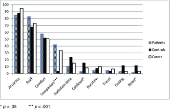
None of the attributes was consistently viewed positively or negatively by all respondents (Figure 2). While patients and carers valued the presence of a companion during the scan this was not important for controls (p < 0.001, Kruskal–Wallis). Controls rated the noise of the scanner as more important and patients gave a lower rating to feeling confined in the scanner (both p < 0.05, Kruskal–Wallis) (Figure 2).
Percentage of respondents rating scan attributes as important.

Willingness to pay
Controls did not have a significant scan preference; carers had a marginal preference for SPECT over PET (one sample Wilcoxon signed-rank test, p = 0.05). Nearly three quarters of all respondents with a preference were willing to pay for the preferred scan (independent of the scan preferred). Although the amounts respondents were willing to pay for SPECT (mean £1,632, SD 1,858, median £1,000) were higher than for PET (mean £1,074, SD 2,197, median £100), this was not significantly different (Mann–Whitney U test). In the hypothetical situation that the non-preferred scan proved more accurate, all respondents who had initially expressed a preference stated that they would prefer the more accurate scan. Of those without an initial preference, the majority (93%) would prefer the more accurate scan.
Physiological measures
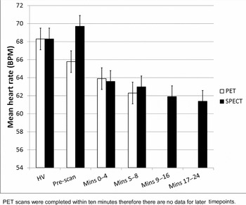
There were some technical difficulties in obtaining pulse rate data; the number of cases used at each data point for analysis ranged from 71 (48 patients and 23 controls) to 92 (66 patients and 26 controls). Since there were no significant differences in mean pulse rate between patients and controls at any data point, data from both groups were combined. While mean pulse rate did not change between the HV and pre-SPECT data points, it decreased significantly (but numerically very slightly) when attending for a PET scan (from 67.9 (SD 11.3) to 65.6 (SD 11.5), p < 0.05, analysis of variance, Figure 3). For both types of scan, pulse rate was significantly lower during the scan than on arrival at the clinic (both p < 0.001, analysis of variance). Compared to beginning of the scan, pulse rate significantly lowered towards the end of the PET scan (p < 0.001, analysis of variance); in contrast, while pulse rate declined gradually throughout the longer SPECT scan, the difference did not reach statistical significance.
Mean heart rate readings at different time points for PET and SPECT scans.

Discussion
This study is the first to compare consumer views of PET and SPECT brain imaging for diagnosis of dementia. Our findings suggest that the two types of scans were equally acceptable to the majority of people with dementia, their carers, and to healthy controls. There was also evidence that, even where a preference for one scan was expressed, diagnostic accuracy over-ruled any initial preferences suggesting that this was more important than other scan characteristics. Economic theory would use the WTP amounts as representative of the strength of preference for each scan. The mean amounts respondents were willing to pay were not significantly different between PET and SPECT again confirming the lack of a strong preference for either scan. Pulse rate data indicated that for most participants, the experience of brain imaging was no more stressful than completing questionnaires in their own homes. Although some statistically significant differences were observed in heart rate, the effect size was very small and not of clinical significance. A previous study similarly found that SPECT and MRI imaging procedures had little impact on heart rate (Sparrow et al., Reference Sparrow, Plein, Jones, Thorley, Hale and Sivananthan2004).
It is of interest that participants’ views and preferences seemed unaffected by the duration of the scan (SPECT scans took over twice as long as PET scans) or the physical surroundings. While SPECT scans took place in a busy hospital department, PET scans took place at a University Research Facility in a quiet clinic with typically only one patient attending at a time. Responses showed that some participants preferred the busier clinic since they enjoyed chatting with other patients whilst waiting for their scan. The importance of individual preferences was also evident on questions relating to comfort during the scan. Up to one-fifth of participants felt cold during scans suggesting that either the ambient temperature needs to be increased or blankets need to be provided. Some participants would have welcomed pillows or eye masks. While blankets and pillows were available, these were not routinely offered to patients. The results highlight the need for a person-centered approach, addressing the needs of individual patients. Staff may also benefit from training in communicating with people with dementia to ensure that patients have a better understanding of the procedures they are to undergo.
A key difference between PET and SPECT scans was that carers were able to remain with the patient throughout SPECT scans, but not during the uptake and scanning phase of PET in order to keep radiation exposure as low as reasonably achievable (ALARA principle). Previous research has shown that a high proportion of older patients, particularly those with dementia, are accompanied to medical consultations (Ishikawa et al., Reference Ishikawa, Roter, Yamazaki, Hashimoto and Yano2006). Having a companion present can reassure both the patient and their carer and may enhance the quality and even feasibility of a successful scan. Over one-third of patients and carers felt having a companion during the scan was important. Guidance allows for “comforters and carers” to legally exceed the normal radiation dose limits, providing that they do so knowingly and willingly (i.e. doses have been estimated and explained to them) (Singleton et al., Reference Singleton, Griffiths, Morrison and Soanes2003). Available evidence indicates that the dose for companions waiting with a patient in the FDG uptake phase is surprisingly low (Singleton et al., Reference Singleton, Griffiths, Morrison and Soanes2003), suggesting scope for increased flexibility regarding the presence of carers throughout PET scans with vulnerable patient groups, such as people with dementia. Alternative approaches to allowing patients to remain in contact with their carer during PET scans, such as intercom systems, could also be considered.
Implications for research
The questionnaires about patient views and experiences of each scan were completed immediately after the scan to minimize problems with recall. While some patients with dementia were not able to recall all details of the scan, they successfully completed the majority of questions, confirming that this approach was acceptable and feasible for people with dementia. While only one study using Q methodology with people with AD has been reported (Forrest, Reference Forrest2000), this approach has some similarities to Talking Mats which can facilitate communication with people with dementia (Murphy et al., Reference Murphy, Gray, van Achterberg, Wyke and Cox2010). The majority of people with dementia successfully completed our simplified version of Q methodology confirming that this can be a useful approach for exploring the views and preferences of people with dementia.
Strengths and limitations of the study
The use of different approaches to data collection allowed us to explore preferences in detail and increases confidence in our findings. Although the questionnaires relating to each scan were identical, different members of staff administered the questionnaires after PET and SPECT and this could potentially have influenced the results. Participants in the study were not attending for routine scans but had volunteered to take part in a research study; their views towards the scans may potentially be more positive than those of patients attending for diagnostic purposes. The different settings for PET and SPECT scans in the present study could have influenced our results (although there was no evidence of a consistent preference for either setting). Future studies could explore patient views on PET and SPECT scans conducted in more similar environments. There were some problems with obtaining readings from the fingertip pulse oximeter. There is, however, no reason to suggest that the participants with missing data differed systematically from those with complete pulse rate readings. Although we did not adjust for multiple comparisons, this was to ensure that we captured any differences between participant views of PET and SPECT and increases our confidence in concluding that, from the perspective of participants, there are minimal differences between the two imaging procedures.
Conclusion
The majority of participants viewed the experience of PET and SPECT scans as roughly equivalent although carers had a marginal preference for SPECT. Diagnostic accuracy was prioritized over other characteristics of scans (e.g. time taken, comfort on the scanner). The presence of a companion was important to some people with dementia and carers and may explain carers’ preference for SPECT. Pulse rate data indicated that for most participants, the experience of brain imaging was no more stressful than completing standardized questionnaires in their own homes. Possible ideas for service development include a more person-centered approach to improve comfort during scans and a more flexible approach to the presence of carers during PET scans. Overall, our findings indicate that the decision whether to use PET or SPECT scans in dementia should be based on diagnostic accuracy rather than other considerations.
Conflict of interest
MF reports grants from National Institute for Health Research (NIHR), JOB reports grants from GE Healthcare and Lilly, and other from GE Healthcare, Lilly, Bayer Healthcare, TauRx, and Cytox outside submitted work.
Description of authors’ roles
CB contributed to study conception, design, obtaining funding, data analysis, data interpretation, and writing of the manuscript.
KO contributed to recruitment and data collection.
CDa contributed to recruitment, data collection, and analysis.
NB contributed to recruitment, data collection, and project management of the study.
JL contributed to study design and management.
DW contributed to study design, obtaining funding, organization, and management of the project.
MF contributed to study design, data analysis, and data interpretation.
HM contributed to data analysis and data interpretation.
CDo contributed to study conception, design, obtaining funding, and data interpretation.
JO’B contributed to study conception, design, obtaining funding, organization and management of the project, data analysis, and data interpretation.
All authors contributed to the drafting of this paper.
Acknowledgments
This paper summarizes independent research funded by the NIHR under its Research for Patient Benefit Programme (Grant reference number NIHR RfPB PB-PG 1207 13105). The study was also supported by the NIHR Biomedical Research Centre in Ageing and Chronic Disease and Biomedical Research Unit in Lewy-body Dementia (Newcastle upon Tyne Hospitals NHS Foundation Trust and Newcastle University) and the Biomedical Research Centre and Unit in Dementia (Cambridge University Hospitals NHS Foundation Trust and the University of Cambridge). We thank the Dementia and Neurodegenerative Diseases Research Network (DeNDRoN) for valuable support with recruitment. The views expressed are those of the authors and do not necessarily reflect those of the RfPB programme, NIHR, NHS, or the Department of Health.