INTRODUCTION
The ecology of Vibrio cholerae Pacini continues to receive scientific attention because cholera remains a global health problem (e.g. Colwell, Reference Colwell1996; Cottingham et al., Reference Cottingham, Chiavelli and Taylor2003). Cholera re-emerged in 1991 in South America and has caused seven outbreaks in Argentina, all of which were produced by an O1 El Tor strain (Pichel et al., Reference Pichel, Rivas, Martin, Ibarra and Binsztein2003). In previous studies, we have focused attention on the viable but non-culturable (VNC) forms of V. cholerae and its association with plankton organisms (Binsztein et al., Reference Binsztein, Costagliola, Pichel, Jurquiza, Ramírez, Akselman, Vacchino, Huq and Colwell2004), taking into account the ecological significance of the VNC state as a means of survival during inter-epidemic periods (Colwell & Huq, Reference Colwell, Huq, Wachsmuth, Blake and Olsvik1994). Binsztein et al. (Reference Binsztein, Costagliola, Pichel, Jurquiza, Ramírez, Akselman, Vacchino, Huq and Colwell2004) reported the presence of VNC V. cholerae O1 in marine zooplankton and microplankton samples from the Argentine shelf of the Atlantic Ocean. These authors reported that while various copepods (Corycaeus amazonicus Dahl, Centropages furcatus Dana and Ctenocalanus vanus Giesbrecht) were shown to be associated with VNC V. cholerae O1, the bacterium could not be found attached to any phyto- nor protozooplankton species. Consequently, the aim of this study was to analyse phyto- and protozooplankton species which could potentially act as reservoirs of VNC V. cholerae in marine waters, focusing only on the microplanktonic fraction.
MATERIALS AND METHODS
During eight cruises conducted on 16 May, 12 June, 2 July, 17 September, 3 October, 4 November, 3 December and 18 December 2003, samples were collected at a fixed-position station on the Argentine continental shelf (38°28′S–57°41′W, 48 m depth). Temperature and salinity were measured in situ with a CTD Seabird SBE 19. Microplankton net samples (25 μm mesh aperture, vertical hauls) were collected and analysed for culturable V. cholerae (González Fraga et al., Reference González Fraga, Pichel, Costagliola, Cecilia, Jurquiza, Peressutti, Caffer, Aulet, Hozbor, Tracanna, Villagra de Gamundi, Hernández, Ramírez, Akselman and Binsztein2007) and for VNC V. cholerae O1 by direct fluorescent antibody (DFA)-direct viable count (DVC) technique (Chowdhury et al., Reference Chowdhury, Xu, Montilla, Hasan, Huq and Colwell1995) and by polymerase chain reaction (PCR) extracting DNA as published by Rivera et al. (Reference Rivera, Lipp, Gil, Choopun, Huq and Colwell2003). For each concentrated plankton sample (final volume, 20 ml), 10 ml was divided into three portions that were placed into vials immediately after sampling. One vial containing 0.025% yeast extract and 0.002% nalidixic acid was incubated for 6 to 12 hours at 35°C and fixed with formaldehyde at a final concentration of 3% for analysis by DFA–DVC; material from this vial was stained and examined with cholera DFA kits under an epifluorescence microscope. The second vial was preserved with formaldehyde at a final concentration of 4%; material from this vial was used to identify and isolate phytoplankton species in order to be stained and examined by the same procedures. The last vial was stored at –20°C for PCR. Three PCRs were performed on each sample as described above with the addition of 400 ng μl−1 of bovine seroalbumin (BSA) (Sigma, St Louis, MO): (1) V. cholerae: (Chun et al., Reference Chun, Huq and Colwell1999); (2) serogroup O1 (Rivera et al., Reference Rivera, Lipp, Gil, Choopun, Huq and Colwell2003) with an annealing of 50°C; and (3) Multiplex ctxA-tcpA El Tor (Rivera et al., Reference Rivera, Chun, Huq, Sack and Colwell2001). The remaining 10 ml of the plankton samples was enriched by addition of alkaline peptone broth for isolation of V. cholerae by conventional culture methods. An inverted Olympus IX-70 epifluorescence microscope connected to a system for capturing and analysing images was used to examine material stained with cholera DFA kits and to identify and isolate microplankton species by micromanipulation.
RESULTS AND DISCUSSION
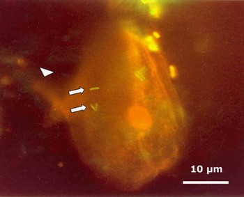
No V. cholerae O1 could be isolated during this period, which agrees with previous results obtained during a four-year study in a nearby area (Costagliola et al., Reference Costagliola, López, Malaspina, Guerrero, Medina, Odizzio, Abelenda and Friss de Kereki2000). However, VNC V. cholerae O1 was identified in samples isolated between May and December 2003, detected by PCR or by both PCR and DFA–DVC during each of the eight cruises. PCR also showed the presence of genes that encode the cholera toxin ctxA and the toxin co-regulated pilus subunit tcpA allele El Tor. VNC V. cholerae O1 was detected by PCR in the eight microplanktonic samples (100%). One sample (2 July) revealed bacterial cells attached to microplankton via immunofluorescence. Therefore, several specimens of each species from the 2 July net sample were isolated, identified and stained. These species were the athecate, not photosynthetic dinoflagellate Noctiluca scintillans (Macartney) Kofoid & Swezy and the diatoms Odontella sinensis (Greville) Grünow, Lauderia annulata Cleve, Proboscia alata (Brightwell) B.G. Sundström, Pseudo-nitzschia sp., Paralia sulcata (Ehrenberg) Cleve, Pleurosigma normanii Ralfs and Thalassiosira spp. While no diatom showed V. cholerae O1 cells attached to their frustules, positive stained bacteria were observed adhering to the cell membrane of N. scintillans (Figure 1). As can be seen in Figure 1, various bacterial cells were attached to the N. scintillans host cell. In July 2003 microplankton composition and relative species abundance indicated a dominance of diatoms. Analysis of quantitative samples from the water column (R. Akselman, unpublished data) showed total microplankton concentration values of 17,000 to 24,000 cells l−1 and N. scintillans concentrations of <100 cells l−1, showing that even though there was a low concentration of N. scintillans in comparison with other species, this dinoflagellate was still colonized by VNC V. cholerae O1 cells. Vibrio cholerae cells attached to N. scintillans were registered associated with temperature and salinity values of 12.25°C and 33.82 psu, in physical conditions of a nearly homogeneous water column.
Photomicrograph by epifluorescence of a Noctiluca scintillans cell showing Vibrio cholerae O1 attached to the cell membrane. Arrows: V. cholerae O1 cells; arrowhead: tentacle of N. scintillans.

Noctiluca scintillans is present worldwide in marine neritic and estuarine waters and is a well-known species that can generate red-tide phenomena owing to its large mass aggregations at sea surface (e.g. Uhlig & Sahling, Reference Uhlig and Sahling1990; Murray & Suthers, Reference Murray and Suthers1999), including the shelf waters of Argentina (Akselman et al., Reference Akselman, Benavides, Negri and Carreto1986). Its large geographical distribution as well as its abundance in littoral waters raises the question of the significance of N. scintillans as a reservoir of V. cholerae. The seasonal periodicity of maximal abundances observed in the summer months for N. scintillans (e.g. Uhlig & Sahling, Reference Uhlig and Sahling1990; Dela-Cruz et al., Reference Dela-Cruz, Ajani, Lee, Pritchard and Suthers2002) and the fact that some cholera outbreaks also occur during these months adds further interest to the present findings. In this context, it must be noted that experimental evidence demonstrates that V. cholerae can proliferate in the free-living state at the expense of dissolved organic matter present during algal dinoflagellate blooms (Mouriño-Pérez et al., Reference Mouriño-Pérez, Worden and Azam2003). Also, in vitro experiments made by Mueller et al. (Reference Mueller, McDougald, Cusumano, Sodhi, Kjelleberg, Azam and Bartlett2007) showed different abilities of various viable cultivated wild and mutant V. cholerae strains to colonize the autotrophic dinoflagellate Lingulodinium polyedrum (F. Stein) J.D. Dodge. Noctiluca scintillans should therefore be added to the list of other aquatic freshwater and marine species (Tamplin et al., Reference Tamplin, Gauzens, Huq, Sack and Colwell1990; Islam et al., Reference Islam, Drasar, Sack, Drasar and Forrest1996, Reference Islam, Rahim, Alam, Begum, Moniruzzaman, Umeda, Amako, Albert, Sack, Huq and Colwell1999) that can act as reservoirs of viable but non-culturable V. cholerae, which helps to explain the survival of this bacterium during inter-epidemic periods. Although further studies are necessary to learn more about the colonization mechanisms and dynamics of permanence of V. cholerae on its hosts, it is significant that N. scintillans could be a reservoir organism for VNC forms of V. cholerae considering the frequently high abundance of this dinoflagellate and its extensive geographical and temporal distribution in marine littoral environments.
ACKNOWLEDGEMENTS
We thank R.M. Negri for providing CTD data, and the crews of the RVs ‘Capitán Oca Balda’ and ‘Capitán Cánepa’ of the Instituto Nacional de Investigación y Desarrollo Pesquero (INIDEP) for cooperation during oceanographic cruises. This work was supported by INIDEP, ANLIS ‘C.G. Malbrán’ and grant PICTR2000-00010 from ANPCyT, SECyT, Argentina.