INTRODUCTION
In the Península Valdés (PV) calving ground, the southern right whale population size has been reported to have an annual increasing rate ranging between 4.57 and 6.2% during the last 15 years (Crespo et al., Reference Crespo, Pedraza, Dans, Coscarella, Svendsen and Degrati2014). Whale calf mortality has nonetheless increased (430 dead calves from 2003 to 2011) in this area during recent years (Rowntree et al., Reference Rowntree, Uhart, Sironi, Chirife, Di Martino, La Sala, Musmeci, Mohamed, Andrejuk, McAloose, Sala, Carribero, Rally, Franco, Adler, Brownell, Seger and Rowles2013), a phenomenon which has been hypothesized to be a consequence of the deleterious effects derived from biotoxins, infectious diseases, kelp gulls’ harassment or a decrease in food abundance for adult females (Uhart et al., Reference Uhart, Rowntree, Sironi, Chirife, Mohamed, Pozzi, Musmeci, Franco, McAloose, Doucette, Sastre and Rowles2009; IWC, 2011; Rowntree et al., Reference Rowntree, Uhart, Sironi, Chirife, Di Martino, La Sala, Musmeci, Mohamed, Andrejuk, McAloose, Sala, Carribero, Rally, Franco, Adler, Brownell, Seger and Rowles2013; Thomas et al., Reference Thomas, Uhart, McAloose, Sironi, Rowntree, Brownell, Gulland, Moore, Marón and Wilson2013). Calf mortality is of scientific and public concern to governmental authorities as well as to managers of whale-watching companies due to its relevance for the species conservation in this region.
PV is the southern-most calving ground for the South-western Atlantic population of E. australis. The other calving ground is located on the coastal waters of Santa Catarina in Brazil. The first individuals annually arrive at PV in late autumn (May) and stay throughout winter and spring (June–December), the maximum number of individuals being recorded in August–September (Crespo et al., Reference Crespo, Pedraza, Dans, Coscarella, Svendsen and Degrati2014). The first births generally occur in August and the last ones in late October (Bastida et al., Reference Bastida, Rodríguez, Secchi and Da Silva2007). Females typically give birth every 3 years and stay in the calving ground with their calves for 2–3 months after birth (Payne, Reference Payne1986; Best & Rüther, Reference Best and Rüther1992; Cooke et al., Reference Cooke, Rowntree and Payne2001). The mother-calf pairs, which stay in the area longer than other groups of whales, are the last ones to leave the area (Rowntree et al., Reference Rowntree, Payne and Schell2001).
In mid-December almost all individuals of E. australis leave PV to spend summer in their feeding grounds located at mid and high latitudes of the South Atlantic and Subantarctic regions (Payne, Reference Payne1986; Rowntree et al., Reference Rowntree, Valenzuela, Franco Fraguas and Seger2008; Valenzuela et al., Reference Valenzuela, Sironi, Rowntree and Seger2009). In these areas, E. australis primarily feeds on euphausiids south of 50°S, on copepods north of 40°S, and on a mixture of euphausiids and copepods between these latitudes (Tormosov et al., Reference Tormosov, Mikhaliev, Best, Zemsky, Sekiguchi and Brownell1998).
Although it was formerly believed that E. australis feeds only in feeding grounds rather than in calving grounds (IWC, 1998), the current general consensus is that southern right whales do also feed in their calving grounds of PV. Adults and juveniles opportunistically begin to filter zooplankton mainly by skimming at the sea surface or by diving to greater depths in late September and October, when denser zooplankton patches follow the spring phytoplankton bloom (Sironi, Reference Sironi2004; Menéndez et al., Reference Menéndez, Berasategui, Lindner, Diodato, Fernández-Severini and Hoffmeyer2007; Hoffmeyer et al., Reference Hoffmeyer, Lindner, Carribero, Fulco, Menéndez, Fernández Severini, Diodato, Berasategui, Biancalana and Berrier2010). These zooplankton patches are composed mainly of large calanid copepods, such as Calanus australis (Brodsky, 1959) and Calanoides carinatus (Krøyer, 1848) as well as developmental stages of euphausiids (Hoffmeyer et al., Reference Hoffmeyer, Lindner, Carribero, Fulco, Menéndez, Fernández Severini, Diodato, Berasategui, Biancalana and Berrier2010). Calanus australis is the most abundant calanid species along the southern coasts of Argentina and is distributed in the inner and middle shelf waters of southern Patagonia (Ramírez & Sabatini, Reference Ramírez and Sabatini2000). In this sector, C. australis usually shows high densities on the middle shelf within the upper 100 m (Sabatini et al., Reference Sabatini, Ramírez and Martos2000). North of 46°S, its distribution partially overlaps with that of C. carinatus. The latter is very abundant in spring and summer in the waters south of PV, between 42–45°S (Ramírez & Sabatini, Reference Ramírez and Sabatini2000; Sabatini et al., Reference Sabatini, Ramírez and Martos2000). Nonetheless, previous research carried out in Nuevo Gulf (NG) in winter reported that C. australis was more abundant than C. carinatus (Menéndez et al., Reference Menéndez, Biancalana, Berasategui, Fernández Severini, Hoffmeyer and Esteves2011), the same being observed with mesozooplankton samples collected in NG during the winter and spring of 2012 and 2013 (M. Hoffmeyer & V. D'Agostino, personal observation). Similarly, mesozooplankton abundance in San José Gulf (SJG) during spring is dominated by C. australis, followed by C. carinatus (M. Hoffmeyer & V. D'Agostino, personal observation).
The copepod integument is formed of a strong cuticle composed of chitin (Mauchline, Reference Mauchline1998). Mandibular gnathobases (MG) are extremely hard because they are composed of chitin and silica (Sullivan et al., Reference Sullivan, Miller, Peterson and Soeldner1975). This explains why MG are resistant to digestive processes and can be found in their predators’ faeces (Stone et al., Reference Stone, Kraus, Prescott and Hazard1988; Menéndez et al., Reference Menéndez, Berasategui, Lindner, Diodato, Fernández-Severini and Hoffmeyer2007). As mandibles’ shape and their morphological features are very important species-specific characters (Itoh, Reference Itoh1970; Sullivan et al., Reference Sullivan, Miller, Peterson and Soeldner1975), they are useful to identify the species of origin. The width of the mandibular gnathobasis edge (WMG) is assumed to be related to copepod body size, this relationship being useful to estimate the size of prey ingested by the predator (Karlson & Båmstedt, Reference Karlson and Båmstedt1994; Saito & Kiørboe, Reference Saito and Kiørboe2001).
Although the microscopic inspection of food remains in faeces is a useful tool to determine the diet of predators, particularly, to examine food ingested by large-sized planktivorous species, such as baleen whales, it has some limitations. For example, if whales feed on soft-body prey species, these easily disintegrate in the stomach during digestion and their remains are no longer identifiable. In spite of this, microscopic inspection was carried out on faecal samples of southern (Menéndez et al., Reference Menéndez, Berasategui, Lindner, Diodato, Fernández-Severini and Hoffmeyer2007) and northern (Stone et al., Reference Stone, Kraus, Prescott and Hazard1988; Leandro et al., Reference Leandro, Rolland, Roth, Lundholm, Wang and Doucette2010) right whales.
Taking into account the current knowledge about the diet of the southern right whales in PV during spring, it has been hypothesized that the calanid copepod C. australis plays a key role in the whales’ food spectrum. Therefore, the main purposes of the present study were to (1) determine the diet of southern right whales that feed in their calving ground of PV, and (2) assess which copepod species and which developmental stages are the main prey items for whales in this area. To this end, plankton remains in faecal samples of live and stranded southern right whales were analysed.
MATERIALS AND METHODS
Sample data
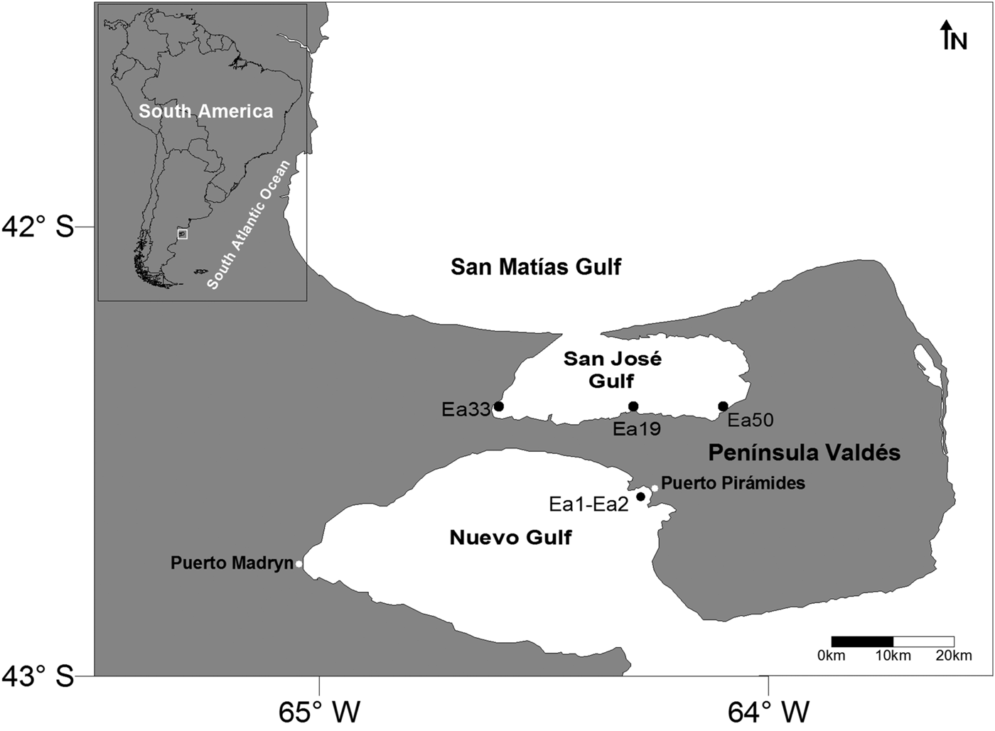
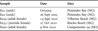
A total of five faecal samples were collected in PV (Figure 1, Table 1). Two samples (Ea1 and Ea2) were fresh faeces from live individuals collected using a 300 µm mesh net attached to a stick by personnel working on whale-watching boats in Pirámides Bay (NG) during the spring of 2004 and 2005. The other three samples were collected within the framework of the Southern Right Whale Health Monitoring Program (SRWHMP) which has been carried out since 2003 in this region and is dedicated to performing post-mortem examinations of right whales that die in PV (Rowntree et al., Reference Rowntree, Uhart, Sironi, Chirife, Di Martino, La Sala, Musmeci, Mohamed, Andrejuk, McAloose, Sala, Carribero, Rally, Franco, Adler, Brownell, Seger and Rowles2013). They were obtained from stranded whales (Ea19, Ea33 and Ea50) in SJG beaches during the period from September to November 2010. Samples were preserved either in 70% alcohol or in 5% buffered formalin.
Location of stranded whales (Ea33, Ea19, Ea50) in San José Gulf and collection site of fresh faecal samples from live whales in Nuevo Gulf (Ea1, Ea2).

Faecal samples of Eubalaena australis: collection dates and sites. unid.: unidentified developmental stage/sex; NG, Nuevo Gulf; SJG, San José Gulf.
Processing and analysis of food remains
Three subsamples (3 mL each) of each faecal sample were cleaned for the identification and quantification of plankton remains in the whale faeces collected. After a good homogenization of the entire faecal sample, each subsample was extracted and washed with distilled water onto a 67 µm Nylon mesh filter. The fraction retained on the filter was mixed with 15 mL distilled water and 1.5 mL sodium hypochlorite (5.5% active chlorine) holding it for 1 h 30 min to remove the organic matter. The material was washed again onto a 67 µm Nylon mesh filter and mixed with 10 mL distilled water. Two millilitres of methylene blue were subsequently added for the staining of microcrustacean remains. A final volume of 12 mL (treated faecal subsample + 10 mL distilled water + 2 mL staining agent) was obtained for each subsample. The remains were examined under a stereo microscope Nikon SMZ1500 or 645 (Nikon, Japan), identified and counted in the three subsamples pooled.
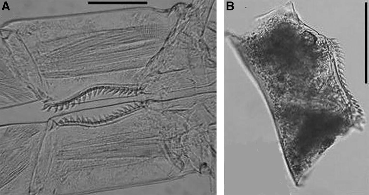
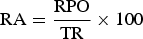
The food remains were classified into the following groups: copepod prosomes (CP), copepod mandibular gnathobases (MG), serrated coxae of fifth legs of copepods (SC) (Figure 2), planktonic organism segments (POS), unidentified plankton remains (UI), and parts or entire frustules of centric planktonic diatoms (D). Abundance of planktonic organism remains per faecal sample was expressed as relative abundance (RA) in per cent. The latter was calculated for each food sample as:
 $${\rm RA} = \displaystyle{{{\rm RPO}} \over {{\rm TR}}} \times 100$$
where RPO = abundance of each type of planktonic organism remains observed in each whale faecal sample (∑ three subsamples = 9 mL) and TR = abundance of total remains found in each faecal sample (9 mL).
Serrated coxae of the fifth legs from Calanus australis, (A) obtained by dissection from a preserved specimen, (B) found in fresh faecal samples from live Eubalaena australis. Scale bars = 100 µm (B), 50 µm (A).
Taking into account the taxonomic diagnostic characters established by different researchers (Mazzochi et al., Reference Mazzochi, Zagami, Ianora, Guglielmo, Crescenti, Hure, Guglielmo and Ianora1995; Bradford-Grieve et al., Reference Bradford-Grieve, Markhaseva, Rocha, Abiahy and Boltovskoy1999; Ramírez & Sabatini, Reference Ramírez and Sabatini2000; Razouls, Reference Razouls, de Bovée, Kouwenberg and Desreumaux2005–2014), SC were identified and categorized as coxae from C. australis. The SC found in whale faeces could have been erroneously identified as coxae from Calanus simillimus (Giesbrecht, 1902) which also has coxae of fifth legs with a serrated inner margin. Still, the differences between C. simillimus and C. australis lie in the shape and number of spines of the serrated inner margin of the coxae. On the other hand, this copepod, which is not as common as C. australis in plankton in PV, has been reported to seasonally inhabit the south of San Matías Gulf (Ramírez, Reference Ramírez1996) and its concentration has been observed to increase towards the shelf-break in spring and summer–early autumn (Ramírez & Sabatini, Reference Ramírez and Sabatini2000).
Processing and identification of mandibular gnathobases
A great similarity between the mandibles found in the faecal samples analysed and those of C. australis could be observed through our optical microscopy analysis. Taxonomic confirmation of copepods whose mandibles were found in faecal material was possible by comparing the mandibles found in the faecal samples collected with those from preserved C. australis specimens. Ten copepodites 5 (C5) and adults (C6) were sorted from formalin-preserved mesozooplankton samples collected by Dr M. Sironi (Instituto de Conservación de Ballenas (ICB), Buenos Aires, Argentina, and SRWHMP) in SJG during an E. australis feeding event in November 2009 (M. Sironi, personal communication). Specimens were dissected under a stereomicroscope using tungsten needles of 0.1 and 0.2 mm in diameter and glycerin as dissecting fluid. Both mandibles were removed (N = 20 per copepod developmental stage) for identification purposes. In parallel, free MG were randomly extracted from the faecal samples (N = 20 per sample).
Both the MG dissected and those extracted from whale faeces were prepared for scanning electron microscopy (SEM) observations. They were dehydrated in a series of graded ethanol baths (70, 80, 90, 96 and twice 100%, 15 min each). Once ethanol was removed, hexamethyldisilazane (HMDS) (Sigma-Aldrich Chemie GmbH) was added to dry the MG (Hazrin-Chong & Manefield, Reference Hazrin-Chong and Manefield2012). Mandibles were subsequently mounted onto double-sided adhesive tape and sputter-coated with gold. Observations and microphotographs were obtained using a Jeol JSM-6460 LV field emission SEM (JEOL, Japan). For the confocal laser scanning microscopy (CLSM) analyses, 10 C5 and C6 of C. australis from the same mesozooplankton samples were dissected to extract their mandibles and also 10 MG were randomly taken from faecal samples after cleaning. Mandibles were mounted on slides using glycerin and were observed and photographed under a Leica TCS SP2LSCM microscope (Leica, Germany).
In order to avoid confusion, MG from preserved C. australis specimens were morphologically compared with preserved C. carinatus specimens, another large calanid copepod in NG and SJG (V. D'Agostino, unpublished results). This comparative analysis revealed (i) differences in MG size as well as in teeth shape and size between both copepods, and (ii) absence of a small dorsal tooth typically located on the inner margin of the ventral tooth in C. carinatus.
Inference of Calanus australis copepod developmental stages ingested by whales
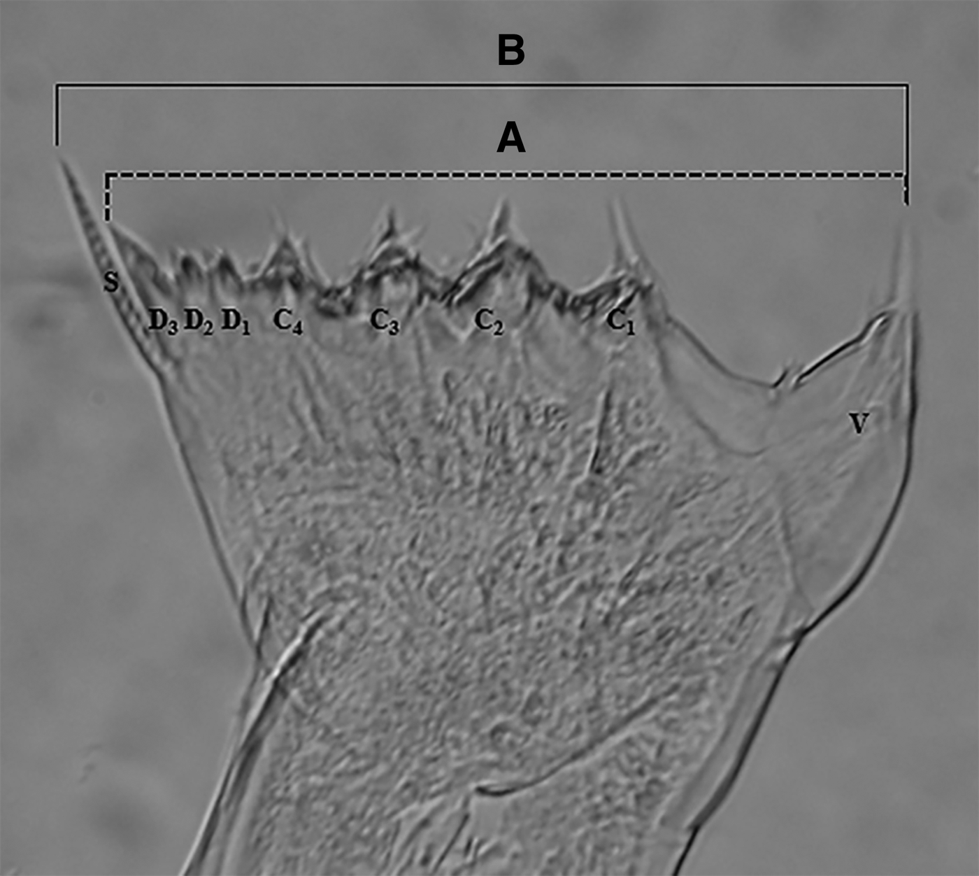
Specimens of C. australis (10 C6 females, 10 C6 males, 10 C5 females, 10 C5 males and 10 C4 without discerning sex) were sorted from the preserved mesozooplankton samples. Their total length (TL) was measured under a Nikon SMZ645 stereomicroscope (Nikon, Japan). The MG obtained by dissections as well as 36–66 MG of different sizes obtained from the faecal samples analysed were photographed and the MG from the faecal samples collected were identified using a Carl Zeiss STD 18 optical microscope (Carl Zeiss, Germany) and a Canon Power Shot G12 camera (Canon, USA). From the images of MG, morphometric measurements of their toothed edges were carried out using ImageJ 1.46r software (Wayne Rasband, National Institute of Health, USA). WMG from the preserved specimens were measured from the ventral tooth (V) to the dorsal seta (S) (Figure 3).Taking into account that the majority of the MG found in the faecal samples analysed had no dorsal seta (i.e. it was broken), the WMG from the preserved specimens were also measured from the ventral tooth (V) to the last dorsal tooth (D3) (Figure 3) following Karlson & Båmstedt (Reference Karlson and Båmstedt1994).
Optical microscope micrograph of a mandibular gnathobasis from Calanus australis, (A) measurements made on the mandibular gnathobases found in Eubalaena australis faecal samples and (B) measurements made on the mandibular gnathobases obtained by dissection from preserved C. australis specimens. V: Ventral teeth. C1-C2-C3-C4: Central teeth. D1-D2- D3: Dorsal tooth. S: Dorsal seta.
In order to estimate the TL of C. australis ingested by E. australis taking into account the WMG values from their MG found in the faecal samples analysed, back-calculation regressions were constructed using TL and WMG with and without the dorsal seta of the different preserved developmental stages of C. australis. To compare slopes of both regressions, a t-test (P < 0.05) was performed.
RESULTS
Diet components of southern right whales’ food and importance of Calanus australis remains
The plankton remains found in the faecal samples analysed and their RA are shown in Figure 4. Copepod fragments, including MG, CP, SC and segments included in the group of plankton organism segments (POS) (Figure 4A–E) were found in all the samples analysed. They were also the most abundant remains in all samples (Figure 4).
Top: Relative abundance (in %) of plankton remains observed in Eubalaena australis faecal samples. Bottom: micrographs of (A) MG: copepod mandibular gnathobases; (B) CP: copepod prosomes; (C) SC: serrated coxa of the fifth legs of a copepod; (D) UI: unidentified plankton remains; (E) POS: plankton organism segments; (F) D: entire or parts of frustules of centric planktonic diatom. Scale bars = 200 µm (D, F), 100 µm (A, B, C), 50 µm (E).
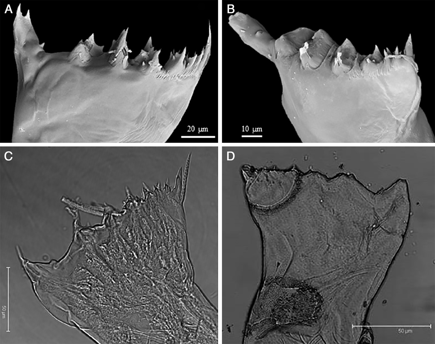
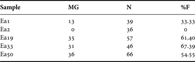
The MG found in the faecal samples analysed and those dissected from specimens of C. australis showed the same structure and morphology (Figure 5). They presented either eight teeth and a dorsal seta (S) or remains of the latter in the majority of the MG from faecal samples (Figure 5B, D). The presence of a small tooth in the inner border of the ventral tooth (V) of the MG found in the faecal samples analysed was the main feature that permitted the identification of C. australis. MG of C. australis were identified in faecal remains of four of the five whales considered (Table 2). They were also observed to be more abundant in the faecal samples of stranded whales (54.55–67.39%F, Table 2) than in those of live whales.
Scanning electron micrographs of mandibular gnathobases from Calanus australis; (A) mandible obtained by dissection of a preserved C. australis specimen; (B) mandible observed in a faecal sample of Eubalaena australis. Confocal laser scanning micrographs of mandibular gnathobases from C. australis; (C) mandible obtained by dissection of a preserved C. australis specimen; (D) mandible observed in a faecal sample of E. australis.
Number of mandibular gnathobases (MG) of Calanus australis present in Eubalaena australis faecal samples. N, total number of copepod MG from faecal samples; %F, per-cent frequency of occurrence of MG of C. australis.
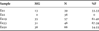
MG of C. australis ranged from 33.33 to 67.39%F (per-cent frequency) of the total MG found in the faecal samples analysed except in one of them (Table 2). In the latter, the majority of MG were observed to be smaller than the rest and they could not be correctly identified as a result of their partial degradation stage. It could nonetheless be assumed that they corresponded either to immature copepodites of C. australis and C. carinatus or to other small-sized species. In addition, whereas serrated coxae of fifth legs (SC) of C. australis were found in the faecal samples from live whales (Ea1 and Ea2, Figure 2b), they were not found in the faecal samples from stranded animals. Interestingly, the only sample without C. australis mandible remains (live whale Ea2) was observed to have SC belonging also to C. australis. The presence of SC only in the samples from live whales could be due to their better conservation conditions with respect to those of the faecal samples from stranded whales.
Unidentified planktonic organism remains (UI) as well as other planktonic organism segments (POS) (Figure 4d, e) which could be from microcrustaceans including copepods present in zooplankton patches of PV were found. In all the faecal samples analysed, either parts or entire frustules of centric planktonic diatoms (D) (Figure 4f) were also found. They were more abundant in the faecal samples from stranded whales than in those from live whales.
Calanus australis copepodite 5, a key food item for Eubalaena australis
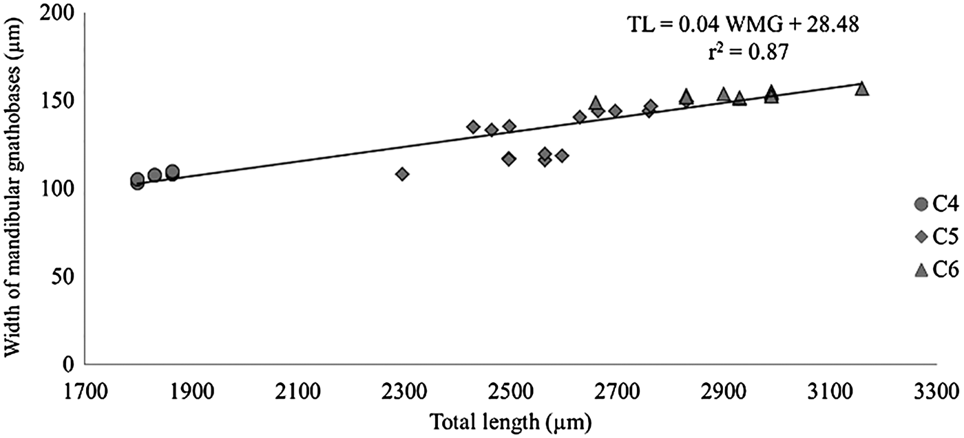
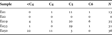
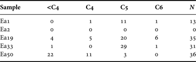
The two back-calculation regression lines found between WMG and TL of C. australis adults and copepodites showed a positive relationship (WMG with dorsal seta: N = 41, r 2 = 0.86, P < 0.0001; WMG without dorsal seta: N = 41, r 2 = 0.87, P < 0.0001). No significant differences were found between the slopes of both regression lines (t = −8.87 × 10−06, P = 1, N = 82). Therefore, the regression line between WMG without dorsal seta and TL (TL = 0.04 WMG + 28.48) was used to infer the developmental stages of C. australis ingested by E. australis (Figure 6). Although the number of faecal samples analysed in the present study was low, it could be observed that C5 of C. australis was the main food item for the majority of E. australis individuals whose faeces were analysed (Table 3). Only in the Ea50 faecal sample (stranded whale), the majority of the developmental stages of C. australis identified were either C4 or smaller (<C4).
Lineal regression relationship between width of mandibular gnathobasis edge (WMG) without dorsal seta (μm) and total length (TL) (μm) in C4: copepodites 4, C5: copepodites 5, and C6: adults of the copepod Calanus australis.
Copepodite stages of Calanus australis ingested by Eubalaena australis. Developmental stages and total length (TL) of copepodites of C. australis, <C4 (stages lower than copepodite 4): <1.79 mm; C4 (copepodite 4): 1.79–1.86 mm; C5 (copepodite 5): 2.3–2.83 mm and C6 (adult): 2.83–3.16 mm. N: total number of MG from C. australis identified in E. australis faecal samples.
DISCUSSION
Although our study material is limited to five faecal samples, the information obtained is most valuable in providing new insights into the diet and feeding ecology of the southern right whale in its calving ground of PV. The major achievement of the present study lies in having been able to assess that C5 of the large calanid copepod C. australis is the numerically dominant food item for most of these whales.
Diet components of southern right whales’ food and importance of Calanus australis remains
Results from the present study demonstrate for the first time that southern right whales feed mainly on copepods during their calving season in both NG and SJG. Similarly, Eubalaena glacialis (Borowski, 1781) was observed to have a dietary preference for large copepod Calanus finmarchicus (Gunnerus, 1770) in the feeding grounds of the western North Atlantic Ocean (Watkins & Schevill, Reference Watkins and Schevill1976; Mayo & Marx, Reference Mayo and Marx1990; Wishner et al., Reference Wishner, Schoenherr, Beardsley and Chen1995; Beardsley et al., Reference Beardsley, Epstein, Chen, Wishner, Macaulay and Kenney1996; Woodley & Gaskin, Reference Woodley and Gaskin1996; Baumgartner & Mate, Reference Baumgartner and Mate2003; Leandro et al., Reference Leandro, Rolland, Roth, Lundholm, Wang and Doucette2010).
Our findings agree with those derived from Menéndez et al.'s qualitative analysis (Reference Menéndez, Berasategui, Lindner, Diodato, Fernández-Severini and Hoffmeyer2007) of faecal material of a live southern right whale from NG (October, 2004) carried out with light microscopy. They found mandibles and prosomes which seemed to be from large calanid copepods, such as C. australis and C. carinatus, and other remains of crustaceans that could have probably belonged to euphausiids. Unlike Menéndez et al.'s (Reference Menéndez, Berasategui, Lindner, Diodato, Fernández-Severini and Hoffmeyer2007) research, in our study the food remains present in whale faecal samples were quantified. Both our results and those from Menéndez et al.'s (Reference Menéndez, Berasategui, Lindner, Diodato, Fernández-Severini and Hoffmeyer2007) study clearly agree with the availability of meso- and macrozooplankton prey that was documented by Hoffmeyer et al. (Reference Hoffmeyer, Lindner, Carribero, Fulco, Menéndez, Fernández Severini, Diodato, Berasategui, Biancalana and Berrier2010) in Pirámides Bay (NG) in October 2005.
Two types of faecal samples were analysed in the present study, namely fresh faeces from live individuals (NG) and faeces from stranded whales (SJG). In spite of their different origin, it was possible to verify the occurrence of C. australis remains in all the samples examined through their MG or SC. In C. australis, MG and SC have both been considered to be the most important diagnostic structures, thus allowing the unequivocal taxonomic identification at species level (Ramírez & Sabatini, Reference Ramírez and Sabatini2000).
Remains of diatom frustules were also found in all the faecal samples analysed. These remains were more abundant in the faecal samples from stranded whales, a phenomenon which could be related to the different sample collection techniques used and not to the abundance of diatoms in the area. The faecal samples from stranded whales were directly collected from the intestine of the animal whereas the fresh samples were collected with nets (300 µm mesh) from the water as faecal material floats after defecation (Rolland et al., Reference Rolland, Hamilton, Kraus, Davenport, Gillett and Wasser2006). Abundance of these plankton remains could thus be underestimated.
The analysis of food remains in faecal samples of large consumers, such as whales, has limitations as a result of the degradation of soft food preys and it is not convenient particularly when whales feed mostly on soft-body prey, such as ctenophores and medusae. It has nonetheless been demonstrated to be useful for trophic studies on right whales when crustaceans dominate in the food available (Stone et al., Reference Stone, Kraus, Prescott and Hazard1988; Menéndez et al., Reference Menéndez, Berasategui, Lindner, Diodato, Fernández-Severini and Hoffmeyer2007; Leandro et al., Reference Leandro, Rolland, Roth, Lundholm, Wang and Doucette2010 and the present study). Ctenophores have been reported to be a food item for right whales on the Argentine shelf (Bastida & Rodríguez, Reference Bastida and Rodríguez2009). The ctenophore Mnemiopsis leidyi (Agassiz, 1860) has been recently observed as an occasional food item for whales in San Matías Gulf (H. Castello, personal communication) and in PV area during spring and at the beginning of the calving season during winter (27 June 2013) in Pirámides Bay (P. Fioramonti, personal communication).
In PV, the dominant prey items for whales are usually meso- and macrocrustaceans which have higher carbon biomasses than soft-body preys (this is a nutritional advantage) and chitin in the whole body. These features make them unalterable remains and more easily recognizable in whale faeces. However, gelatinous plankton should not be disregarded as food items for southern right whales in this region. Alternative techniques to study their diet should therefore be considered and tested, such as analysis of stomach contents of recently stranded specimens providing samples of more freshly ingested food items that are likely to be more easily recognizable and identifiable.
Calanus australis copepodite 5, a key food item for Eubalaena australis
Mesozooplankton abundance and biomass are lower in the North Patagonian gulfs during winter and the beginning of spring (Esteves et al., Reference Esteves, Solís, Gil, Santinelli, Sastre, Gonzalez Raies, Hoffmeyer, Commendatore and Marcovecchio1996; Ramírez, Reference Ramírez1996; Hoffmeyer et al., Reference Hoffmeyer, Lindner, Carribero, Fulco, Menéndez, Fernández Severini, Diodato, Berasategui, Biancalana and Berrier2010 and references therein; Menéndez et al. Reference Menéndez, Biancalana, Berasategui, Fernández Severini, Hoffmeyer and Esteves2011; M. Hoffmeyer, personal observation) than in the frontal zone located in the inner and middle shelf adjacent to PV (Sabatini & Martos, Reference Sabatini and Martos2002; Derisio, Reference Derisio2012; Spinelli et al., Reference Spinelli, Pájaro, Martos, Esnal, Sabatini and Capitanio2012) and than in the southern feeding grounds of E. australis during summer and autumn (Ramírez & Sabatini, Reference Ramírez and Sabatini2000; Sabatini et al., Reference Sabatini, Ramírez and Martos2000, Reference Sabatini, Akselman, Reta, Negri, Lutz, Silva, Segura, Gil, Santinelli, Sastre, Daponte and Antacli2012; Atkinson et al., Reference Atkinson, Whitehouse, Priddle, Cripps, Ward and Brandon2001; Sabatini, Reference Sabatini2008; Padovani et al., Reference Padovani, Viñas, Sánchez and Mianzan2012). However, C. australis is numerically important in mesozooplankton assemblages in both NG and SJG during mid-spring (Ramírez, Reference Ramírez1996; Hoffmeyer et al., Reference Hoffmeyer, Lindner, Carribero, Fulco, Menéndez, Fernández Severini, Diodato, Berasategui, Biancalana and Berrier2010; M. Hoffmeyer & V. D'Agostino, personal observation), whereas C. carinatus and juveniles of euphausiids are found in lower abundances (M. Hoffmeyer & V. D'Agostino, personal observation). This relatively high food availability found in discrete patches significantly dominated by C. australis may explain why this copepod is likely to be a key food item for whales in PV.
Ours is the first study conducted to date on the prey size of the southern right whale in its calving ground in PV. The significantly positive relationship observed between WMG and TL has made it possible to infer that E. australis individuals feed mainly on C5 of C. australis. Our findings on E. australis’ feeding behaviour in PV agree with observations of E. glacialis in the northern hemisphere. As is the case with C. australis, E. glacialis feeds mainly on C5 of C. finmarchicus, a large-sized calanid copepod of the northern hemisphere (Baumgartner, Reference Baumgartner2003; Baumgartner & Mate, Reference Baumgartner and Mate2003). Copepodite 5 of C. finmarchicus contains higher caloric content with respect to any other stage of free-living copepods (Davies et al., Reference Davies, Ryan and Taggart2012), which means high-quality food for whales. Large calanid copepods accumulate mainly triacylglycerols and wax esters, two types of storage lipids (Michaud & Taggart, Reference Michaud and Taggart2007). Using these energy reserves, they can cope with unfavourable feeding conditions during winter, when they undertake ontogenetic migrations to greater depths to overwinter at reduced metabolic rates. In addition, although wax esters have been found to be the dominant lipids in C. finmarchicus, their presence has been recorded, though in low amounts, in northern right whale faecal samples (Swaim et al., Reference Swaim, Westgate, Koopman, Rolland and Kraus2009), thus indicating that right whales assimilate these lipids from copepods. A similar process may occur in E. australis when it feeds on C5 of C. australis in PV during spring. The presence of oil sacs full of lipids in specimens of C. australis, mainly C5 and adult females, in October–November over North Patagonian gulfs (V. D'Agostino, personal observation) suggests that this copepod may contain new lipid reserves by feeding on the spring phytoplankton bloom. Likewise, the fact that in the Southern Patagonian shelf C. australis population which is mostly dominated by C5 and adult females has been observed to carry large reserves of lipids in late summer, suggests that copepods could be ready to overwinter (Sabatini, Reference Sabatini2008).
Opportunistic foraging in a calving ground is a known behaviour in right whales as observed in the E. australis population in South African waters (Best & Schell, Reference Best and Schell1996) and the northern right whale population in the Bay of Fundy and Cape Cod Bay (Kenney et al., Reference Kenney, Hyman, Owen, Scott and Winn1986; Kraus et al., Reference Kraus, Prescott, Knowlton and Stone1986). Eubalaena australis also forages in the calving ground of PV, this being a behaviour which has been increasing over the years (Sironi, Reference Sironi2004; Hoffmeyer et al., Reference Hoffmeyer, Lindner, Carribero, Fulco, Menéndez, Fernández Severini, Diodato, Berasategui, Biancalana and Berrier2010; V. D'Agostino, personal observation). This ‘off-season feeding’ may allow whales not only to stay long enough in their calving ground so that their calves reach full developmental stage before migrating to feeding grounds but also to improve the physical condition of adults at the end of spring (Hoffmeyer et al., Reference Hoffmeyer, Lindner, Carribero, Fulco, Menéndez, Fernández Severini, Diodato, Berasategui, Biancalana and Berrier2010).
The above-mentioned increase in feeding observed among southern right whales in PV could be due to a decrease in food abundance in their feeding grounds, which was proposed as one of the explanations to calf mortality in PV. Rowntree et al. (Reference Rowntree, Uhart, Sironi, Chirife, Di Martino, La Sala, Musmeci, Mohamed, Andrejuk, McAloose, Sala, Carribero, Rally, Franco, Adler, Brownell, Seger and Rowles2013) suggest that the El Niño–Southern Oscillation event causes an increase in superficial temperature off South Georgia, which is part of the feeding area of E. australis, thus producing a decline in Antarctic krill (Euphausia superba Dana, 1850) abundance. This decrease in food availability could consequently lead to poor nutrition and reproductive failure in adult females.
CONCLUSIONS
This study provides the first quantitative analysis of the diet of southern right whale Eubalaena australis in PV during the calving season in austral spring, with copepods as well as other zooplankton and centric diatoms making up the bulk of recognizable prey items ingested.
This study further demonstrates that in PV calving ground, E. australis feed mainly on C5 of Calanus australis, based on a detailed analysis of mandibular gnathobases and fifth legs coxae recovered from faecal samples. Given the limited samples of whales, further research on this issue would be necessary in order to develop a more complete approach to the diet of right whales during their calving season in PV.
These findings improve our understanding of the feeding ecology of this whale population during their visit to the coastal waters of PV to calve and mate, providing baseline information on their diet and contributing to their conservation.
ACKNOWLEDGEMENTS
We are very grateful to the Southern Right Whale Health Monitoring Program (SRWHMP), to Instituto de Conservación de Ballenas (ICB), and to C. Marón and M. Sironi for providing the faecal samples from stranded whales and preserved zooplankton samples which were analysed in this study. Thanks are due to the staff of Whales Argentina Co. for collecting fresh faecal samples and giving us their help during sampling in Nuevo Gulf. Thanks are also due to Laboratorio de Mamíferos Marinos-Centro Nacional Patagónico (CENPAT-CONICET), Laboratorio de Ecología y Taxonomía de Zooplancton – Instituto Argentino de Oceanografía (IADO) (CCTBB, CONICET), Laboratorio de Parasitología (CENPAT-CONICET), ALUAR Co., and especially to J. Groizard for his help and facilities for SEM observations. Thanks are also due to the Dirección General de Conservación de Fauna y Flora and Subsecretaría de Áreas Protegidas de la Provincia del Chubut for their permission to carry out this study within the Natural Reserve of Península Valdés in Chubut, Argentina. Finally, we also thank Dr M. Sabatini, Dr M.S. Dutto and Dr F.G. Dellatorre for their critical reading and suggestions on the first version of this manuscript as well as two anonymous reviewers whose suggestions have substantially improved this manuscript.
FINANCIAL SUPPORT
This study was supported by grant PIT2096 (ANPCYT) to MSH and own funds. VCD was supported by doctoral fellowships from CONICET.