Introduction
The neurodevelopmental basis of schizophrenia refers to abnormal brain development before the onset of illness and is supported by compelling evidence of dysmaturation in the size of the brain (Wright et al. Reference Wright, Rabe-Hesketh, Woodruff, David, Murray and Bullmore2000), medial temporal lobe and hippocampi (Woodruff et al. Reference Woodruff, Wright, Shuriquie, Russouw, Rushe, Howard, Graves, Bullmore and Murray1997; Giedd et al. Reference Giedd, Blumenthal, Jeffries, Rajapakse, Vaituzis, Liu, Berry, Tobin, Nelson and Castellanos1999), cerebral ventricular enlargement (Giedd et al. Reference Giedd, Blumenthal, Jeffries, Rajapakse, Vaituzis, Liu, Berry, Tobin, Nelson and Castellanos1999; Woodruff et al. Reference Woodruff, Wright, Shuriquie, Russouw, Rushe, Howard, Graves, Bullmore and Murray1997), and less subcortical grey and white matter (see reviews by Wright et al. Reference Wright, Rabe-Hesketh, Woodruff, David, Murray and Bullmore2000 and Shenton et al. Reference Shenton, Dickey, Frumin and McCarley2001).
Kanaan et al. (Reference Kanaan, Kim, Kaufmann, Pearlson, Barker and McGuire2005) recently reviewed the application of diffusion tensor imaging (DTI) to investigate white matter structural integrity in schizophrenia. They observed that fractional anisotrophy (FA) was frequently reported to be decreased in patients compared to healthy controls, particularly in the corpus callosum (Foong et al. Reference Foong, Maier, Clark, Barker, Miller and Ron2000, Reference Foong, Symms, Barker, Maier, Miller and Ron2002; Ardekani et al. Reference Ardekani, Nierenberg, Hoptman, Javitt and Lim2003; Hubl et al. Reference Hubl, Koenig, Strik, Federspiel, Kreis, Boesch, Maier, Schroth, Lovblad and Dierks2004; Kalus et al. Reference Kalus, Slotboom, Gallinat, Federspiel, Gralla, Remonda, Strik, Schroth and Kiefer2005), cingulum bundle (Kubicki et al. Reference Kubicki, Westin, Nestor, Wible, Frumin, Maier, Kikinis, Jolesz, McCarley and Shenton2003, Reference Kubicki, Park, Westin, Nestor, Mulkern, Maier, Kikinis, Jolesz, McCarley and Shenton2005; Sun et al. Reference Sun, Wang, Cui, Breeze, Du, Wang, Cong, Zhang, Li, Hong and Zhang2003; Hubl et al. Reference Hubl, Koenig, Strik, Federspiel, Kreis, Boesch, Maier, Schroth, Lovblad and Dierks2004), striatum (Hubl et al. Reference Hubl, Koenig, Strik, Federspiel, Kreis, Boesch, Maier, Schroth, Lovblad and Dierks2004; Kubicki et al. Reference Kubicki, Park, Westin, Nestor, Mulkern, Maier, Kikinis, Jolesz, McCarley and Shenton2005; Szeszko et al. Reference Szeszko, Ardekani, Ashtari, Kumra, Robinson, Sevy, Gunduz-Bruce, Malhotra, Kane, Bilder and Lim2005), frontal and temporal gyri (Ardekani et al. Reference Ardekani, Nierenberg, Hoptman, Javitt and Lim2003; Szeszko et al. Reference Szeszko, Ardekani, Ashtari, Kumra, Robinson, Sevy, Gunduz-Bruce, Malhotra, Kane, Bilder and Lim2005), arcuate fasciculi (Hubl et al. Reference Hubl, Koenig, Strik, Federspiel, Kreis, Boesch, Maier, Schroth, Lovblad and Dierks2004; Kubicki et al. Reference Kubicki, Park, Westin, Nestor, Mulkern, Maier, Kikinis, Jolesz, McCarley and Shenton2005) and hippocampal/parahippocampal region (Ardekani et al. Reference Ardekani, Nierenberg, Hoptman, Javitt and Lim2003; Kalus et al. Reference Kalus, Buri, Slotboom, Gralla, Remonda, Dierks, Strik, Schroth and Kiefer2004, Reference Kalus, Slotboom, Gallinat, Federspiel, Gralla, Remonda, Strik, Schroth and Kiefer2005). Other DTI-related studies of water diffusivity (Shin et al. Reference Shin, Kwon, Ha, Park, Kim, Hong, Moon, Lee, Kim, Kim and Chung2006), grey matter (Rose et al. Reference Rose, Chalk, Janke, Strudwick, Windus, Hannah, McGrath, Pantelis, Wood and Mowry2006) and symptom correlates (Hubl et al. Reference Hubl, Koenig, Strik, Federspiel, Kreis, Boesch, Maier, Schroth, Lovblad and Dierks2004) also indicate disruption in fronto-temporo-limbic microstructure in the disorder. Together with aberrant cortical cell migration in the prefrontal, anterior cingulate and entorhinal cortices (Benes, Reference Benes1993; Eastwood & Harrison, Reference Eastwood and Harrison2003), evidence of widespread regional dysconnectivity of the brain in schizophrenia is accumulating. However, Kanaan et al. (Reference Kanaan, Kim, Kaufmann, Pearlson, Barker and McGuire2005) added the important caveat that all DTI studies so far have involved neuroleptic-treated patients, small samples, and were methodologically heterogeneous. This could explain inconsistencies across studies (Steel et al. Reference Steel, Bastin, McConnell, Marshall, Cunningham-Owens, Lawrie, Johnstone and Best2001; Foong et al. Reference Foong, Symms, Barker, Maier, Miller and Ron2002; Price et al. Reference Price, Bagary, Cercignani, Altmann and Ron2005), making evaluation of larger numbers of neuroleptic-naive samples essential to definitively address the issue of white matter structural dysconnectivity in schizophrenia.
DTI detects brain structural changes overlooked on conventional magnetic resonance imaging (MRI) (Moseley, Reference Moseley2002) by measuring the degree of non-randomness (FA) of water diffusion in the brain, from 0 (perfectly isotropic) to 1 (perfectly anisotropic) (Hoptmann et al. Reference Hoptman, Ardekani, Butler, Nierenberg, Javitt and Lim2004). In healthy children, FA values increase in a region-specific manner in the brain and are related to childhood cognitive achievement (Deutsch et al. Reference Deutsch, Dougherty, Bammer, Siok, Gabrieli and Wandell2005; Schmithorst et al. Reference Schmithorst, Wilke, Dardzinski and Holland2005). Therefore, FA represents an innovative approach to evaluate the brain where region-specific dysmaturation is suspected. We recently reported generalized cerebral morphological abnormalities in grey and white matter and cerebrospinal fluid in never-medicated, first-episode schizophrenia, with notable subcortical abnormalities in the brain (Chua et al. Reference Chua, Cheung, Cheung, Tsang, Chen, Wong, Cheung, Yip, Tai, Suckling and McAlonan2007). In the present study, we anticipated that DTI would demonstrate generalized structural dysconnectivity, including the subcortical region. We used a region-of-interest (ROI) analysis to independently confirm our method.
Method
Subjects
A consecutive series of patients presenting to our hospital were screened for eligibility to join the study. Inclusion criteria were: age 18–45 years; no previous antipsychotic treatment; first-ever experience of psychotic symptoms (i.e. hallucinations and/or delusions and/or thought disorder); no significant mood/organic disorder, with decline in daily functioning and fulfilling criteria for a diagnosis of DSM-IV schizophrenia (APA, Reference Andreasen, Arndt, Swayze, Cizadlo, Flaum, O'Leary, Ehrhardt and Yuh1994) after independent assessments by two specialists in psychiatry. Healthy controls from the local community were recruited by advertisement after screening for good physical health with no history of psychiatric or neurological disorder. Exclusion criteria were: any history of neurological problems; loss of consciousness; persistent headaches; head trauma; electroconvulsive therapy; psychostimulant use; history of neuroleptic use; and special school attendance. The study received the approval of the Institutional Review Board of the hospital. Patients were screened on the first day of presentation to hospital, and provided full informed written consent to participate. Symptoms were rated by a specialist psychiatrist with clinical responsibility for the care of the patients using the Positive and Negative Syndrome Scale (PANSS), a 30-item scale with positive, negative and general psychopathology items demonstrating high reliability and validity (Kay et al. Reference Kay, Fiszbein and Opler1987). Duration of psychosis was assessed by the Interview for the Retrospective Assessment of Schizophrenia (IRAOS; Hafner, Reference Hafner1992). According to clinical need, patients were prescribed the following medications as required: an anxiolytic (lorazepam 0.5 mg bd prn) for agitation or a hypnotic (zopiclone 7.5 mg nocte prn) for sleep. An MRI scan was performed according to the protocol below before initiating antipsychotic medication. We have successfully followed the patients who participated in the study for a minimum of 1 year and confirmed the diagnosis of schizophrenia.
MRI data acquisition
A GE Signa 1.5 T system (General Electric, Milwaukee, WI, USA) was used to acquire DTI and T2-weighted/PD (proton density) datasets. A 5-min DTI sequence followed Leung et al. (Reference Leung, Ooi, Kwong, Chan, Cao and Khong2004) and comprised single-shot, spin–echo, echo–planar imaging with TR=10 000 ms, TE=100 ms, acquisition matrix=128×128, and field of view=28 cm, slice thickness of 5 mm with a 1.5-mm gap. Diffusion-sensitizing gradient encoding (Basser et al. Reference Basser, Mattiello and LeBihan1994; Basser, Reference Basser1995) was applied in 25 directions using a diffusion-weighted factor b=1200 s/mm2, with one image, b0 (non-diffusion-weighted image), acquired without a diffusion gradient, b=0 s/mm2. Diffusion gradient directions were determined using minimal potential energy arrangements of points on a sphere (Hardin et al. Reference Hardin, Sloane and Smith1997), and 26 images were obtained at each section yielding 468 or 494 images. A T2/PD sequence provided dual-echo, fast spin–echo data sets aligned to AC-PC across the whole brain, 3 mm thick, Nex=1, TR 5 s, TE 20/80 ms, matrix 256×192.
Imaging processing and analysis
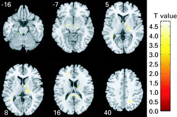
FA maps for each subject were calculated according to the method of Basser & Pierpaoli (Reference Basser and Pierpaoli1996) using DTIstudio (Radiology Department, Johns Hopkins University, USA). All subsequent image pre-processing and statistical manipulations were performed on SPM2 (Wellcome Department of Cognitive Neurology, Institute of Neurology, UK) running in matlab 6.5 (The MathWorks, Inc., Natick, MA, USA). Images were processed based on optimized voxel-based morphometry (VBM) methods (Basser & Pierpaoli, Reference Basser and Pierpaoli1996): first, a customized template of each brain tissue class was made by averaging linearly normalized, multi-spectral segmented T2 and PD weighted image pairs of all patients and controls, and smoothed with an 8-mm full-width half-maximum (FWHM) three-dimensional Gaussian kernel. Multi-spectral segmentation was used to enhance tissue classification accuracy (Styner et al. Reference Styner, Charles, Park and Guerig2002). Second, the b0 of each subject was segmented and spatially normalized (Ashburner & Friston, Reference Ashburner and Friston2000) to the custom template to correct for spatial distortion in the FA maps (Szeszko et al. Reference Szeszko, Ardekani, Ashtari, Kumra, Robinson, Sevy, Gunduz-Bruce, Malhotra, Kane, Bilder and Lim2005). As the current study focused on white matter, normalization parameters were determined by normalizing the white matter map to the custom white matter template (Specht et al. Reference Specht, Minnerop, Muller-Hubenthal and Klockgether2005). These transformation parameters were then applied to the FA maps so that the complete DTI dataset of each subject was transformed into MNI space (Hubl et al. Reference Hubl, Koenig, Strik, Federspiel, Kreis, Boesch, Maier, Schroth, Lovblad and Dierks2004; Leung et al. Reference Leung, Ooi, Kwong, Chan, Cao and Khong2004; Jones et al. Reference Jones, Symms, Cercignani and Howard2005). All MNI coordinates were converted to Talairach using a standard non-linear algorithm (Brett, Reference Brett2002). A white matter mask was used to limit the FA analysis to white matter only (Hubl et al. Reference Hubl, Koenig, Strik, Federspiel, Kreis, Boesch, Maier, Schroth, Lovblad and Dierks2004; Leung et al. Reference Leung, Ooi, Kwong, Chan, Cao and Khong2004; Szeszko et al. Reference Szeszko, Ardekani, Ashtari, Kumra, Robinson, Sevy, Gunduz-Bruce, Malhotra, Kane, Bilder and Lim2005) and the normalized FA maps were smoothed with a 6-mm FWHM Gaussian kernel for group comparison using a two-sample t test. Contrasts were used to detect FA changes between patients and controls, and results were thresholded at false discovery rate (FDR)-corrected p<0.05 (see Fig. 1 and Table 1). FDR is the proportion of false positives (incorrect rejections of the null hypothesis) among those tests for which the null hypothesis is rejected (Genovese et al. Reference Genovese, Lazar and Nichols2002). The FDR method is more appropriate to apply to neuroimaging data than the more conservative Bonferroni approach when group differences are likely to be present in multiple pixels (Genovese et al. Reference Genovese, Lazar and Nichols2002).
Fractional anisotropy (FA) deficits in patients versus healthy controls. Relative deficit clusters (in yellow) in white matter FA in never-medicated patients with first-episode schizophrenia (n=25) compared to controls (n=26). The maps are orientated with the right side of the brain shown on the right side of the panel. The z coordinate for each axial slice in the standard space of Talairach and Tournoux is given in millimetres.

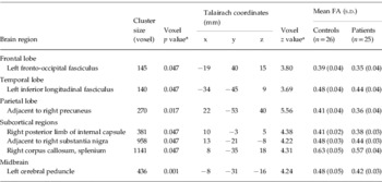
Significant fractional anisotropy (FA) differences between patient and healthy control groups

FDR, False discovery rate; s.d., standard deviation.
a All brain regions have their voxel z value significant at the FDR-corrected level of 0.05.
Region-of-interest (ROI) analysis
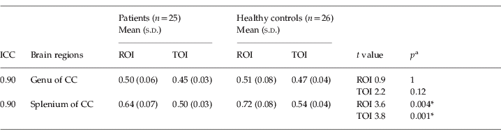
ROIs of radius 3 mm were placed on the FA map by an operator who was ‘blind’ to subject group membership. We selected ROIs on the corpus callosum as it is relatively easy to define and avoids the risk of contamination from the cerebrospinal fluid due to registration errors in voxel-base studies (Kanaan et al. Reference Kanaan, Kim, Kaufmann, Pearlson, Barker and McGuire2005). The splenium was found post hoc to have significantly reduced FA in the patients from our voxel-based analysis, and the genu acted as a control ROI on the same principal interhemispheric tract. The first ROI for genu and splenium was placed 6 mm above the lowest (i.e. inferior-most) axial slice on the z axis when the genu or the splenium was first barely seen. It was located at the midline of the genu or the splenium within the central boundary of the genu or splenium. The second ROI for genu and splenium was placed on the axial slice 6 mm above the first ROI, and also located in the midline and centrally within the boundaries of the genu or the splenium. Twenty-five patients and 26 healthy subjects were traced ‘blind’ to subject group membership. Ten randomly selected subjects’ scans were also measured independently by another operator also ‘blind’ to subject group membership. The ICC for the ROIs was 0.90 for genu and 0.90 for splenium. Mean FA values within the ROIs were calculated by using dTV software (Masutani et al. Reference Masutani, Aoki, Abe, Hayashi and Otomo2003; Masutani, Reference Masutani2007) for between-group comparison (see Table 2).
ROI analysis by group: mean fractional anisotropy (FA) values for each group in the genu and splenium of the corpus callosum (CC)
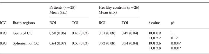
ICC, Intra-class correlation coefficient; ROI, region of interest; TOI, target of interest.
ICC is also shown for independent ROI tracing ‘blind’ to group membership.
a Two-tailed significance, Bonferroni corrected.
* p<0.005.
Results
Characteristics of the sample
Of a total of 30 patients eligible to enter the study, four either declined or were not stable enough to participate in the opinion of their responsible clinician-in-charge. We recruited 26 healthy controls from the local community through advertisement. Thus, the final sample comprised 25 patients and 26 healthy controls, balanced on age (patients: mean=28.5 years, s.d.=9.4; controls: mean=28.2 years, s.d.=9.2; independent samples t test, p=0.90, two-tailed), gender (13 male, 13 female patients; 11 male, 14 female controls, χ2=0.206, two-tailed), handedness (right-handed patients 87%, right-handed controls 95%, χ2=0.135, two-tailed) best paternal socio-economic status (SES) (patients mean value=3.0, s.d.=1.2; controls mean value=4.3, s.d.=3.5, independent samples t test, p=0.20, two-tailed) and ethnicity (all Chinese). For patients, PANSS positive symptom mean score=20.4, s.d.=3.7, PANSS negative symptom mean score=14.6, s.d.=6.7, PANSS general psychopathology mean score=39.1, s.d.=10.7. All patients were neuroleptic-naive and the median duration of untreated psychosis (DUP) was 180 days.
Group differences in FA score
FA values were significantly lower in the patients than in healthy controls in the left fronto-occipital fasciculus, left inferior longitudinal fasciculus, white matter adjacent to right precuneus, splenium of corpus callosum, right posterior limb of internal capsule, white matter adjacent to right substantia nigra, and left cerebral peduncle (voxel level significant at the FDR-corrected level of 0.05) (Fig. 1, Table 1). There were no regions where FA values were significantly higher in the patients than in the healthy controls.
Discussion
This is the first DTI study of white matter integrity in neuroleptic-naive patients, experiencing their first episode of schizophrenia. We found that FA values were significantly lower in patients than in healthy controls in the white matter of the following regions: left fronto-occipital fasciculus (adjacent to anterior cingulate gyrus, association tract linking orbital and lateral frontal cortices to occipital cortex), left inferior longitudinal fasciculus (adjacent to middle temporal gyrus, association tract linking occipital and temporal lobes), white matter adjacent to right precuneus, splenium of corpus callosum, right posterior limb of internal capsule, white matter adjacent to right substantia nigra, and left cerebral peduncle. There were no regions where FA values were significantly higher in the patients than in the healthy controls.
Our group differences largely concur with a recent comprehensive review (Kanaan et al. Reference Kanaan, Kim, Kaufmann, Pearlson, Barker and McGuire2005) of 16 studies using DTI in schizophrenia patients, 14 of which reported reduced fractional anisotropy (i.e. FA or the degree to which diffusion of water is directionally hindered or anisotropic; Basser, Reference Basser1995) in patients with schizophrenia. This was most commonly located in the white matter of the anterior cingulum and corpus callosum (Kanaan et al. Reference Kanaan, Kim, Kaufmann, Pearlson, Barker and McGuire2005). When restricted to the five studies reviewed that used voxel-based analysis, other regions affected by schizophrenia included the white matter of the prefrontal cortex (Burns et al. Reference Burns, Job, Bastin, Whalley, Macgillivray, Johnstone and Lawrie2003), middle and superior temporal gyrus (Ardekani et al. Reference Ardekani, Nierenberg, Hoptman, Javitt and Lim2003; Szeszko et al. Reference Szeszko, Ardekani, Ashtari, Kumra, Robinson, Sevy, Gunduz-Bruce, Malhotra, Kane, Bilder and Lim2005), arcuate fasciculus (superior longitudinal fasciculus) (Burns et al. Reference Burns, Job, Bastin, Whalley, Macgillivray, Johnstone and Lawrie2003), and uncinate fasciculus (anterior temporo-frontal fibre tracts) (Hubl et al. Reference Hubl, Koenig, Strik, Federspiel, Kreis, Boesch, Maier, Schroth, Lovblad and Dierks2004); one study reported no differences (Foong et al. Reference Foong, Symms, Barker, Maier, Miller and Ron2002). Our findings partly overlap with some recent voxel-based studies of DTI in schizophrenia that have confirmed reduced white matter connectivity in the fronto-temporo-parietal lobes and cerebral peduncle (midbrain) (Hao et al. Reference Hao, Liu, Jiang, Gong, Liu, Tan, Kuang, Xu, Yi and Zhang2006), and adjacent to the anterior cingulate cortex (Hao et al. Reference Hao, Liu, Jiang, Gong, Liu, Tan, Kuang, Xu, Yi and Zhang2006; Kumra et al. Reference Kumra, Ashtari, Cervellione, Henderson, Kester, Roofeh, Wu, Clarke, Thaden, Kane, Rhinewine, Lencz, Diamond, Ardekani and Szeszko2005). We note that direct comparison between different laboratories is limited when results are reported in grey matter rather than white matter terminology, partly because of the current lack of convention or the lack of an atlas displaying white matter tracts (Kanaan et al. Reference Kanaan, Kim, Kaufmann, Pearlson, Barker and McGuire2005; Catani, Reference Catani2006). In contrast to conventional MRI studies, which evaluate morphological change, diffusion-weighted scanning may present a more sensitive tool for determining individuals at risk for developing schizophrenia (DeLisi et al. Reference DeLisi, Szulc, Bertisch, Majcher, Brown, Bappal, Branch and Ardekani2006). However, it is important to note that all other DTI studies so far have evaluated chronic patients who have been exposed to years of neuroleptic treatment and these tended to identify widespread white matter association tract reduced FA such as in the arcuate fasciculus (superior longitudinal fasciculus that links perisylvian, frontal, parietal, temporal lobes) and uncinate fasciuli (connects orbito-frontal to anterior and medial temporal lobes) (Hubl et al. Reference Hubl, Koenig, Strik, Federspiel, Kreis, Boesch, Maier, Schroth, Lovblad and Dierks2004; Catani & ffytche, Reference Catani and ffytche2005; Kubicki et al. Reference Kubicki, Park, Westin, Nestor, Mulkern, Maier, Kikinis, Jolesz, McCarley and Shenton2005). With regard to patients in their first episode of illness, a recent study of adults comprising a modest sample of 10 patients newly diagnosed with schizophrenia or schizo-affective disorder, most of whom had been medicated for a median of only 2 weeks (Szeszko et al. Reference Szeszko, Ardekani, Ashtari, Kumra, Robinson, Sevy, Gunduz-Bruce, Malhotra, Kane, Bilder and Lim2005), demonstrated internal capsular, frontal and superior temporal white matter lower FA than in healthy volunteers. From the same laboratory, a study of 26 children and adolescents with early-onset schizophrenia who had been ill for some 2 years and were receiving maintenance neuroleptic treatment showed lower FA in the left anterior cingulate in the proximity of the left caudate nucleus (Kumra et al. Reference Kumra, Ashtari, Cervellione, Henderson, Kester, Roofeh, Wu, Clarke, Thaden, Kane, Rhinewine, Lencz, Diamond, Ardekani and Szeszko2005). These two studies indicate basal ganglia changes, which is of interest because our own neuroleptic-naive, first-episode sample also showed subcortical FA deficits (in addition to the more generalized abnormalities in cortico-cortical association and commissural tracts throughout the brain white matter). It is conceivable that lower subcortical FA values might represent a biomarker for illness onset or future relapse and we consider that this warrants a longitudinal study.
We found lower white matter ‘connectivity’ in the splenium of the corpus callosum of the patients. The corpus callosum is the principal interhemispheric white matter tract and myelinates rostro-caudally (Thompson et al. Reference Thompson, Vidal, Giedd, Gochman, Blumenthal, Nicolson, Toga and Rapoport2001) from birth to adolescence (Gilmore et al. Reference Gilmore, Zhai, Wilber, Smith, Lin and Gerig2004) in a linear fashion (Giedd et al. Reference Giedd, Blumenthal, Jeffries, Rajapakse, Vaituzis, Liu, Berry, Tobin, Nelson and Castellanos1999), attenuating after puberty (Thompson et al. Reference Thompson, Giedd, Woods, MacDonald, Evans and Toga2000). It surges in growth at around age 7, and can be used to assess anomalous neurodevelopment in childhood (Giedd et al. Reference Giedd, Blumenthal, Jeffries, Rajapakse, Vaituzis, Liu, Berry, Tobin, Nelson and Castellanos1999). The splenium connects homologous temporo-occipital cortices and showed lower FA in the patients, similar to previous work (Foong et al. Reference Foong, Maier, Clark, Barker, Miller and Ron2000; Price et al. Reference Price, Cercignani, Parker, Altmann, Barnes, Barker, Joyce and Ron2007). During neurodevelopment, the splenium has consistently higher FA values than, and myelinates before, the genu (Schneider et al. Reference Schneider, Il'yasov, Hennig and Martin2004), and our data might suggest early inefficiency in white matter connectivity between left and right temporo-occipital lobes.
We also identified decreased FA in the right substantia nigra, which is of importance, given the classical view that increased dopamine release from the midbrain can sensitize the prefrontal-limbic cortical targets to lead to psychosis (Laruelle, Reference Laruelle2000). For the same reason, dopaminergic drugs for treatment of Parkinson's disease boost dopaminergic transmission in the nigro-striatal system, but unfortunately can induce psychosis (Rascol et al. Reference Rascol, Payoux, Ory, Ferreira, Brefel-Courbon and Montastruc2003). Our finding of FA decrease in the left posterior limb of the internal capsule is close to that reported recently in the vicinity of the left caudate nucleus in minimally treated schizophrenia patients (Szeszko et al. Reference Szeszko, Ardekani, Ashtari, Kumra, Robinson, Sevy, Gunduz-Bruce, Malhotra, Kane, Bilder and Lim2005). The internal capsule abuts onto the basal ganglia, which are rich in dopamine receptors involved in antipsychotic response, so it would be reasonable to expect that local post-treatment change in interdendritic connections may also be associated with change in FA in this region. Furthermore, the posterior limb of the internal capsule connects the motor cortex by long association tracts to the cerebral peduncle, and it is therefore not surprising that FA in both regions is similarly decreased in our sample. Concerning the thalamus, it is a crucial gateway for sensory input to the cortex, and smaller thalamic volumes have been reported in schizophrenia (Andreasen et al. Reference Andreasen, Arndt, Swayze, Cizadlo, Flaum, O'Leary, Ehrhardt and Yuh1994; Gaser et al. Reference Gaser, Nenadic, Buchsbaum, Hazlett and Buchsbaum2004). It is thought to contribute to ventricular enlargement, which is already apparent in never-medicated patients in their first episode of psychosis (Chua et al. Reference Chua, Cheung, Cheung, Tsang, Chen, Wong, Cheung, Yip, Tai, Suckling and McAlonan2007).
We found lower FA in the left fronto-occipital fasciculus and left inferior longitudinal fasciculus. This suggests that patients have lower antero-posterior white matter connectivity between left orbito-frontal and left middle temporal cortices on the one hand, and the left occipital cortex on the other. This is compatible with the evidence of fronto-temporo-limbic disruption in the disorder (Weinberger & Lipska, Reference Weinberger and Lipska1995; Ardekani et al. Reference Ardekani, Nierenberg, Hoptman, Javitt and Lim2003). During normal development, FA increases during the second trimester of uterine life and is thought to correspond to neuronal migration from the germinal matrix (Gupta et al. Reference Gupta, Hasan, Trivedi, Pradhan, Das, Parikh and Narayana2005). It reaches near-adult white matter FA values by age 3, and stabilizes towards late adolescence (Schneider et al. Reference Schneider, Il'yasov, Hennig and Martin2004). Subsequently, the fronto-striatal maturation pattern is normal reduction in frontal lobe grey matter and increased myelination (Sowell et al. Reference Sowell, Thompson, Holmes, Jernigan and Toga1999). This suggests that risk of illness might be predicted by lower FA in the fronto-striatal regions (Sowell et al. Reference Sowell, Thompson, Holmes, Jernigan and Toga1999). Although these FA age-related changes reflect critical time periods prior to late adolescence, we think that they are not readily applicable to our own sample. Kumra et al. (Reference Kumra, Ashtari, Cervellione, Henderson, Kester, Roofeh, Wu, Clarke, Thaden, Kane, Rhinewine, Lencz, Diamond, Ardekani and Szeszko2005) reported a subtle association of FA with age, but they studied adolescents who had been ill for 2 years and medicated. We have not identified a relationship between FA and age/duration of illness, possibly because FA tends to stabilize after late adolescence, or because of sample size. We believe that a neurodevelopmental interpretation cannot be made, but our study is valuable because it is the first to show widespread FA deficits independent of chronicity and medication, and this might lend tentative support to a disconnectivity theory of schizophrenia (Bullmore et al. Reference Bullmore, Woodruff, Wright, Rabe-Hesketh, Howard, Shuriquie and Murray1998; Wright et al. Reference Wright, Sharma, Ellison, McGuire, Friston, Brammer, Murray and Bullmore1999).
Strengths and weaknesses of study
We recruited patients who had been carefully diagnosed and closely ethnically and demographically balanced with the healthy controls. The major strength of our study was that all our patients were neuroleptic-naive and experiencing their first episode of psychosis. Therefore, the results from studies incorporating more chronically ill patients in receipt of antipsychotic treatment, or those with an admixture of neuroleptic-naive and neuroleptic-treated patients, may well differ from ours. We also screened out drug abuse and excluded patients younger than 18. This may explain why our patients were some years older than other first-episode psychotic populations and had been ill for about 1 year. Although there was no significant intergroup difference in sex, and the number of females was similar between the groups, there was a slight excess of healthy control females in our sample. Females are more likely to have increased left-sided frontal asymmetry with higher FA values (Szeszko et al. Reference Szeszko, Vogel, Ashtari, Malhotra, Bates, Kane, Bilder, Frevert and Lim2003). Thus, having fewer females in the schizophrenia group could indirectly accentuate any left-sided frontal deficits in FA value. However, we did not observe any left frontal asymmetry in FA value in the voxel-based results, nor in the ROI analysis, which showed no group difference in FA values in the genu of the corpus callosum. Accordingly, we do not think that the between-group sex difference was a substantial confounder of our findings. Finally, we adopted a voxel-based approach not constrained by prior hypotheses and included an ROI analysis of the genu and splenium of the corpus callosum as a post hoc confirmatory analysis and also because this is a readily traceable and prime inter-hemispheric tract. At the time of study commencement, the DTI sequence used relatively thick, non-contiguous slices and for the sake of data continuity this protocol remained throughout the sample acquisition. Partial volume artefacts can arise from this, and other inherent methodological problems in the DTI voxel-based approach [e.g. heterogeneity of white matter FA from differential tract coherence and spatial registration problems adjacent to the ventricles (Leung et al. Reference Leung, Ooi, Kwong, Chan, Cao and Khong2004), relatively poor spatial resolution, and smoothing filter size differences (Jones et al. Reference Jones, Symms, Cercignani and Howard2005)], can also contribute to pre-processing and interpretation challenges. To limit these problems, we confined our analysis to the area delineated by the white matter (WM) mask, and we used a relatively small filter size (6 mm), which has been reported to be helpful in reducing the likelihood of false-positive results (Jones et al. Reference Jones, Symms, Cercignani and Howard2005). The future plan is to repeat the DTI scan after antipsychotic treatment to evaluate brain connectivity changes and determine whether this can assist in the prediction of early treatment response.
In conclusion, DTI represents a useful new tool to assess the brain in schizophrenia. We have found that evaluation of white matter connectivity early in the illness and before drug treatment commences suggests that schizophrenia is associated with significant FA decrease throughout the brain, including the subcortical region. In a future extension of this study, we plan to examine post-treatment brain changes and outcome. We hope that DTI might eventually provide a potentially useful tool to predict early outcome or even guide treatment choices in newly psychotic patients.
Acknowledgements
This work was supported by a research grant to Dr Chua from the CRCG, the University of Hong Kong. We thank our colleagues for assistance in subject recruitment. We thank M. K. Leung for her assistance with the ROI measurements.
Declaration of Interest
None.