Implications
This study provides information about polyethylene glycosylated (PEGylated) porcine glucagon-like peptide-2 (pGLP-2), a long-acting form of pGLP-2, on repair of intestinal damage in weaning piglets challenged with Escherichia coli lipopolysaccharide. It therefore has implication for the use of this peptide in farm animal, which is applied in medical science for inflammatory intestinal disease. The protective effects of PEG-pGLP-2 on intestinal function were possibly associated with the release of GLP-2R mediator (keratinocyte growth factor) and the decrease in the expressions of intestinal pro-inflammatory cytokines.
Introduction
In weaning, pigs suffer from social, environmental and dietary stress (Lallès et al., Reference Lallès, Boudry, Favier, Floc’h, Luron, Montagne, Oswald, Pié, Piel and Sève2004), resulting in gut mucosal injury (Touchette et al., Reference Touchette, Carroll, Allee, Matteri, Dyer, Beausang and Zannelli2002; Pié et al., Reference Pié, Lallès, Blazy, Laffitte, Sève and Oswald2004; Yi et al., Reference Yi, Carroll, Allee, Gaines, Kendall, Usry, Toride and Izuru2005); gut mucosal injury then causes diarrhoea and poor growth. In the small intestine, a pro-glucagon gene in the enteroendocrine L cells undergoes specific post-translational cleaving, thereby releasing glucagon-like peptide-2 (GLP-2), a 33-amino acid peptide. GLP-2 elicits many effects (Drucker et al., Reference Drucker, Erlich, Asa and Brubaker1996; Tsai et al., Reference Tsai, Hill, Asa, Brubaker and Drucker1997; Wojdemann et al., Reference Wojdemann, Wettergren, Hartmann, Hilsted and Holst1999; Benjamin et al., Reference Benjamin, McKay, Yang, Cameron and Perdue2000; Au et al., Reference Au, Gupta, Schembri and Cheeseman2002) mainly related to the regulation of gastrointestinal growth and functions, such as stimulation of mucosal growth, enhancement of nutrient absorption, reduction of intestinal permeability, inhibition of gastric emptying and secretion of gastric acid. The effects of GLP-2 are also transduced by a single G protein-coupled receptor (GLP-2R) predominantly expressed in the gastrointestinal tract (Munroe et al., Reference Munroe, Gupta, Kooshesh, Vyas, Rizkalla, Wang, Demchyshyn, Yang, Kamboj, Chen, McCallum, Sumner-Smith, Drucker and Crivici1999).
The intestinal repair effects of GLP-2 provide a therapeutic potential for diseases associated with intestinal damage in weaning and diarrhoeal diseases affecting piglets. However, the short half-life (t 1/2=8.4±0.9 min) of intact porcine GLP-2 (pGLP-2) caused by the cleavage of dipeptidyl peptidase-IV (DPP-IV) (Pedersen et al., Reference Pedersen, Hjollund, Johnsen, Orskov, Rosenkilde, Hartmann and Holst2008) requires frequent high-dosage administration in clinical applications. This shortage limits the development of pGLP-2 for the treatment of intestinal diseases of weaning piglets because frequent injection induces stress. For the first time, our team designed and purified polyethylene glycosylated (PEGylated) pGLP-2, a long-acting form of pGLP-2. This derivative has longer half-life (t 1/2) than pGLP-2 in DPP-IV and alleviates colonic injury in murine colitis (Qi et al., Reference Qi, Wu, Wan, Men and Xu2014).
Whether PEG-pGLP-2 is sufficient to repair intestinal dysfunction and damage in weaning piglets remains unclear. The activities of mucosal disaccharidases, such as sucrase, lactase and maltase, were used as indicators of intestinal mucosal maturation and digestive function (Pi et al., Reference Pi, Liu, Shi, Li, Odle, Lin, Zhu, Chen, Hou and Leng2014). However, reports concerning the effects of GLP-2 on intestinal digestive enzymes and nutrient transport have presented conflicting results (Brubaker et al., Reference Brubaker, Izzo, Hill and Drucker1997; Petersen et al., Reference Petersen, Hartmann, Schmidt, Holst and Sangild2000). In addition to intestinal digestive enzymes, pro-inflammatory cytokines are implicated in gut injury. For instance, the overproduction of pro-inflammatory cytokines adversely affects gut integrity and epithelial function. Furthermore, the molecular mechanisms of the pleiotropic actions of GLP-2 in the gut mucosa remain obscure (Drucker and Yusta, Reference Drucker and Yusta2014). This study evaluated the therapeutic effects of PEG-pGLP-2 on intestinal digestive function (brush border enzyme activity), anti-inflammatory function (mRNA expression of pro-inflammatory cytokines and myeloperoxidase (MPO) activity) and possible mechanisms (mRNA expression of GLP-2R, the per cent of GLP-2R, Ki-67 and Bcl-2-positive cells of total 200 cells in the ileum, and serum keratinocyte growth factor (KGF) content) of PEG-pGLP-2 in weaning piglets challenged with Escherichia coli lipopolysaccharide (LPS).
Material and methods
Preparation of mono-PEGylated pGLP-2
Mono-PEGylated pGLP-2 conjugates were prepared and purified according to previously described procedures (Qi et al., Reference Qi, Wu, Wan, Men and Xu2014). In brief, 1 mg/ml pGLP-2 and pGLP-2-(1-35)-amide (>98% purity) were synthesised by GL Biochem Ltd (Shanghai, China) in a 50 mmol/l Tris-HCl buffer solution at pH 8.5. The resulting solution was reacted with 4 mol of excess mPEG5k-SPA (Beijing Kaizheng Biotech Development Co., Ltd, Beijing, China) at room temperature for 30 min. The reaction was quenched using 1% trifluoroacetic acid/deionised water. PEGylated pGLP-2 mixtures were separated and purified by the ion-exchange chromatography with CM Sepharose FF resin (GE Healthcare Bio-Science AB, Uppsala, Sweden). The equilibration phase was 20 mmol/l acetate buffer at pH 4.0, and the elution phase was 20 mmol/l acetate buffer+1 mol/l NaCl at pH 4.0. After washing was completed, a linear gradient of elution buffer was applied from 0% to 60% for 60 min at a flow rate of 1 ml/min to elute the PEGylated pGLP-2 products. Eluates were then monitored at 215 nm. PEGylated products were collected, desalted and concentrated at a cutoff of 3 kDa (Millipore Corporation, Billerica, MA, USA). Afterwards, products were unfractionated and lyophilised to produce powder before they were further characterised.
Animals and experimental protocol
Animal experiments were conducted in accordance with the guidelines of the Zhejiang Farm Animal Welfare Council of China and approved by the ethics committee of Zhejiang Academy of Agricultural Sciences. A total of 18 female weaning piglets at 21 days of age (Duroc) with an initial average BW of 5.0 to 5.5 kg were grouped into three treatments: control, LPS, and LPS+PEG-pGLP-2. Each treatment has six replicates of one piglet. The piglets were housed on plastic slatted floors with feeders and automatic stainless nipple waterers. Feeds and water were available ad libitum. Temperature was maintained at 25°C to 28°C and a cycle of 16-h light and 8-h dark was set. The piglets from the LPS+PEG-pGLP-2 group were injected with 10 nmol PEG-pGLP-2/kg at 5 to 7 days of the trials daily. On 8th day, the piglets in the LPS and LPS+PEG-pGLP-2 groups were intraperitoneally administered with LPS (100 µg/kg BW, E. coli serotype O55: B5; Sigma Chemical Inc., St Louis, MO, USA). The control group was administered with the same volume of saline solution.
Tissue collection
Blood samples were collected from the anterior vena cava before LPS was injected and 3 h after LPS was injected on 8th day of the trial. Afterwards, blood samples were centrifuged at 3000 rpm for 10 min at 4°C to obtain the plasma. Plasma samples were stored at −80°C until analysis. The piglets were anesthetised and sacrificed to collect intestinal samples 3 h after LPS was injected. The small intestine was cut from its mesentery and immediately placed in an ice bath. The segment from the pylorus to the ligament of Trietz was considered as the duodenum, the proximal segment of the remaining parts of the small intestine was considered as the jejunum and the distal segment at 10 cm proximal to the ileocoecal junction was considered as the ileum. The duodenum, the jejunum and the ileum of each sample were thoroughly washed with ice-cold physiological saline solution. Each segment (1 cm) was collected to isolate RNA or perform histological analysis. To isolate RNA, we immediately placed the segments in 1 ml of RNA locker after dissection was performed. To conduct histological analysis, we fixed the segments in 10% (v/v) buffered formalin for 48 h; we then embedded these segments in paraffin in accordance with standard procedures. Intestinal segments (5 cm in length) were longitudinally opened and the contents were flushed with ice-cold physiological saline solution. Mucosae were collected by scraping the intestinal segments with a sterile glass microscope slide, rapidly frozen in liquid nitrogen and stored at −80°C until analysis.
Intestinal mucosal disaccharidase activities
Frozen intestinal mucosal samples were powdered in liquid nitrogen and homogenised in ice-cold 0.9% NaCl solution by using a homogeniser (1 g of sample/9 ml of 0.9% NaCl). The homogenates were centrifuged at 3000 rpm for 15 min at 4°C to obtain the supernatant fluid. The protein concentrations (mg protein/ml) of the homogenates were determined using a BCA protein assay kit (Pierce Biotechnology, Inc., Rockford, IL, USA) according to the manufacturer’s instructions. Lactase, sucrase and maltase activities were measured using lactase, sucrase and maltase activity assay kits (Nanjing Jiancheng Bio-Engineering Institute, Nanjing, China) according to the manufacturer’s instructions (Wang et al., Reference Wang, Hou, Yi, Wang, Ding, Chen, Long, Liu and Wu2013). Lactase, sucrase and maltase activity units were defined as nanomoles of disaccharide hydrolysed per ml of tissue homogenates (U/ml protein, 37°C, pH=6.0); these units were calculated as follows:
 $${\rm Activity}\,\left( {{\rm U}/{\rm ml}\,{\rm protein}} \right)={{{{A{\rm sample}\,{\minus}\,A{\rm control}} \over {A{\rm standrad}\,{\minus}\,A{\rm blank}}}{\times}{\rm standard}\,{\rm concentration}\,(5.55\,{\rm mmol/l})} \over {{\rm reaction}\,{\rm time} \,(20\,\min )}}{\times}1000}$$
Real-time PCR
Total RNA was isolated using RNAiso Plus kits (TaKaRa Biotechnology (Dalian) Co., Ltd, Dalian) according to the manufacturers’ instructions. Approximately 2 µl of aliquot of the total RNA was used to synthesise first-strand cDNA by utilising PrimeScript® RT-reagent kit (TaKaRa Biotechnology (Dalian) Co.,Ltd, Dalian) with a gDNA eraser. Real-time PCR was performed using ABI plus one (Life Technologies, Carlsbad, CA, USA) and SYBR Premix Ex Taq
TM (TaKaRa Biotechnology (Dalian) Co., Ltd, Dalian). The primer pairs used in this experiment were listed as follows: zonula occludens-1 (ZO-1) forward primer GACTTAAAGCTGCCTCAACAGA and ZO-1 reverse primer GGTTTGTTTCAGGCGAAAGG; occludin (OCCL) forward primer CCATGGCCTACTACTCGTCCAA and OCCL reverse primer CGGCACCGGTGTTGATTTAT; claudin-1 (CLAU) forward primer TTCTGGGAGGTGCCCTACTT and CLAU reverse primer TGGATAGGGCCTTGGTGTTG; interleukin-8 (IL-8) forward primer TTCGATGCCAGTGCATAAATA and IL-8 reverse primer CTGTACAACCTTCTGCACCCA; IL-10 forward primer TGATGGGGAGGATATCAAGG and IL-10 reverse primer TGGAGCTTGCTAAAGGCACT, interferon-γ (INF-γ) forward primer CAAAGCCATCAGTGAACTCATCA and INF-γ reverse primer TCTCTGGCCTTGGAACATAGTCT; tumour necrosis factor-α (TNF-α) forward primer: ATGGATGGGTGGATGAGAAA and TNF-α reverse primer TGGAAACTGTTGGGGAGAAG; GLP-2R forward primer TGTCCTACGTGTCGGAGATGTC and GLP-2R reverse primer TAATTGGCGCCCACGAA; and GADPH forward primer TCCACCACCCTGTTGCTGTAG and GADPH reverse primer GACCACAGTCCATGACATCACT. RT-PCR analysis was performed using an ABI Step One Plus Real-time PCR system (Life Technologies, Carlsbad, CA, USA) and SYBR SYBR® Premix Ex Taq
TM. PCRs were performed in triplicate under the following conditions: 95°C for 30 s and 40 cycles of 95°C for 5 s and 60°C for 30 s. Fluorescence data were collected at the end of each cycle and quantified. Melting curve analysis was performed at 55°C to 95°C to verify single product generation at the end of the assay. Standard curves were generated on the basis of the data obtained from the standards of 2 to 2−6 dilution series template. Standard curves were then analysed using Step One Software (v2.2, Applied Biosystems, Inc.). Data were normalised to GADPH by using the
 $$2^{{{\minus}\Delta \Delta \,C_{t} }} $$
quantification method.
MPO activity and KGF content
The MPO activity in the intestinal mucosa was evaluated using commercially available kits (Nanjing Jiancheng Bioengineering Institute, Nanjing, China) and expressed in relative absorbance units. The values were corrected for total protein content by conducting BCA protein assay. Serum KGF content was also evaluated using a porcine KGF ELISA kit (Nanjing Jiancheng Bioengineering Institute).
Immunohistochemistry
Paraffin-embedded colonic sections were cut at a thickness of 4 µm, deparaffinised and rehydrated. Antigens were then retrieved using a microwave with 10 mmol/l citrate buffer (pH 6.0) for 2 min. Afterwards, the sections were blocked in 3% H2O2 phosphate buffered saline (PBS) for 10 min to suppress endogenous peroxidase activity. These sections were washed for 3 to 5 min in PBS and incubated for 1 h at room temperature with a primary antibody (GLP-2R, AB9179, 1 : 150, Millipore Corporation; Ki-67/HRP, clone PC6, 1 : 100, Dako Denmark A/S, Glostrup, Denmark; Bcl-2, N19, 1 : 80, Santa Cruz Biotechnology, CA, USA) or PBS (as a negative control treatment). After washing for 3 to 5 min in PBS, we incubated the sections with an HRP-labelled polymer anti-rabbit kit (EnVisionTM Kits, Dako) at room temperature for 40 min and then with 3,3'-diaminobenzidine chromagen for 5 min. Subsequently, the sections were counterstained with Harris’s haematoxylin. The bright-field images were photographed under a BX20 microscope (Olympus Ltd, Tokyo, Japan). Semi-quantitative analysis was performed on the coloured sections using a computer imaging analysis system (Leica QWin V3 image analysis software; Leica Microsystems, Heidelberg, Germany). Briefly, 8 to 10 digital images were captured randomly from each slide using a 40× objective. GLP-2R, Ki-76 and Bcl-2 immunostaining were quantified according to the intensity of the color and the percentage of positively stained area. Brown areas were considered as positive. The intensity of the color was graded as 0 (no color); 1 (light yellow); 2 (light brown) and 3 (brown), and the percentage of positively stained area was graded as 0 (<5%); 1 (5% to 25%); 2 (25% to 50%); 3 (51% to 75%) and 4 (>75%). The two grades were added together to give a final score which represented the extent of GLP-2R, Ki-76 and Bcl-2 immunostaining.
Data analysis
Data were expressed as mean±s.d. Statistical differences between groups were determined by ANOVA with Duncan’s multiple comparison. P<0.05 was considered statistically significant.
Results
Enzyme activities
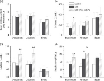
LPS treatment decreased lactase activity (U/ml protein) in the duodenum (P<0.01, Figure 1c) compared with the control treatment. By contrast, LPS+PEG-pGLP-2 treatment increased lactase activity (U/ml protein) in the duodenum (P<0.01, Figure 1c) and the jejunum (P<0.05, Figure 1c). LPS+PEG-pGLP-2 also increased sucrase activity (U/ml protein) in the jejunum (P<0.01, Figure 1d). No significant differences in the total protein content (Figure 1a) and maltase activities (Figure 1b) in the jejunum or ileum were observed among the three groups. Nevertheless, LPS+PEG-pGLP-2 treatment improved maltase activity in the duodenum compared with the LPS treatment (Figure 1b).
Total protein content (a), maltase activity (b), lactase activity (c) and sucrase activity (d) in the duodenum, jejunum and ileum of the piglets (n=6) treated with control (saline solution), LPS (100 µg LPS/kg) or LPS+PEG-pGLP-2 (10 nmol/kg PEG-pGLP-2+100 µg LPS/kg). Data are expressed as mean±s.d., & P<0.05 compared with the control group; # P<0.05 compared with the LPS group. LPS=lipopolysaccharide; PEG-pGLP-2=polyethylene glycosylated porcine glucagon-like peptide-2.

RT-PCR
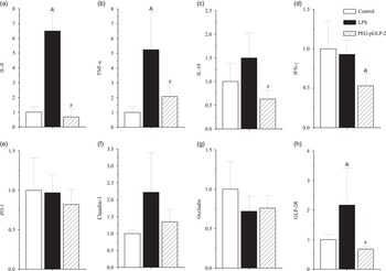
To ascertain whether or not LPS+PEG-pGLP-2 treatment modifies the LPS induction of intestinal cytokines, we analysed the mRNA levels of TNF-α, IL-8, IL-10 and INF-γ in the ileum. The relative expression levels of pro-inflammatory cytokines in the ileum of the piglets from the control, LPS and LPS+PEG-pGLP-2 groups are shown in Figure 2a to d. LPS treatment increased the mRNA expression levels of IL-8 (P<0.01, Figure 2a), TNF-α (P<0.01, Figure 2b) and IL-10 (P>0.05, Figure 2c) compared with the control treatment. However, PEG-pGLP-2 therapy reduced these effects during the experimental period. LPS+PEG-pGLP-2 treatment decreased the mRNA expression levels of IL-8 (P<0.01, Figure 2a), IL-10 (P<0.05, Figure 2c) and TNF-α (P<0.01, Figure 2b) compared with LPS treatment. The relative expression levels of the tight junction proteins, including ZO-1 (Figure 2e), CLAU-1 (Figure 2f) and OCCL (Figure 2g), in the ileum of the three groups showed no significant differences. The mRNA expression of CLAU-1 was increased by LPS treatment (P=0.14, Figure 2f) but reduced by LPS+PEG-pGLP-2 treatment (P=0.14, Figure 2f). The relative expressions of GLP-2R in the ileum of the piglets from the control, LPS and LPS+PEG-pGLP-2 groups are shown in Figure 2h. LPS treatment increased the mRNA expression levels of GLP-2R (P<0.05, Figure 2h) compared with the control treatment. However, LPS+PEG-pGLP-2 treatment reduced this effect during the experimental period. LPS+PEG-pGLP-2 treatment decreased the mRNA expression levels of GLP-2R (P<0.05, Figure 2h) compared with LPS treatment.
Relative expressions IL-8 (a), TNF-α (b), IL-10 (c), INF-γ (d), ZO-1 (e), claudin-1 (f), occludin (g) and GLP-2R (h) of the ileum of piglets (n=6) treated with control (saline solution), LPS (100 µg LPS/kg) and LPS+PEG-pGLP-2 10 nmol/kg PEG-pGLP-2+100 µg LPS/kg). Data are expressed as mean±s.d., & P<0.05 compared with the control group; # P<0.05 compared with the LPS group. IL-8=interleukin; TNF-α=tumour necrosis factor-α; INF-γ=interferon-γ; ZO-1=zonula occludens-1; GLP-2R=glucagon-like peptide-2 receptor; LPS=lipopolysaccharide; PEG-pGLP-2=polyethylene glycosylated porcine glucagon-like peptide-2.

MPO activity
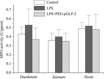
No significant differences in MPO activities (Figure 3) in the duodenum, jejunum or ileum were observed among the three groups.
MPO activity in the duodenum, jejunum and ileum of piglets (n=6) treated with control, LPS and LPS+PEG-pGLP-2. MPO=myeloperoxidase; LPS=lipopolysaccharide; PEG-pGLP-2=polyethylene glycosylated porcine glucagon-like peptide-2.

KGF content
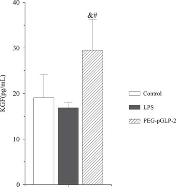
No significant differences in serum KGF content (Figure 4) were observed between the LPS group and the control group (P>0.05, Figure 4). However, LPS+PEG-pGLP-2 treatment significantly increased KGF content.
KGF contents of the serum of piglets (n=6) treated with control, LPS and LPS+PEG-pGLP-2. Data are expressed as mean±s.d., & P<0.05 compared with the control group; # P<0.05 compared with the LPS group. KGF=keratinocyte growth factor; LPS=lipopolysaccharide; PEG-pGLP-2=polyethylene glycosylated porcine glucagon-like peptide-2.

Immunohistochemistry
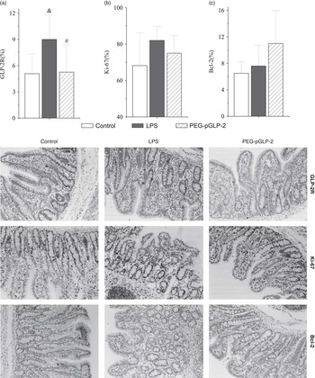
Compared with the control treatment, LPS treatment tended to increase the content of GLP-2R in the ileum (P=0.06, Figure 5a). LPS+PEG-pGLP-2 treatment tended to decrease the levels of GLP-2R (P=0.06, Figure 5a) compared with LPS treatment. No significant differences were observed in Ki-67 (Figure 5b) and Bcl-2 (Figure 5c) contents in the ileum of the three groups.
Immunohistochemistry staining for GLP-2R, Ki-67 and Bcl-2 proteins in the ileum of piglets treated with Control, LPS and LPS+PEG-pGLP-2 (n=6). Data are expressed as means±s.d., & P<0.05 compared with the control group; # P<0.05 compared with the LPS group. GLP-2R=glucagon-like peptide-2 receptor; LPS=lipopolysaccharide; PEG-pGLP-2=polyethylene glycosylated porcine glucagon-like peptide-2.

Discussion
This study mainly aimed to evaluate the effects on intestinal digestive function (brush border enzymes activity), anti-inflammatory function (mRNA expression of pro-inflammatory cytokines and MPO activity) and possible mechanism (mRNA expression of GLP-2R, percentages of GLP-2R, Ki-67 and Bcl-2-positive cells in the ileum and serum KGF content) of PEGylated pGLP-2 in weaning piglets challenged with E. coli LPS. Thus, a pig model of gut injury was established by injecting E. coli LPS. LPS is a potent endotoxin found in the cell membrane of all gram-negative bacteria. LPS induced intestinal morphological injury, enhanced mucosal permeability and bacterial translocation and caused symptoms of acute bacterial infection, including anorexia, hypersomnia and fever (Mercer et al., Reference Mercer, Smith, Cross, Russell, Chang and Cacioppo1996). These effects of LPS were associated with its ability to stimulate the rapid synthesis and release of pro-inflammatory cytokines. In the present study, LPS induced these acute symptoms and increased the production of pro-inflammatory cytokines (e.g., IL-8, TNF-α and IL-10) in the ileum.
Digestive function
The activities of mucosal disaccharidases, including sucrase, lactase and maltase, have been used as indicators of intestinal mucosal maturation and digestive function (Pi et al., Reference Pi, Liu, Shi, Li, Odle, Lin, Zhu, Chen, Hou and Leng2014). Studies had shown that GLP-2 increased brush border digestive enzyme activity in mice, rats and pre-mature and term-delivered neonatal piglets (Brubaker et al., Reference Brubaker, Izzo, Hill and Drucker1997; Kitchen et al., Reference Kitchen, Fitzgerald, Goodlad, Barley, Ghatei, Legon, Bloom, Price, Walters and Forbes2000; Petersen et al., Reference Petersen, Hartmann, Schmidt, Holst and Sangild2000). However, other studies had demonstrated that digestive enzyme activities were not affected by GLP-2 treatment. Petersen et al. (Reference Petersen, Hartmann, Schmidt, Holst and Sangild2000) demonstrated that GLP-2 (96 µg/kg per day for 6 days) does not stimulate foetal mucosal growth and elicits limited effects on brush border enzyme activities in utero. Petersen et al. (Reference Petersen, Burrin and Sangild2001) demonstrated that the enzymes sucrase-isomaltase and lactase-phloridzin hydrolase were unaffected by GLP-2 in late-gestation foetal and parenterally fed neonatal piglets when GLP-2 (12.5 nmol/kg) was administered twice daily for 6 days. In another study, newborn total parenteral nutrition-fed pigs were subjected to daily single administration of teduglutide for 7 days; as a result, small bowel growth was increased and minimal effect was elicited on digestive enzyme expression or nutrient absorption (Thymann et al., Reference Thymann, Stoll, Mecklenburg, Burrin, Vegge, Qvist, Eriksen, Jeppesen and Sangild2014a). However, GLP-2 treatment resulted in adverse outcomes, including decreased tissue maltase and sucrose in both sham and small bowel resection piglets (Pereira-Fantini et al., Reference Pereira-Fantini, Nagy, Thomas, Taylor, Sourial, Paris, Holst, Fuller and Bines2008). These different effects of GLP-2 on small intestinal brush border function may be dependent on gestational age at birth (Petersen et al., Reference Petersen, Elnif, Schmidt and Sangild2002), units of enzyme activities (Brubaker et al., Reference Brubaker, Izzo, Hill and Drucker1997) or GLP-2 analogues (Thymann et al., Reference Thymann, Le Huërou-Luron, Petersen, Hedemann, Elinf, Jensen, Holst, Hartmann and Sangild2014b). GLP-2-treated mice for 10 days exhibit significantly increased activities of brush border enzymes. No significant differences could be detected between vehicle- and GLP-2-treated mice (Brubaker et al., Reference Brubaker, Izzo, Hill and Drucker1997) when GLP-2 was expressed as units of enzyme per 5 cm of duodenum and data were normalised for the increased protein content (U enzyme activity per g of protein). The activity of brush border enzymes was also increased by injecting a long-acting acylated GLP-2 analogue (25 μg/(kg BW 12 h)) (Thymann et al., Reference Thymann, Le Huërou-Luron, Petersen, Hedemann, Elinf, Jensen, Holst, Hartmann and Sangild2014b). In the current experiment, LPS+PEG-pGLP-2-treated pigs exhibited a significantly increased lactase activity (U enzyme activity per ml of tissue homogenates) in the duodenum and the jejunum mucosa compared with the LPS-treated pigs. Saccharase activity was also significantly increased in the jejunum mucosa of weaning piglets compared with the LPS-treated pigs. In conclusion, GLP-2 may improve gut function in weaning pigs. However, the effects may be significant only when pigs suffer from stress or diarrhoea; these effects may also be significant if the time of exposure to GLP-2 is extended using long-acting analogues.
Anti-inflammatory function
The effects of LPS were associated with its ability to stimulate rapid pro-inflammatory cytokine synthesis and release (Pi et al., Reference Pi, Liu, Shi, Li, Odle, Lin, Zhu, Chen, Hou and Leng2014). However, studies had yet to be conducted to investigate the regulation of intestinal pro-inflammatory cytokines by administering PEG-GLP-2 in LPS-treated piglets. In the present study, the mRNA expressions of IL-8 and TNF-α significantly increased in the ileum of the LPS-challenged group. By contrast, the mRNA expressions of IL-8, IL-10, and TNF-α significantly decreased in the LPS+PEG-pGLP-2 treatment group compared with the LPS treatment group; this result was similar to that in previous reports regarding dextran sulphate-induced colitis (Drucker et al., Reference Drucker, Yusta, Boushey, DeForest and Brubaker1999; Sigalet et al., Reference Sigalet, Wallace, Holst, Martin, Kaji, Tanaka and Sharkey2007). Hence, LPS+PEG-GLP-2 treatment possibly reduced gut mucosal injury partially by suppressing pro-inflammatory cytokine production.
MPO was an enzyme predominantly found in neutrophils and has been used as an effective quantitative index of inflammation (Boushey et al., Reference Boushey, Yusta and Drucker1999). In mice with experimental colitis, 166.7 nmol/kg (i.p.) PEG-pGLP-2 was administered once in 8 days; as a result, the normal level of MPO activity was significantly recovered (Qi et al., Reference Qi, Wu, Wan, Men and Xu2014). In the present study, no significant differences were observed in the MPO activities in the intestinal mucosa of piglets in the three groups. This difference occurred possibly because the weaning piglets in the control group were suffering from stress or infection.
Possible mechanism
To explore the possible mechanism by which PEG-pGLP-2 attenuated intestinal inflammatory response and recovered digestion function, we examined the mRNA expression levels of GLP-2R and the percentage of positive staining for GLP-2R in the ileum. The effects of GLP-2 were mediated by GLP-2R, a G protein-coupled receptor (Munroe et al., Reference Munroe, Gupta, Kooshesh, Vyas, Rizkalla, Wang, Demchyshyn, Yang, Kamboj, Chen, McCallum, Sumner-Smith, Drucker and Crivici1999), which was highly selective for GLP-2, reacted only weakly following exposure to equimolar concentrations of structurally related peptides, such as glucagon and GLP-1 (DaCambra et al., Reference DaCambra, Yusta, Sumner-Smith, Crivici, Drucker and Brubaker2000). In the present study, LPS treatment increased the mRNA content of GLP-2R in the ileum compared with the control treatment. LPS+PEG-pGLP-2 treatment significantly decreased GLP-2 levels compared with LPS treatment. Petersen et al. (Reference Petersen, Hartmann, Holst, Le Huerou-Luron, Bjørnvad and Sangild2003) found that the highest mRNA content of small intestinal GLP-2R was detected at birth; this content decreased when foetal, suckling and weaned pigs consumed enteral food; thus, enteral feeding transiently increased plasma GLP-2 concentrations and decreased the mRNA levels of small intestinal GLP-2R during the development of pigs. Similarly, the mRNA expression of GLP-2R was downregulated in various models after GLP-2 was endogenously increased with feeding or after rodent models were treated with exogenous GLP-2 (Lovshin et al., Reference Lovshin, Yusta, Iliopoulos, Migirdicyan, Dableh, Brubaker and Drucker2000; Kaji et al., Reference Kaji, Tanaka, Redstone, Wallace, Holst and Sigalet2009; Sigalet et al., Reference Sigalet, de Heuvel, Wallace, Bulloch, Turner, Wales, Nation, Wizzard, Hartmann, Assad and Holst2014). The downregulation of this receptor after exposure to a receptor ligand was a common phenomenon, as in the case of epidermal growth factor receptors (Canesi et al., Reference Canesi, Malatesta, Battistelli, Ciacci, Gallo and Gazzanelli2000).
KGFs had been identified as targets of GLP-2 action. Ørskov et al. (Reference Ørskov, Hartmann, Poulsen, Thulesen, Hare and Holst2005) suggested that GLP-2 stimulated colonic growth via KGF, which was released by subepithelial myofibroblasts with GLP-2 receptors. However, whether GLP-2 increased KGF gene expression and circulating or intestinal levels of bioactive KGF remained unknown (Drucker and Yusta, Reference Drucker and Yusta2014). In the present study, the serum KGF content of the weaning piglets was unchanged after LPS was injected, but LPS+PEG-pGLP-2 treatment significantly increased serum KGF content. Hence, GLP-2 possibly stimulated the release of mediators, such as KGF, in weaning piglets.
Normal intestinal morphology is dependent on the balance of epithelial cell turnover. Decreased cell proliferation and increased apoptosis may be the main mechanisms responsible for intestinal mucosal injury. GLP-2 could stimulate intestinal growth by inhibiting apoptosis and increasing cell proliferation (Burrin et al., Reference Burrin, Stoll, Jiang, Petersen, Elnif, Buddington, Schmidt, Holst, Hartmann and Sangild2000). Sigalet et al. (Reference Sigalet, de Heuvel, Wallace, Bulloch, Turner, Wales, Nation, Wizzard, Hartmann, Assad and Holst2014) found the proliferation rate of crypt cells (Ki-67 staining) increased in the GLP-2-treated animals, and the rate of apoptosis (caspase-3) decreased. However, GLP-2 (3-33) decreased the crypt Ki67-positive cells in the small intestines of diet-induced obesity models; GLP-2 also increased the per cent of caspase-3-positive cells possibly by reducing endogenous GLP-2 (Baldassano et al., Reference Baldassano, Amato, Cappello, Rappa and Mulè2013). Ki-67 was considered as a marker of proliferating cells, but we found no significant difference in cell proliferation in the ileum; this result was similar to that of Benight et al. (Reference Benight, Stoll, Olutoye, Holst and Burrin2013). One possible explanation was that the rate of proliferation was maximally stimulated by enteral feeding and limited any effect of GLP-2. We also determined the abundance of Bcl-2, which inhibited caspase-3-mediated cell death. The Bcl-2 protein content was increased by LPS+PEG-pGLP-2 treatment, and this result was similar to that of Burrin et al. (Reference Burrin, Stoll, Guan, Cui, Chang and Hadsell2007). However, the mechanisms of this change should be further investigated.
Conclusions
In the present study, lactase and saccharase activities in the intestinal mucosa and serum KGF increased by LPS+PEG-pGLP-2 treatment. The mRNA expressions of IL-8 and TNF-α and the content of GLP-2R in the ileum were simultaneously downregulated in PEG-pGLP-2-treated piglets after these piglets were subjected to LPS challenge. Therefore, the protective effects of PEG-pGLP-2 on intestinal function (brush border enzyme activities) were possibly associated with the release of GLP-2R mediator (KGF) and the decrease in the expressions of intestinal pro-inflammatory cytokines.
Acknowledgements
This study was supported by grants from the Earmarked Fund for Modern Agro-industry Technology Research System (Grant No. CARS-36), the National Natural Science Funds of China (Grant No. C170105/31101724) and the National Key Technology R&D Program (Grant No. 2012BAD39B03-04). The authors report no conflicts of interest.