Introduction
Clinical evidence and experimental studies have reported that the influence of environmental factors early in life is associated with alterations in the gene expression, and that these changes modify the programmed health–disease life pattern of adult individuals. Reference Silveira, Portella, Goldani and Barbieri1,Reference Beydoun and Saftlas2 Studies involving the Developmental Origins of Health and Disease (DOHaD) concept are aimed at investigating the relationship among environmental variations occurring at critical moments of development, and during the development of diseases in adulthood. Reference Silveira, Portella, Goldani and Barbieri1
During development, the dynamic interaction among genome, epigenome, and stochastic and environmental factors contributes to the role of individual cells in the formation of functional organs. It also allows a critical balance to be maintained on a continuous basis between cell proliferation and death, and between cell regeneration and repair. Reference Ho, Cheong and Adgent3 Gestation is considered as a period of high vulnerability due to developmental events, such as germ cell specialization and migration, and morphogenesis of the gonads, which are structures of the reproductive tract and external genitalia. Reference Ho, Cheong and Adgent3 Moreover, prenatal stress exerts a negative impact on the development of the offspring throughout its childhood and may also persist into adulthood. Reference Beydoun and Saftlas2,Reference Eriksson4 These critical periods of development should be considered not only as vulnerable stages, but also as cycles of opportunity, considering that an insult caused during this period can lead to permanent changes. Reference Bouret5
In a circadian system, the light–dark (LD) cycle is the most reliable and efficient external signal that synchronizes the biological rhythms with the environment. In mammals, photopic information is perceived by specialized retinal photoreceptors, and is transmitted directly to the suprachiasmatic nucleus in the hypothalamus. Thus, the circadian information is transmitted to other organs through hormonal signals or the nervous system. Reference Hastings, Reddy and Maywood6 The circadian rhythm is involved in several physiological processes, such as that of sleep/wake, Reference Dijk, Duffy, Riel, Shanahan and Czeisler7 regulation of body temperature, Reference Van Someren8 hormone secretion, Reference Copinschi and Van Cauter9 tissue repair, Reference Janich, Meng and Benitah10 and cardiovascular functions. Reference Cooke-Ariel11 However, the fetus does not respond directly to the effects of light during development. Circadian information is transferred through the mother to the fetus. Reference Reppert and Schwartz12,Reference Parraguez, Valenzuela, Vergara, Ducsay, Yellon and Serón-Ferré13
The effect of exposure to endocrine disrupters during gestation has been extensively studied under DOHaD lines, given the importance of estrogen and androgen in this period. Reference Ho, Cheong and Adgent3 However, information on intrauterine exposure to other stressors and on lifestyle is still unclear. In this scenario, the aim of the present study was to evaluate whether changes in the circadian cycle of pregnant rats exposed to constant light can alter the development of the reproductive system in the male offspring. Our hypothesis is that the alteration in the maternal circadian cycle during the gestational period can impair the development of the male reproductive system, with consequences during adulthood.
Material and methods
Ethical approval
The experimental protocol was approved by the Institutional Animal Care and Use Committee of the University of Maringá (CEUA/UEM protocol number 4504080715).
Animals and experimental conditions
Rattusnovergicus of the Wistar lineage (males and females, aged between 70 and 80 days of age) were obtained from the Sectorial Room of the Laboratory of Cellular Biology of Secretion, linked to the Department of Cell Biology and Genetics of the State University of Maringá. The animals were housed at the same place, in polypropylene cages (45 cm/30 cm/15 cm) under light-controlled conditions with a 12 h LD cycle (07:00–19:00) and a temperature of 22.0 ± 2°C. After 1 week of adaptation, rats were mated at a ratio of three females to each male. Pregnancy (around postnatal day (PND) 90) was confirmed by the presence of sperm in a vaginal smear, and pregnant dams were individually housed and separated into two groups: LD (group exposed to normal light and dark cycle – 12 h/12 h; n = 10) and light–light group (LL) (group exposed to constant light – 24 h; n = 10). For the control of the light and dark cycle, the pregnant rats were kept in a specific rack (Master-One®, RibeirãoPreto) for studying circadian rhythms that control the exposure to light. In the first compartment was the LD group with normal LD cycle (light phase from 07:00 to 19:00) and in the second compartment was the LL group that were kept at constant exposure to light (24 h of light phase; ≅ 200 lux, measured by a lux meter).
After birth, mothers and offspring were kept in normal lighting conditions (12 h light/12 h dark; light period from 07:00 to 19:00) cycle and controlled room temperature (22 ± 2°C). The litter size was standardized to eight pups per female and no alterations in newborns’ weight and size were observed. At 21st PND, one male of each offspring (n = 10/group) were kept in polypropylene cages (four rats per cage). During all experimental periods, the rats had free access to water and food (Nuvital®, Curitiba, Brazil) and dams of both groups did not show alteration in body weight gain.
Reproductive organs weight
At PND 90, male rats were anesthesized (thiopental, 150 mg/kg intraperitoneally) and euthanized by decapitation. Testis, epididymis, vas deferens, and seminal vesicle (without the coagulating gland, full and empty of seminal fluid) were removed and their weights (absolute and relative to body weights) were determined. Testis and epididymis were used for sperm counts (n = 7/group), histopathological analysis (n = 5 rats per group), and oxidative stress analyses (n = 10). Spermatozoa from the vas deferens were analyzed for morphological analysis and sperm motility (n = 10/group).
Sperm number, daily sperm production per testis, and sperm transit time through epididymis
Testicles decapsulated and epididymis (divided into caput/corpus and cauda) were weighed and homogenized as described previously by Robb et al., Reference Robb, Amman and Killian14 with the adaptations described by Siervo et al. Reference Siervo, Vieira and Ogo15 After dilution (1:10 for testis; 1:20 for each epididymal portion) of the homogenized, a small sample was transferred to Neubauer chamber and four fields were counted per animal sample) for counting homogenization-resistant spermatids (stage 19 of the spermatogenesis) in the testis, and spermatozoa in epididymis. To calculate the daily sperm production (DSP), the concentration of spermatids per testis was divided by 6.1, which is the number of days in which mature spermatids are present in the seminiferous epithelium. To calculate sperm transit time through the epididymis, the number of sperm in each portion was divided by DSP.
Morphometric and histopathological analyses
The testicles and epididymis were removed, fixed in methacarn, Reference Puchtler, Waldrop, Meloan, Terry and Conner16 and embedded in Paraplast (SIGMA Life Science). Histological cross sections (5 µm) were stained with hematoxylin–eosin (HE) and examined for general histopathological, morphometric, and stereological analysis features as described by Favareto et al. Reference Favareto, De Toledo and Kempinas17
Histopathological analysis of the testis
One-hundred random testicular cross sections per rat were examined under Opton microscope at a magnification of 1000×. The seminiferous tubules were categorized as normal or abnormal. Abnormal tubules were identified as those with immature germ cells in the lumen, acidophilic cells, and vacuolization. Reference Siervo, Vieira and Ogo15 Leydig cells and the interstitial tissue were observed for the presence of inflammatory or degenerative alterations.
Nuclei of Sertoli cells were counted in 10 seminiferous tubules (3 nonconsecutive testis cross sections) per rat (n = −5 animals/group) at stage VII of spermatogenesis, under a light microscope (Opton), at 1000× magnification.
Morphometric analysis in the testis: seminiferous tubule diameters and seminiferous epithelium height
Ten random testicular cross sections were examined per rat in stage IX of the seminiferous epithelium cycle. Seminiferous tubule diameters were measured using an Opton photomicroscope at a magnification of 400× and BELView version 6.2.3.0 for Windows; BEL Engineering. The height of the seminiferous epithelium was measured using the same tubules and methodology described above. The mean of four measures of diameter and height was calculated for each seminiferous tubule and used for statistical analyses. Reference Siervo, Vieira and Ogo15
Kinetics of spermatogenesis
One-hundred random seminiferous tubular sections per rat were classified into one of the four categories of the seminiferous epithelium cycles (stages I–VI, VII–VIII, IX–XIII, and XIV) according to Leblond and Clermont, Reference Leblond and Clemont18 under a light microscope (Opton) at magnifications of 100× and 400×.
Histopathological and stereological analysis in the epididymis
In the histopathological analysis, epididymal cross sections (caput and cauda) were evaluated qualitatively using an Opton microscope (100× and 400× magnification). For stereological analysis, 10 random cross-sections per animal of caput (Region 2A) and cauda (Region 5A/B) epididymis Reference Miller and Killian19 were captured using a photomicroscope (Opton) and BELView version 6.2.3.0 (BEL Engineering) for Windows at a magnification of 400× and analyzed. This analysis was performed using Weibel’s multipurpose graticule with 168 points Reference Weibel20 to quantify the relative proportions of the epididymal components (epithelium, stroma, and lumen).
Sperm morphology
The right side vas deferens was removed and washed internally with 1.0 ml of 10% formol saline. Smears of histological slides were prepared from this solution and observed under a photomicroscope (Opton) at a magnification of 400×. Two-hundred spermatozoas were analyzed per rat. The morphological analysis was classified into three general categories: (1) normal morphology; (2) head abnormalities, that is spermatozoa without the characteristic curvature or in an isolated form (i.e. no tail attached); and (3) tail abnormalities, namely broken tail, tail rolled into a spiral, and isolated (i.e. no head attached). This evaluation was conducted as described previously. Reference Siervo, Vieira and Ogo15
Sperm motility
To obtain sperm, left vas deferens were rinsed with 1.0 ml of modified HTF medium with gentamicin (Human Tubal Fluid, Irvine Scientific1), at 34°C–37°C. A warmed (34°C–37°C) Makler counting chamber (Sefi-Medical, Haifa, Israel) was loaded with a small aliquot of sperm solution (10 ml). Sperm motility was assessed by visual estimation (100 spermatozoas per animal, in duplicate) under a light microscope (Motic) at 100× magnification and was done by the same person throughout the study. Spermatozoa were classified as mobile or immobile. Reference Siervo, Vieira and Ogo15
Hormonal analysis
Blood plasma was obtained via centrifugation at 3000 g for 15 min at 4°C and stored at −20°C until assayed via immunoassay. The total testosterone present in the plasma was measured via chemiluminescence (2nd Generation Testosterone Architect System, Abbott, Wiesbaden, Germany), according to the manufacturer’s recommendations. The intra-assay coefficient of variation and minimum sensitivity of the assay were 4.6% and 0.15 nmol/l, respectively.
Biomarkers of oxidative stress
The testes and epididymis were separately homogenized in Tris-HCl buffer (pH 7.4) in a 1:20 ratio and centrifuged at 12,000 g for 10 min at 4ºC. The protein quantification of the samples was determined by the Bradford method, using bovine serum albumin as the standard. Reference Bradford21 This homogenate sample was diluted to 1 mg of protein.ml−1, and an analysis of oxidative stress were performed.
Lipoperoxidation (LPO) was measured with the aim of indirectly quantifying the peroxides that were produced. The result reflects the intensity of lipid peroxidation. Reference Lushchak, Kubrak, Louzinsky, Storey, Storey and Lushchak22 The measurements were performed using the thiobarbituric acid reactive substances (TBARS) method at an absorbance of 535 nm Reference Buege and Aust23 and with a comparison to a standard curve for malondialdehyde (MDA), which is the main byproduct of cellular lipid peroxidation. For the preparation of the sample, a 0.33 mg.ml−1 aliquot of the sample protein was added in 6.7% trichloroacetic acid (TCA) and in a final volume of 180 μl was vortexed, left in an ice bath for 5 min, and centrifuged at 12,000g for 5 min at 4ºC. For the measurement of the TBARS, the following substances were added to a microplate: 40 μl of the supernatant and different concentrations of MDA in triplicate, and a reaction medium containing 21.42 mM of thiobarbituric acid (TBA), 17.86 mM of NaOH (used for TBA solubilization), 0.73 M of TCA, 0.032 mM of butylated hydroxytoluene (BHT), and 3% ethanol (used for BHT solubilization) in PBS buffer. The reading of the reaction was performed at 22ºC, after 60 min of incubation at 60ºC. The lipid peroxidation was estimated from the MDA curve, and the results were expressed as nmol of TBARS.mg−1 of protein.
The antioxidant enzyme superoxide dismutase (SOD) was quantified according to the method originally proposed by Crouch et al. Reference Crouch, Gandy, Kimsey, Galbraith, Galbraith and Buse24 with some modifications described below. The principle of this analysis was to quantify the complex formed between superoxide and nitro blue tetrazolium (NBT), which was measured at 560 nm during a 1 h period. An aliquot of 0.75 mg.ml−1 of protein in 25% ethanol was prepared in a total volume of 800 μl. The sample was centrifuged at 12,000 g for 20 min at 4ºC. In a 96-well microplate, the supernatant was pipetted in triplicate; a final volume of 200 μl (0.1 mg of protein.ml−1), 0.09 mM of NBT, 0.015 mM of EDTA, 34.78 mM of hydroxylamine sulfate, and 79 mM of sodium carbonate buffer (pH 10.2), and the plate was read at 22ºC. The values of the activity of antioxidant enzymes were expressed as U mg−1 of protein. One unit of SOD activity was defined as the amount of enzyme that inhibits the oxidation reaction of NBT by 50% of the maximum inhibition.
The aim of the analysis of glutathione transferase (GST) is that the enzymatic activity of GST catalyzes the conjugation of GSH with the synthetic substrate CDNB, which produces a conjugate detected at 340 nm. Reference Habig, Pabst and Jakoby25 During the assay, the enzymatic activity was proportional to the production rate of the conjugate compound. The assay was performed in triplicate in a microplate, and the final concentration of the sample was 0.020 mg of protein.ml−1. The reaction medium produced final concentrations of 0.94 mM of CDNB and 0.94 mM of GSH. The reading was done at 22ºC. The molar extinction coefficient of the GSH/CDNB compound was 9.6 mM−1.cm−1, and the unit was expressed as mmol.min-1.mg-1 of protein.
Glutathione reductase (GR) catalyzed the reduction of glutathione disulfide (GSSG) through the oxidation of NADPH; the decrease in absorbance was measured at 340 nm. Reference Sies, Koch, Martino and Boveris26 The assay was performed in duplicate in a microplate. The final concentrations within the reaction medium were 0.138 mM of NADPH, 3.81 mM of GSSG, and 3.75 mM of EDTA. The reaction was performed at 22ºC. The molar extinction coefficient of NADPH was 6.22 mM−1.cm−1. The unit was expressed as mmol.min−1.mg−1 of protein.
Statistical analysis
All parameters were submitted to the Shapiro–Wilk test for normality and thus classified into parametric and nonparametric data. Parametric results were statistically analyzed by unpaired t-test. Nonparametric results were analyzed by the Mann–Whitney test. Differences were considered significant when P < 0.05. The statistical analyses were performed with GraphPad Prism (version 5.0 – GraphPad Software, La Jolla, California, USA) program.
Results
Reproductive organs weight
The weight of the reproductive organs is listed in Table 1. A reduction in epididymis weight was observed in the LL versus the LD group. In contrast, the weight of other organs remained unchanged between these two groups.
Weight of reproductive organs

LD, light/dark group; LL, light/light group. *indicate groups that differ statistically (P < 0.05, unpaired t-test). Data are the mean ± standard error of mean (SEM). Shapiro–Wilk normality test; P > 0.05.
Sperm number, daily sperm production, and sperm transit time through caput/corpus regions of epididymis
Exposure to continuous light during pregnancy did not change the sperm number and the sperm concentration in both testis and epididymis. In addition, the DSP of the testes and sperm transit time in caput/corpus and cauda epididymis of male offspring were not altered (Table 2).
Sperm count

LD, light/dark group; LL, light/light group. *indicate groups that differ statistically (P < 0.05, unpaired t-test). Data are the mean ± SEM. Shapiro–Wilk normality test; P > 0.05.
Sperm morphology and motility
The results of sperm morphology are presented in Table 3. A decrease in the number of spermatozoa with normal morphology was observed in group LL. Consequently, an increase in the number of abnormal sperm heads was noted in the group exposed to continuous light in relation to the control group (P = 0.0001). The main abnormality observed in the spermatozoa was that their heads lacked the characteristic curvature. Continuous light exposure during the gestation period did not alter sperm motility (mobile sperm number: LD = 67.44 ± 3.31, LL = 63.88 ± 2.07, vs. immobile sperm number: LD = 32.56 ± 3.31, LL = 36.13 ± 2.07; mean ± SEM, P = 0.3900) in male offspring.
Sperm morphology

LD, light/dark group; LL, light/light group. *indicate groups that differ statistically (P < 0.05, Mann–Whitney test). Data are the median [Q1–Q3]. Shapiro–Wilk normality test; P < 0.05.
Histopathological analysis and spermatogenesis kinetics
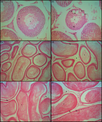
Exposure to continuous light during gestation caused an increase in the number of seminiferous tubules with vacuolization in the male offspring (P = 0.04) (Table 4 and Fig. 1a and b). There was a significant reduction in the Sertoli cell number per seminiferous tubule at stage VII of the spermatogenesis cycle in the LL group (Table 4). The Leydig cells and the interstitial connective tissue seemed to be uniform in size and shape in both groups.
Histopathological analysis of the testes

LD, light/dark group; LL, light/light group. *indicate groups that differ statistically (P < 0.05, unpaired t-test). Data are the mean ± SEM. Shapiro–Wilk normality test; P > 0.05.
Histopathological analysis of seminiferous tubules of testes, caput, and cauda epididymis. Photomicrograph of seminiferous tubules in testes (a and b), caput epididymis (c and d), and cauda epididymis (e and f). Sections from LD group (a, c, and e) and LL group (b, d, and f). (a) seminiferous tubules normal aspect, (b) observed vacuolization. (c, d, e, and f) Epithelium, lumen, and stroma shows normal aspect. Arrows in (b) indicates vacuolization. Ep, epithelium; It, interstitial tissue; L, lumen; LD, light/dark group; LL, light/light group. Hematoxilin and eosin stain. Magnification 100.

Analysis of the kinetics of spermatogenesis revealed no significant difference in the number of seminiferous tubules in the spermatogenesis stages between the groups (mean ± SEM): I–VI (LD = 36.8 ± 4.04, LL = 41.0 ± 2.19), VII–VIII (LD = 47.2 ± 4.09, LL = 42.2 ± 3.91), IX–XIII (LD = 9.60 ± 4.39, LL = 10.4 ± 8.90), and XIV (LD = 6.40 ± 1.07, LL = 5.60 ± 1.28).
Morphometric analysis of the testes
Exposure to continuous light during pregnancy caused a significant reduction in the diameter of the seminiferous tubule of the offspring in the LL versus the LD group (P = 0.02). However, no significant difference was noted in the height of the seminiferous epithelium (Table 5).
Morphometric analysis of the testis

LD, light/dark group; LL, light/light group. *indicate groups that differ statistically (P < 0.05, unpaired t-test). Data are the mean ± SEM. Shapiro–Wilk normality test; P > 0.05.
Histopathological analysis and stereological analyses in the epididymis
The histopathological analysis revealed no cellular alterations in the epididymal tissue in either of the experimental groups (Fig. 1c–d). Results of the stereological analyses are presented in Table 6. Specifically, a significant increase in the epithelial compartment was detected. This was followed by a decrease in the luminal compartment of the caput (2A region) and the compartment of the cauda epididymis (5A/B region) in the LL versus the LD group.
Stereological analysis of the epididymis

LD, light/dark group; LL, light/light group. * indicate groups that differ statistically (P < 0.05, Mann–Whitney test). Number of points in each epididymal portion. Data are the median [Q1 ± Q3]. Shapiro–Wilk normality test; P < 0.05.
Hormonal analysis
Disrupting the circadian cycle caused a significant reduction in plasma testosterone levels in the male offspring in the LL versus the LD group (Fig. 2).
Hormonal analysis: testosterone plasma levels. Data are the mean ± SEM. *P > 0.05, Mann–Whitney test. LD, light/dark group; LL, light/light group.

Biomarkers of oxidative stress
There was no significant difference between the experimental groups in relation to the lipid peroxidation levels in the testis and epididymis. An increase in the enzymatic activity glutathione peroxidase (GPx) and glutathione s-transferase (GST) was observed in the testis of animals exposed to continuous light during the intrauterine period. However, the other SOD and GR enzymes remained unchanged in the testis. In the epididymis, continuous light caused a reduction in GR levels in relation to the LD group. The other SOD, GPx, and GST enzymes remained unchanged (Table 7).
Biomarkers of oxidative stress in the testes and epididymis

LD, light/dark group; LL, light/light group. *indicate groups that differ statistically (P < 0.05, unpaired t-test). Data are the mean ± SEM. Shapiro–Wilk normality test; P > 0.05.
Discussion
The present study shows that exposure to continuous light during pregnancy impairs the reproductive system of the male offspring in adulthood, evidenced by significant abnormal spermatozoa, vacuolization, a reduction in seminiferous tubule diameter, epididymal weight, and luminal compartment, and by an increase in the epithelial compartment. In addition, the damage caused by changes in the circadian rhythm can be associated with reduced testosterone levels, but not with oxidative stress. Although studies show that circadian alteration impairs fetal development; none of them have reported its relationship with the intrauterine development of the male reproductive system, and its effects in adulthood, as the present does.
The maternal circadian rhythm is involved in the programming of fetal and neonatal circadian clocks Reference Reiter, Tan, Korkmaz and Rosales-Corral27 and it can also influence the pineal-defining transcriptome, which is established prior to the neonatal period. Reference Hartley, Coon, Savastano, Mullikin, Fu and Klein28 Disorders of the circadian system in fetal life have been associated with long-term metabolic and behavioral consequences in the offspring. Reference Cisternas, Compagnucci, Conti, Ponce and Vermouth29,Reference Voiculescu, Rosca, Zeca, Zagrean and Zagrean30 The fetal circadian clock begins to develop in the second half of pregnancy Reference Sladek, Sumová, Kováciková, Bendová, Laurinová and IIInerová31 and epigenetics could be associated with prenatal stress and fetal programming, and could impact the offspring adversely throughout its childhood and persist during adulthood. Reference Beydoun and Saftlas2,Reference Eriksson4 Therefore, stress conditions during gestation can compromise testicular and epididymal functions in adult life since specific events in the intrauterine development of the male reproductive system are considered extremely important. Reference Ho, Cheong and Adgent3
In relation to the oxidative stress assay, previous studies have reported increased levels of lipoperoxidation, and reduced levels of antioxidant biomarkers in the brain, liver, and kidneys of 3-month-old male rats exposed to continuous light for 26 days. Reference Escribano, Moreno, Tasset and Túnez32 Verma et al. Reference Verma, Singh and Ibrahim Rizvi33 observed an increase in oxidative stress levels in the erythrocytes of male rats (150 ± 20 g) exposed to continuous light for 10 days. However, there are no studies on how this deleterious event may affect the reproductive organs of male offspring undergoing circadian rhythm alteration during gestation. It has been reported that oxidative stress is associated with a decline in the energy metabolism of the sperm and consequently, in motility viability sperm as well in DNA fragmentation. Reference Baumber, Vo, Sabeur and Ball34,Reference Bilodeau, Blanchette, Cormier and Sirard35 The alterations evidenced in the GST and GPx antioxidant enzymes in the testis and GR antioxidant enzymes in the epididymis indicate that the organism against oxidative damage was maintained, thus enabling protection of the cell membrane and attempting to maintain the redox homeostasis of the reproductive organs. Therefore, the non-alteration of the oxidative stress biomarkers found may be associated with the maintenance of sperm motility. During the period of intrauterine development, the seminiferous cords are in formation. At puberty, spermatogenesis begins under the hormonal influence, and these cords open and develop in the seminiferous tubules for the production and passage of spermatozoa. Comparatively, in the present study, the alteration in the intrauterine circadian cycle affected the correct opening of these tubules, as evidenced by the reduction in the seminiferous tubule diameter, showing a stagnation in the development of seminiferous tubules
In the current study, increased vacuolization in the seminiferous tubules was related to the activity and number of Sertoli cells, and to a reduction in testosterone levels. According to Zambrano et al., Reference Zambrano, Guzmán, Rodríguez-González, Durand-Carbajal and Nathanielsz36 Sertoli cells begin to differentiate between the 13th and 14th day of gestation and thus, the alteration in the circadian cycle in this period affected the Sertoli cell differentiation, leading to permanent changes in the testicle of the offspring. However, despite this event, the sperm count in the testes remained unaltered in adulthood.
Exposure to continuous light during the gestational period resulted in a change in sperm morphology, with an increase in sperm head changes. The alterations in sperm morphology of the offspring evidenced after exposure to continuous light during the gestational period are associated with a reduction in the testosterone levels and an alteration in the spermiogenesis process. In this process, the nuclear and cytoplasmic compounds undergo a complex series of morphological, histochemical, and biochemical changes, ultimately ending with the reproduction of highly differentiated and specialized germ cells called spermatozoa. Reference Guraya37
Despite the reduction in the luminal compartment allowed fewer sperm to pass through the epididymal duct, this reduction did not alter spermatozoa motility, indicating that the epididymal epithelial cells performed the function of sperm maturation correctly.
Light is a powerful element in circadian, neuroendocrine, and neurobehavioral regulation, and has a profound influence on the health and well-being of all mammals, including laboratory animals. Reference Dauchy, Dauchy and Hanifin38 Many hormones and enzymes are essential for life, and are secreted following a circadian pattern, according to the photoperiod; testosterone is one such hormone. The organs of the male reproductive system develop as a result of hormonal action, like that of testosterone. In the present study, the faulty development of the morphology of male organs, such as seminiferous tubules and epididymal ducts, may be attributed to reduced testosterone levels. Despite this reduction, the daily production and motility of spermatozoa was not affected. Thus, the reduction in testosterone caused morphological changes in the organs and spermatozoa, but not in the functioning of the spermatogenic cells, the seminiferous tubules, or the epididymal duct cells.
Testosterone secretion and spermatogenesis are influenced by increased concentrations of glucocorticoids, since it causes a reduction in gonadotropin-releasing hormone (GnRH) and luteinizing hormone (LH) levels. Reference Gore, Attardi and Defranco39 Studies have shown that alteration in the LD cycle may increase corticosterone levels during pregnancy and that corticosterone increases more rapidly in male offspring than in female offspring. Reference Montano, Wang, Even and VomSaal40-Reference Harvey and Chevins43 In addition, the high levels of melatonin during pregnancy Reference Waddell, Wharfe, Crew and Mark44,Reference Reiter, Tan, Korkmaz and Rosales-Corral45 are able to cross the placental membrane and modify the circadian system of the developing fetus. Reference Tain, Huang and Hsu46 Circadian cycle regulation process is based on a group of circadian genes such as the CLOCK gene family that includes the BMAL and CLOCK transcription factors that interruption of this process can result in infertility. Reference Stocker, Macklon, Cheong and Bewley47,Reference Fernandez, Marino and Varcoe48
Maternal melatonin may be involved in the early stage of fetal development Reference Simonneaux49 such as intrauterine growth Reference Mendez, Abarzua-Catalan and Vilches50 and organogenesis. Reference Torres-Farfan, Valenzuela and Mondaca51 Although, in the present study, there were no changes in maternal weight and litter size, evidence indicate that the constant light exposure during the intrauterine period influenced the normal development of the reproductive organs: testis and epididymis, without causing damage to sperm production and sperm motility. In addition, we cannot rule out the increase in glucocorticoids and the decrease in melatonin of models of continuous light exposure, which may have an additional role in the development of offspring although the oxidative stress has not changed.
In conclusion, exposure to continuous light during pregnancy resulted in impairment of male offspring adulthood development, which is shown by the alterations in the morphology that could be partly explained by a reduction in testosterone.
Acknowledgments
This paper represents part of the doctorate thesis by FM Ogo (State University of Londrina – Brazil) under the advisory of GSA Fernandes.
Financial support
This research was supported by CAPES – PROEX (Coordinating Body for the Improvement of Postgraduate Studies in Higher Education, Brazil).
Conflicts of interest
None.
Ethical standards
The authors assert that all procedures contributing to this work comply with the ethical standards of the relevant national guides on the care and use of laboratory animals (Sectorial Room of the Laboratory of Cellular Biology of Secretion, linked to the Department of Cell Biology and Genetics of the State University of Maringá) and has been approved by the Institutional Committee (Ethics Committee on Animal Use of State University of Maringá (CEUA/UEM protocol number 4504080715)).”