Many different devices for percutaneous patent oval foramen closure have been developed over the last 5–10 years; some of them have specific anatomical indications, and some others have been conceived to fit the majority of anatomy – usually the results reported in literature concern a single type of device or different devices implanted without anatomical preferences.Reference Cifarelli, Musto and Parma1–Reference von Bardeleben, Richter and Otto3 We strongly believe that the complication and closure rates are influenced by a strategy, which is not linked with specific patient anatomy.Reference Rigatelli, Cardaioli and Dell’Avvocata4 Thus, we propose a strategy of device type and selection based on careful measurements of inter-atrial septum characteristics. We sought to prospectively evaluate the results of such strategy as regard as immediate success, complications and follow-up closure rate.
Materials and methods
We prospectively enroled 109 consecutive patients with a mean age of 43 plus or minus 15.5 years, 68 females, who had been referred to our centre for catheter-based closure of inter-atrial shunts over a 48-month period. In line with our institutional protocol, all patients were screened with transesopahgeal echocardiography before the intervention. Inclusion criteria for patent oval foramen percutaneous closure included: a concurrent shower or curtain shunt pattern on transcranial DopplerReference Sloan, Alexandrov and Tegeler5 with Valsalva manoeuvre, positive (single or multiple ischaemic foci) cerebral magnetic resonance imaging, previous stroke or Transient ischaemic attack, and medium or large patent oval foramen on transesophageal echocardiography.Reference Lang, Bierig and Devereux6 All patients with patent oval foramen were studied by Transthoracic Echocardiography and Transcranial Doppler, respectively, before Transesophageal Echocardiography. The Hospital Ethical Board approved the study. Written informed consent was obtained from all patients enroled in the study.
Echocardiography protocols
Transesophageal echocardiography was conducted using a GE Vivid 7 (General Electric Corporation, Nowrfolk, VI, USA) with bubble test and Valsalva manoeuvre under local anaesthesia. Patients who met the criteria for patent oval foramen closure were offered an intra-cardiac echocardiography study by using the mechanical 9F 9 megahertz UltraICE catheter (EP Technologies, Boston Scientific Corporation, San Jose, California, United States of America). The intra-cardiac echocardiography study was conducted as previously described,Reference Rigatelli and Hijazi7 by performing a manual pull-back from the superior vena cava to the inferior vena cava through five sectional planes; measurement of diameters of the oval fossa, the entire atrial septum length and rims were obtained with electronic calliper edge-to-edge on the aortic valve plane and the four-chamber plane. Patent oval foramen tunnel length was also measured. Intra-cardiac echocardiographic monitoring of the implantation procedure was conducted on the four-chamber plane. Atrial septal aneurysms were classified according to Olivares et al.Reference Olivares-Reyes, Chan, Lazar, Bandlamudi, Narla and Ong8
Closure protocol
Combined antibiotic therapy, Gentamicin 80 milligrams plus ampicillin 1 gram, or Vancomicin 1 gram if allergy was recorded on anamnesis, was administered intra-venously 1 hour before the procedure. The right femoral vein was catheterised through a eight French sheath and used for pre-operative right cardiac catheterisation and replaced with a 10 or 12 French long sheath for device implantation, whereas the left femoral vein was catheterised through a eight French sheath and replaced with a precurved nine French long sheath for intra-cardiac echocardiography study.
Intra-operative device selection: intra-cardiac echocardiography anatomical features – driven strategy
On the basis of intra-cardiac echocardiography findings and measurements and depending from operators’ experience and preferences, the operators selected the Amplatzer Occluder family (PFO Occluder, Cribriform Occluder, ASD Occluder, AGA Medical Corporation, Golden Valley, Minnesota, United States of America) or the Premere Closure System (St Jude Medical Inc., GLMT). The Amplatzer PFO Occluder was selected when atrial septal aneurysm was bidirectional with moderate (three right-to-left or three left-to-right atrial septal aneurysm, defined as moderate aneurysm with prevalent right or left bulging following Olivares et alReference Olivares-Reyes, Chan, Lazar, Bandlamudi, Narla and Ong8), whereas the Premere occlusion system was chosen when atrial septal aneurysm was absent or in the case of motionless or unidirectional atrial septal aneurysm (one right, two left atrial septal aneurysm defined as mild aneurysm with prevalent right or left bulging following Olivares et alReference Olivares-Reyes, Chan, Lazar, Bandlamudi, Narla and Ong8), when patent oval foramen tunnel length was more or equal to 10 millimetres, and in all case of hypertrophy of the inter-atrial septum rims (thickness more than 10 millimetres). The Amplatzer Cribriform Occluder was selected in the case of multi-perforated atrial septal aneurysm being careful to cross the most central hole in the oval fossa with the guidewire during intra-cardiac echocardiography guidance as previously described.Reference Zanchetta, Rigatelli, Pedon, Zennaro, Carrozza and Onorato9 This kind of device was also selected when atrial septal aneurysm was huge and bidirectional, four to five RL or four to five LR atrial septal aneurysm defined as huge aneurysm with prevalent right or left bulging following Olivares et al,Reference Olivares-Reyes, Chan, Lazar, Bandlamudi, Narla and Ong8 in order to obtain a complete coverage of the oval fossa on both sides of the atrial septum. Care was taken to choose a device with an entire left-disk diameter, which did not exceed the entire atrial septum length on intra-cardiac echocardiography measurement.
Medications and follow-up protocol
In patients without thrombophilia medications protocols after closure included aspirin 100 milligrams once a day for 6 months. In patients with thrombophilia the post-operative regimen included aspirin 100 milligrams once a day in patient with no history of deep vein thrombosis and thrombophilia but no antiphospholipid antibodies, aspirin 100 milligrams indefinitely in patient with thrombophilia but no history of deep vein thrombosis or peripheral arterial thrombosis especially in presence of antiphospholipid antibodies or factor V Leiden mutation, and warfarin in patients with antiphospholipid antibodies and factor V Leiden mutation and history of DVT or peripheral arterial thrombosis.
In patients who underwent closure, transesophageal echocardiography was scheduled at 1 month and repeated at 6 months following closure if there was no more than a trivial shunt, to assess potential erosions or thrombosis and residual shunts more accurately. All the patients also underwent transcranial Doppler ultrasound and magnetic resonance imaging of the brain before and 1 month after the procedure. Transthoracic echocardiography was scheduled at 6 months and 1 year after the transcatheter closure to assess mid-term residual shunt and the effect of the device on atrial and cardiac valve function. At each scheduled ecocardiographic control any residual shunt on bubble test was graded as trivial, small, moderate, or severe as previously described.Reference Boutin, Musewe and Smallhorn10 Clinical examination was scheduled at 1, 6, and 12 months.
Definition
Success was defined as the ability to release the device in a stable position under fluoroscopy and intra-cardiac echocardiography guidance with no more than a trivial shunt on immediate angiography and transesophageal echocardiography 24 hours after closure. Global complications included any degree of groin haematoma, atrial wall perforation, and pericardial effusion, entrapment of device or sheath or intra-cardiac echocardiography equipment through venous valves or embryonic remnants, and air embolism. Aortic erosion or device removal was included in the global complication rate. Pre-discharge occlusion rate was defined as a percentage of complete occlusion (presence of no more than a trivial shunt).
Statistical analysis
Chi-square, corrected Chi-square, and Student’s t-tests were used to compare frequencies and continuous variables: level of significance was set to p-value less than 0.05. Kaplan–Meyer curve was used to evaluate actuarial occlusion rates.
Results
On the basis of transesophageal echocardiography two patients were excluded from the intra-cardiac study due to for lipomatosus septum, whereas on the basis of intra-cardiac echocardiography study and measurements 9 patients were referred for medical treatment (patients with very large atrial septal aneurysm requiring a device that would exceed the length of the inter-atrial septum). The remaining 100 patients (91.7%) underwent attempted transcatheter closure (Table 1).
Demographic and clinical data of the 100 patients who underwent transcatheter closure.

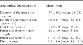
Device selection and measurement results. Intra-cardiac echocardiography measurements of inter-atrial septum components are reported in Table 2. A total of 26 patients have a long channel patent oval foramen, 44 patients had a large atrial septal aneurysm (more than four RL), 24 patients had a moderate atrial septal aneurysm (more than two RL but less than four RL), and 6 patients had hypertrophic rims. According on the criteria discussed above, the Amplatzer PFO (AGA Medical Corp., Golden Valley, Minnesota, USA) Occluder disk mean diameter was 25 millimetres in 20 patients and 35 millimetres in four patients. The Amplatzer ASD Cribriform Occluder was 25/25 millimetres in 41 patients and 30/30 millimetres in three patients. The Premere device size was 25 millimetres in 12 cases and 20 millimetres in 20 cases.
Intra-cardiac echocardiography measurements of anatomical features of the inter-atrial septum (measurements refer to the four-chamber view).

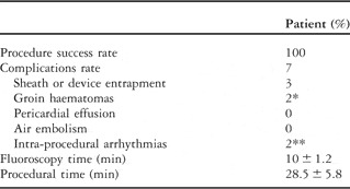
Procedural results (Table 3). In three cases of patent oval foramen the release of the Amplatzer Occluder was difficult and complicated by thoracic pain because it caught up in a venous valve remnant during sheath advancement. Forced and prolonged sheath manipulation during the procedure was judged to be the cause of mild groin haematoma in two patients without need of transfusion. Two patients suffered atrial fibrillation episodes during the procedure and were electrically cardioverted within 48 hours. Hospital staying was 2.0 plus or minus 1.2 days. Complication rate was 7% – all complications were not dangerous including minor haematomas and intra-procedurally cardioverted atrial fibrillation.
Summary of the results in respect to anatomy and implanted device.

Mid-term follow-up (Table 4). Six patients suffered either from supraventricular tachycardia (3 patients) or atrial fibrillation (3 patients) within 1 month of the procedure (6%); the sinus rhythm was restored in all patients with antiarrhythmic drugs. No aortic erosions, device thrombosis, or recurrent ischaemic cerebral events were observed. Pre-discharge and follow-up occlusion rates at a mean of 34 plus or minus 11.1 months were 91% and 96%, respectively. Five patients had a moderate intra-protesic shunt at 6-month follow-up: only three patients had persistent moderate shunt at 36-month follow-up and one patient had a persistent trivial shunt.These patients had moderately hypertrophic rims associated with very large atrial septal aneurysms and were treated with indefinite antiplatelet regimen. The residual shunts depended in these cases on moderate misalignment of the Amplatzer caused by the hypertrophic rims.
Procedural results.

*Not requiring blood transfusion
**Intra-procedurally cardioverted
Discussion
Our study showed that a strategy of careful device selection based upon evaluation of the right atrium and inter-atrial septum major characteristics is very safe and effective, avoiding the rare, but always possible, late complications of patent oval foramen transcatheter closure such as device thrombosis and aortic erosion. As a collateral finding this study confirmed that mid-term outcomes of intra-cardiac echocardiography assisted closure compared very well with the results of transesophageal echocardiography guided series and seems even better in terms of late complications.Reference Hein, Buscheck and Fischer11 In particular, the absence of erosion and device thrombosis in intra-cardiac echocardiography guided procedures seem very attractive compared with transesophageal echocardiography assisted procedure in which erosion and thrombosis appeared to be 0.1% and 2.5%, respectively.Reference Krumsdorf, Ostermayer and Billinger12, Reference Amin, Hijazi, Bass, Cheatham, Hellenbrand and Kleinman13This effect is probably due to the higher accuracy of intra-cardiac echocardiography that allows for a more careful device selection.
Indeed, a careful evaluation of atrial septum, right atrium, and their anatomical parts seem to be of paramount importance for a safe and effective closure of inter-atrial shunt as recently suggested by Proskelj et al.Reference Prokselj, Kozelj, Zadnik and Podnar14 Some “sensible” characteristics seem in our experience impacting of complication and closure rate: the patent oval foramen tunnel length, the thickness of the rims, the oval fossa diameter, and the atrial septal aneurysm severity all need attention because their potential of device misalignment, not proper device configuration, and uncovering of misdiagnosed fenestration.
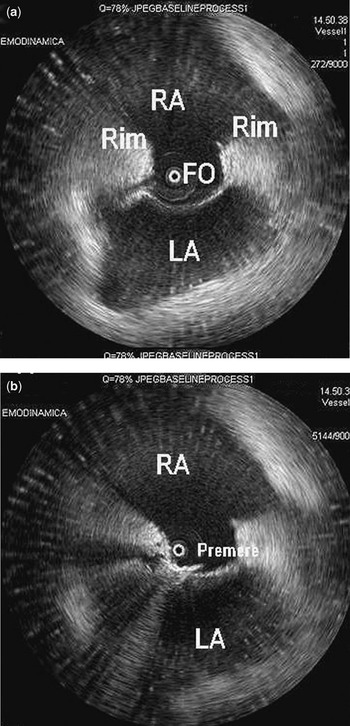
To overcome these potential reasons of failure we propose a strategy based upon identification and measurement by intra-cardiac echocardiography of all sensible characteristics. On the basis of ICE study, we selected in all cases a device with disk diameter that does not exceed the entire inter-atrial septum length on intra-cardiac echocardiography in order to prevent aortic erosion.Reference Amin, Hijazi, Bass, Cheatham, Hellenbrand and Kleinman13 In the same way attention has paid to select an asymmetrically opening device when facing with long channel patent oval foramen in order to minimise device misalignment typical of the Amplatzer device family in such anatomy. Again, we opted for a full-coverage of the oval fossa when facing with large atrial septal aneurysm keeping attention to the inter-atrial septum/device size ratio. Finally, we tried to treat hypertrophic rims patent oval foramen using as soft device such as the Premere, made by a single nitinol wire for both left and right portion of the device with the right portion covered by a tissue patch, selecting larger size compared with the oval fossa diameter to cover the thick rims (Fig 1) and to avoid the not correct device configuration typical of the Amplatzer of Starflex family when facing with hypertrophic rims: a residual shunt is usually caused by the large distance between the two disks. In our experience more hypertrophic rims claim to larger Premere device: with a patent oval foramen tunnel of less of equal to 15 millimetres and rims thickness less of equal to 10–12 millimetres we recommend a 20 millimetres device, whereas for patent oval foramen tunnel length more or equal to 15 millimetres or thickness more or equal 12 millimetres, we would recommend a 25 millimetres devices.
An example of anatomy – driven device selection: (a) very thick oval fossa rims, moderate long channel (13 millimetres), and moderate atrial septal aneurysm on intracardiac echocardiography study in a 60-year-old patient with recurrent stroke and coronary artery disease; (b) despite the oval fossa measure only 18 millimetres, a 25 millimetres Premere instead of a 18 millimetres Amplatzer, or 20 millimetres Premere was selected and implanted in order to cover the thick rims and avoid distorted device configuration. ACO, Amplatzer Cribriform Occluder; GW, guidewire; LA, left atrium; RA, right atrium.

Our study showed that a device selection strategy based upon of measurements of specific right atrium components compared very well with recent standard single or mixed-device series irrespective of anatomy or focused only on atrial septal aneurysm.Reference Cifarelli, Musto and Parma1–Reference von Bardeleben, Richter and Otto3 In particular, we recorded only minor complications and we observed a slightly higher occlusion rate in our study compared with recent series.
Although our own criteria are somewhat empirical and arbitrary, they should be taken in account not only as useful trips in the structural interventionalist armamentarium, but also, because our study suggested that such criteria driven from identification and measurement of the right atrium and inter-atrial septum components results in improved safety and effectiveness, conditions that are mandatory when facing with otherwise healthy subjects, such as the patients with patent oval foramen.