Postpartum dairy cows are often in a state of negative energy balance (NEB) that is induced by a relatively low intake of dry matter and an increased demand for energy to support milk production (McArt et al., Reference McArt, Nydam, Oetzel, Overton and Ospina2013; Song et al., Reference Song, Li, Gu, Fu, Peng, Zhao, Zhang, Li, Wang, Li and Liu2016). NEB initiates fat mobilization and a subsequent increase in the concentration of nonesterified fatty acids (NEFA) and β-hydroxybutyrate (BHB) in the blood (Song et al., Reference Song, Li, Li, Li, Shi, Ding, Zhang, Li, Liu and Wang2014). Therefore, excessive BHB accumulates in the liver and blood and then induces ketosis. Importantly, oxidative stress is observed in the ketotic cows and is positively related to high BHB and NEFA levels (Li et al., Reference Li, Ding, Wang, Feng, Li, Wang, Liu and Li2016a). The hepatic malondialdehyde (MDA) content was significantly increased, but the activities of catalase (CAT), superoxide dismutase (SOD) and glutathione peroxidase (GSH-Px) were markedly decreased in ketotic cows compared with control cows (Du et al., Reference Du, Chen, Huang, Peng, Zhao, Zhang, Zhu, Wang, Li and Liu2017) Furthermore, in vitro evidence showed that high levels of NEFA or BHB treatment markedly induced oxidative stress and apoptosis in cow hepatocytes (Shi et al., Reference Shi, Li, Deng, Li, Sun, Yuan, Song, Wang, Li, Li and Liu2015; Song et al., Reference Song, Li, Gu, Fu, Peng, Zhao, Zhang, Li, Wang, Li and Liu2016). These in vivo and vitro findings indicated that ketotic cows displayed severe oxidative stress.
Cytochrome P4502E1 (CYP2E1) is a crucial molecule in hepatocytes that metabolizes fatty acids and mediates oxidative stress (Butura et al., Reference Butura, Nilsson, Morgan, Morgan, French, Johansson, Schuppe-Koistinen and Ingelman-Sundberg2009; Cederbaum, Reference Cederbaum2010). Osabe et al. (Reference Osabe, Sugatani, Fukuyama, Ikushiro, Ikari and Miwa2008) reported that hepatic CYP2E1 was overexpressed in mice with an oxidative stress state (Osabe et al., Reference Osabe, Sugatani, Fukuyama, Ikushiro, Ikari and Miwa2008). In contrast, inhibition of CYP2E1 effectively blocked the ethanol-mediated oxidative stress and reduced liver injury (Leung and Lu, Reference Leung and Lu2017). These studies both demonstrated that CYP2E1 played an important role in the mediation of oxidative stress. Accordingly, we hypothesized that high levels of NEFA and BHB might influence hepatic CYP2E1 expression and activity and further induce hepatic oxidative stress in dairy cows with ketosis.
Materials and methods
Animal
The Ethics Committee for the Use and Care of Animals at Jilin University approved the study protocol. Lactating Holstein cows with similar number of lactation (mean ± standard deviation; 3 ± 0.5) and days in milk (6 ± 1.5 d) were chosen from a 1000-cow dairy farm located in Jilin City, Jilin Province, China. Ketosis is a common metabolic disease during the transition period, especially in the two weeks after parturition (Du et al., Reference Du, Chen, Huang, Peng, Zhao, Zhang, Zhu, Wang, Li and Liu2017). The basic description of the ketotic and healthy cows is provided in online Supplementary Table S1. Total mixed ration (TMR) was fed ad libitum once per day at 1100 h and it met the animals’ nutritional requirements. The nutrient composition of the diets is presented in online Supplementary Table S2. The nutrient composition is balanced, but the onset of lactation in dairy cows is accompanied by low dry matter intake (Gross et al., Reference Gross, van Dorland, Bruckmaier and Schwarz2011), which is the main reason for the ketosis. The cows were classified as suspected clinical ketosis by veterinarians if feed intake, milk yield, or both were reduced and a nitroprusside test for ketone bodies in milk was positive (Du et al., Reference Du, Zhu, Peng, Cui, Zhang, Shi, Guan, Sha, Shen, Yang, Li, Wang, Li and Liu2018). Subsequently, the blood concentration of BHB in these cows was measured. According to the clinical symptoms and serum BHB concentration (Itle et al., Reference Itle, Huzzey, Weary and von Keyserlingk2015; Du et al., Reference Du, Zhu, Peng, Cui, Zhang, Shi, Guan, Sha, Shen, Yang, Li, Wang, Li and Liu2018), six CK cows with serum BHB concentration higher than 3 mm and eight control cows with serum BHB concentration less than 1.2 mm were randomly selected. Blood samples were collected via coccygeal venipuncture with heparin and centrifuged at 1900 g for 15 min at 4 °C before feeding. The blood glucose, BHB, and NEFA concentrations were detected using a Hitachi 7170 autoanalyzer (Hitachi, Tokyo, Japan) with commercially available kits (Randox Laboratories, Crumlin, UK). The blood concentration of BHB was determined immediately and to ensure the accuracy of diagnosis. Liver samples (approximately 100 mg) were taken from the 11th or 12th right intercostal space by an experienced veterinarian using a liver biopsy needle after blood samples collection and before feeding. The body weight was measured using an electronic scale after selected. The milk production was recorded from the milking apparatus. During the experimental work, the cows were housed in a tie-stall barn.
Isolation of cow primary hepatocytes and treatment
Three 1-day-old calves (Holstein calf, female, 30–40 kg, health, fasting) were sacrificed by bleeding from the carotid artery following intravenous injection with sodium pentobarbital. The liver caudate lobe was obtained through surgical liver excision. The hepatocytes were isolated using the collagenase IV perfusion method as described by Li et al., (Reference Li, Ding, Wang, Liu, Huang, Zhang, Guo, Wang, Li, Liu, Wu and Li2016b) and detailed in online Supplementary File. Isolated hepatocytes were treated with different concentrations of BHB (0, 1.2, 2.4, or 4.8 mm) or NEFA (0, 0.6, 1.2, or 2.4 mm). BHB was used in its sodium salt form of sodium β-hydroxybutyrate (Sigma-Aldrich, St Louis, MO, USA). BHB powder was dissolved in distilled water, sterilized by filtration. NEFA included oleic acid (43.5%), linoleic acid (4.9%), palmitic acid (31.9%), stearic acid (14.4%), and palmitoleic acid (5.3%) purchased from Sigma-Aldrich (St Louis, MO, USA). The medium was supplemented with 2% BSA to bound NEFA. The control group was also treated with 2% BSA.
Quantitative real-time polymerase chain reaction (qRT-PCR) assay
The total RNA was extracted from liver and hepatocytes using RNAiso Plus (TaKaRa Biotechnology Co., Ltd., Dalian, China) according to the manufacturer's instructions. Details are provided in the online Supplementary File.
Western blotting
The total protein of the liver tissue and hepatocytes were extracted using a protein extraction kit (Sangon Biotech Co., Ltd, Shanghai, China) according to the manufacturer's instructions and as detailed in online Supplementary File. All bands were analyzed using Image-pro Plus (Media Cybernetics, Rockville, MD). The full blots are shown in online Supplementary Fig. S1.
CYP2E1 activity analysis
The crushed liver tissue and hepatocytes were lysed with lysis buffer in an ice bath for 30 min. The lysate was then centrifuged for 10 min at 12 000 g at 4 °C, and the supernatant was used to determine the enzyme activity of CYP2E1 using a biochemical kit (Bluegene Biotech Co., Ltd., Shanghai, China). Enzyme activity was expressed as nm/min per mg protein.
Statistical analysis
In the in vivo studies, data from Western blotting, qRT-PCR, and enzyme activity were normally distributed and analyzed with paired t tests; other data were not normally distributed and analyzed with Wilcoxon-signed-rank-test. In the in vitro studies, comparisons among groups were calculated using a one-way ANOVA with subsequent Bonferroni correction. All analyses were performed using SPSS (Statistical Package for the Social Sciences) 16.0 software (IBM, Chicago, IL, USA). Baseline characteristics are expressed as the median and interquartile range, and other data are expressed as the mean ± sem. P < 0.05 was considered statistically significant.
Results
The milk production of clinically ketotic and control cows
As shown in online Supplementary Table S2, the milk production in cows with clinical ketosis was lower than in control cows, but there were no significant statistical difference (P > 0.05).
The blood concentrations of glucose, BHB and NEFA in ketotic and control cows
As shown in online Supplementary Table S2, the blood concentration of glucose was significantly lower in cows with clinical ketosis than in control cows (P < 0.01). However, the blood concentrations of NEFA and BHB were markedly higher in cows with clinical ketosis than in control cows (P < 0.01). These results indicate that cows with clinical ketosis experience energy negative balance and fat mobilization.
The changes in CYP2E1 expression and activity in cows with clinical ketosis
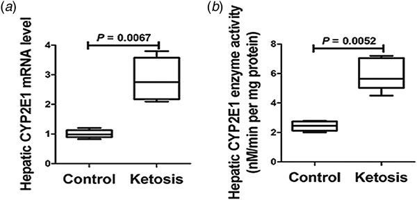
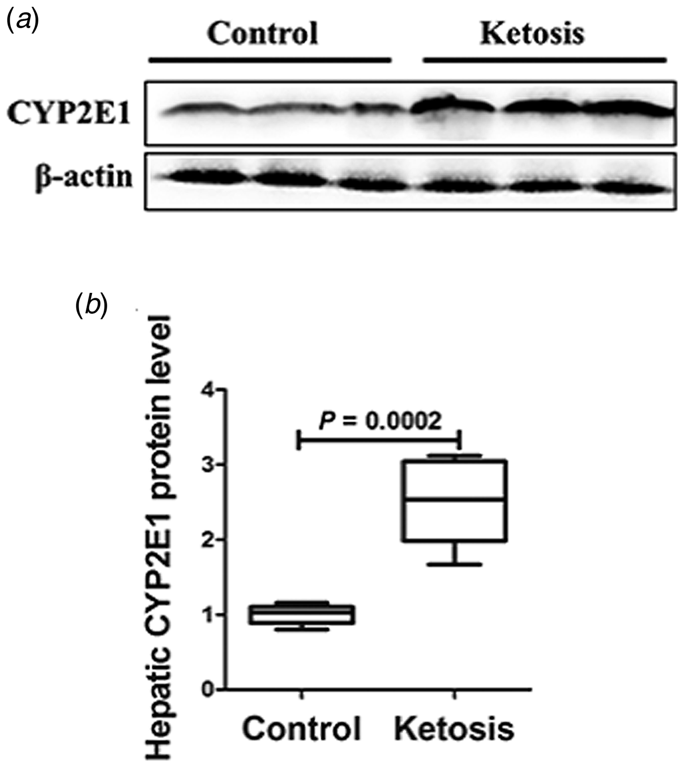
The expression and activity of hepatic CYP2E1 in cows with clinical ketosis and control cows were detected using qRT-PCR, Western blotting, and biochemical kit. The mRNA (Fig. 1a; P < 0.01) and protein expression (Fig. 2; P < 0.01) levels of CYP2E1 were significantly higher in cows with clinical ketosis than in control cows. Furthermore, the activity of CYP2E1 was also markedly augmented in cows with clinical ketosis (Fig. 1b; P < 0.01). These results indicate that the expression and activity of CYP2E1 was overinduced in clinically ketotic cows.
The hepatic mRNA expression and activity of CYP2E1 in cows with clinical ketosis. Dairy cows were divided into clinical ketosis (n = 6) and control (n = 8) groups. (a) The hepatic CYP2E1 mRNA level. (b) The hepatic CYP2E1 enzyme activity. P < 0.05 was considered to be statistically significant. The data are presented as the mean ± sem.

The hepatic protein expression of CYP2E1 in cows with clinical ketosis. Dairy cows were divided into clinical ketosis (n = 6) and control (n = 8) groups. (a) Western blotting results of CYP2E1. (b) The protein level of CYP2E1. The data are presented as the mean ± sem.

The effects of NEFA and BHB on the CYP2E1 expression and activity in cow hepatocytes
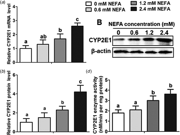
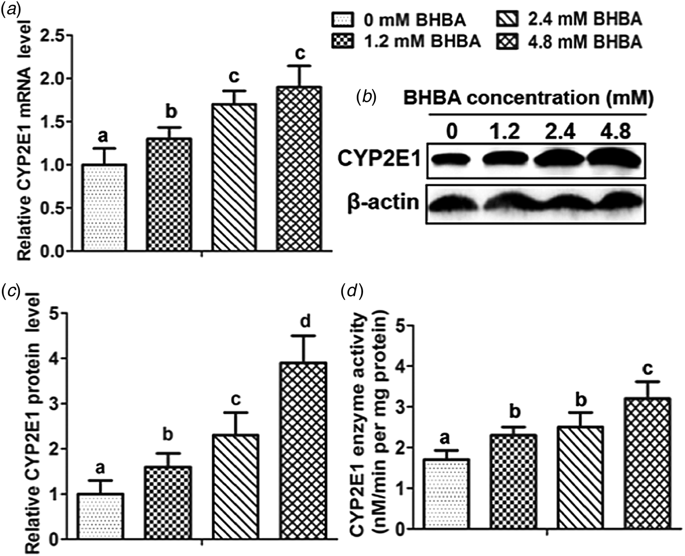
The mRNA and protein levels of CYP2E1 were increased gradually in NEFA treated hepatocytes and were significantly higher at 1.2 and 2.4 mm NEFA dose level than in control group (Fig. 3a–c; P < 0.05). Furthermore, the activity of CYP2E1 showed a similar trend and was also markedly increased in 1.2 and 2.4 mm NEFA treatment level (Fig. 3d; P < 0.05). Similarly, high levels of BHB treatment (1.2, 2.4, and 4.8 mm) also significantly increased the mRNA and protein levels and activity of CYP2E1 in cow hepatocytes (Fig. 4a–d; P < 0.05). These results show that high levels of NEFA or BHB significantly increased the expression and activity of CYP2E1 in cow hepatocytes in vitro.
The effects of NEFA on the CYP2E1 expression and activity in cow hepatocytes. Cow hepatocytes were treated with different concentrations of NEFA (0, 0.6, 1.2, or 2.4 mm). Each treatment was repeated 12 times. (a) The CYP2E1 mRNA level in cow hepatocytes; (b, c) The Western blotting results and protein level of CYP2E1 in cow hepatocytes. (d) The CYP2E1 enzyme activity in cow hepatocytes. The data are presented as the mean ± sem. a, b, and c: the same letter indicates no significant difference (P > 0.05), whereas different letters indicate a significant difference (P < 0.05).

The effects of BHB on the CYP2E1 expression and activity in cow hepatocytes. Cow hepatocytes were treated with different concentrations of BHB (0, 1.2, 2.4, or 4.8 mm). Each treatment was repeated 12 times. (a) The CYP2E1 mRNA level in cow hepatocytes; (b, c) The Western blotting results and protein level of CYP2E1 in cow hepatocytes. (d) The CYP2E1 enzyme activity in cow hepatocytes. The data are presented as the mean ± sem. a, b, and c: the same letter indicates no significant difference (P > 0.05), whereas different letters indicate a significant difference (P < 0.05).

Discussion
Cows experience a series of nutritional, physiological and behavioral changes and become more susceptible to nutritional and metabolic diseases, such as fatty liver and ketosis, during the transition period (Li et al., Reference Li, Huang, Gu, Du, Lei, Yuan, Sun, Wang, Li and Liu2015; Song et al., Reference Song, Li, Gu, Fu, Peng, Zhao, Zhang, Li, Wang, Li and Liu2016). Laboratory investigations have shown that dairy cows with ketosis exhibit low blood concentration of glucose and high blood concentrations of BHB and NEFA (Song et al., Reference Song, Li, Gu, Fu, Peng, Zhao, Zhang, Li, Wang, Li and Liu2016). In this study, we also found that blood concentration of glucose was significantly lower but NEFA and BHB concentrations were markedly higher in cows with clinical ketosis than in control cows, which indicated that fat mobilization in adipose tissue of cows with clinical ketosis was over induced.
Dairy cows with subclinical or clinical ketosis exhibit oxidative stress (Li et al., Reference Li, Ding, Wang, Feng, Li, Wang, Liu and Li2016a). CYP2E1 is a crucial molecule that mediates oxidative stress. Furthermore, Bai and Cederbaum (Reference Bai and Cederbaum2006) reported that transgenic mice overexpressing CYP2E1 had exacerbated oxidative stress (Bai and Cederbaum, Reference Bai and Cederbaum2006). In this study, we found that the hepatic mRNA, protein expression, and activity of CYP2E1 were increased and significantly higher in ketotic cows than in control cows, which indicated that the expression and activity of hepatic CYP2E1 was over-induced in ketotic cows. Considering the role of CYP2E1, over-induced CYP2E1 might be involved in the induction of oxidative stress in cows with clinical ketosis.
BHB is the main form of ketone body in dairy cows and is a result of incomplete oxidation of NEFA in liver cells. Importantly, high levels of NEFA or BHB could cause cell lipotoxicity. In vitro evidence showed that NEFA or BHBA treatment markedly increased the reactive oxygen species (ROS) and MDA contents, inhibited the activities of SOD and GSH-Px in cow hepatocytes, resulting in redox imbalance and oxidative stress-mediated apoptosis in cow hepatocytes (Song et al., Reference Song, Li, Li, Li, Shi, Ding, Zhang, Li, Liu and Wang2014, Reference Song, Li, Gu, Fu, Peng, Zhao, Zhang, Li, Wang, Li and Liu2016). These results led us to speculate that NEFA- or BHB-induced oxidative stress was partly mediated by CYP2E1. In this study, high levels of NEFA or BHB treatment significantly increased the mRNA and protein levels and activity of CYP2E1 in cow hepatocytes. Taken together, the data in vivo and vitro indicate that high levels of NEFA or BHB can over-induce hepatic expression and activity of CYP2E1, which may partly mediate the oxidative stress in cows with clinical ketosis. Overexpression of CYP2E1 resulted in ROS overproduction and induced oxidative damage (Butura et al., Reference Butura, Nilsson, Morgan, Morgan, French, Johansson, Schuppe-Koistinen and Ingelman-Sundberg2009). Excessive ROS induced lipid peroxidation, structural changes in proteins and strand breaks in nucleic acids, damaging cells and organelles by altering cell membrane integrity and function, which induced ‘second hit’ and resulted in apoptosis (Schieber and Chandel, Reference Schieber and Chandel2014). In addition, inhibitors of CYP2E1 prevented the elevation of oxidative stress, lipid peroxidation and liver pathology (Jiang et al., Reference Jiang, Wang, Xue, Wang, Jiang, Liang, Dias, Gregory, Chen and Zhang2017). Therefore, CYP2E1 may be an effective therapeutic target for addressing NEFA and BHB-induced oxidative stress in ketotic cows.
In conclusion, the expression and activity of hepatic CYP2E1 was over-induced in cows with clinical ketosis. High levels of NEFA or BHB could augment the expression and activity of CYP2E1 in cow hepatocytes, which may partially mediated the oxidative stress in cows with clinical ketosis.
Supplementary material
The supplementary material for this article can be found at https://doi.org/10.1017/S0022029919000025
Acknowledgements
This work was supported by the National Key Research and Development Program (China; grant no. 2016YFD0501206), the National Natural Science Foundation of China (China; grant nos. 31572581, 31672621, and 31772810), the Jilin Province Science Foundation for Youths (Changchun, China; grant no. 20160520063JH) and the Jilin Natural Science Foundation (Changchun, China; grant no. 20170101148JC)