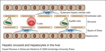
The liver, which is the largest solid organ in the body, plays a central role in the regulation of metabolic homeostasis and participates in many important immunological functions. Hepatocytes comprise about 70% of hepatic cells (Ref. Reference Racanelli and Rehermann1). The remaining 30% is made up of nonparenchymal cells, including hepatic stellate cells (HSCs), Kupffer cells, endothelial cells, cholangiocytes and several subsets of resident lymphocytes (Ref. Reference Racanelli and Rehermann1) (Fig. 1).
The hepatic sinusoid and hepatocytes in the liver. Sinusoids (distensible vascular channels) are lined with sinusoidal endothelial cells. Kupffer cells (resident liver macrophages) reside within the hepatic sinusoids. The space between the endothelium and hepatocytes is called the space of Disse. Hepatic stellate cells are present in this space.

Hepatocytes are organised into epithelial plates and are primarily responsible for metabolic functions in the liver. Between the plates are the sinusoids, which are distensible vascular channels lined with sinusoidal endothelial cells (Fig. 1). Small fenestrations in the sinusoidal linings allow direct cell-to-cell contact and free diffusion of many substances. Kupffer cells are resident liver macrophages that reside within the hepatic sinusoids and act as a first line of defence against antigens passing through the gastrointestinal barrier. Between the endothelium and hepatocytes lies the space of Disse where lymph is collected for delivery to lymphatic capillaries. HSCs are present within this space, store 80% of body vitamin A and participate in hepatic wound healing (Ref. Reference Friedman2), regulation of sinusoidal blood flow (Ref. Reference Friedman2), angiogenesis (Ref. Reference Novo3) and hepatocyte growth (Ref. Reference Ramadori4). The space of Disse also contains basement-membrane-like matrix, which is essential for the differentiated and normal functions of all the hepatic cellular compartments (Ref. Reference Friedman2).
Wound healing is the normal response of tissue to an injury, and liver fibrosis occurs as a result of repeated cycles of injury and repair. The cascade of events that establish hepatic fibrosis is complex, and is influenced by how different cell types in the liver interact in response to injury. Hepatic fibrosis and its end-stage – cirrhosis – are a major cause of mortality and morbidity worldwide. The most common causes of cirrhosis are alcohol abuse, hepatitis B, hepatitis C and, increasingly, obesity, which leads to the metabolic syndrome that can be complicated by non-alcoholic fatty liver disease (NAFLD). Other less-common causes include primary biliary cirrhosis, haemochromatosis, autoimmune hepatitis and primary sclerosing cholangitis.
Research over the past decade has greatly improved our knowledge of the cellular and molecular biology of fibrosis in the liver. Several high-profile papers have shed new light on the important roles that hepatic neuroendocrine pathways play in the processes of wound healing and regeneration in the liver. These functions are also being increasingly recognised in different organs (Refs Reference Xu5, Reference Bigliardi-Qi6, Reference Kawamoto and Matsuda7). The hepatic neuroendocrine system is upregulated in the liver following injury and can regulate the pattern of wound healing and hepatic regeneration in different ways. In this review, we will highlight some of the molecular mechanisms underlying hepatic wound healing and address recent advances in understanding the role of the neuroendocrine system in liver fibrosis. The fact that many modulators of neuroendocrine factors are already in widespread clinical use makes this field particularly exciting. There is now, more than ever, a need to generate effective antifibrotic therapies in a world where mortality from liver disease will see exponential growth as a result of the current obesity epidemic, as well as increasing burdens from alcohol abuse and viral hepatitis.
Hepatic wound healing
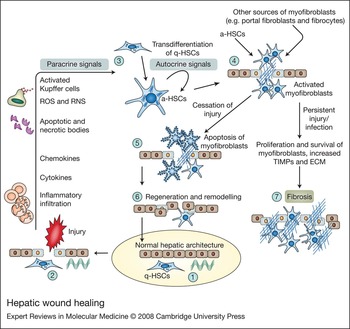
The normal resolution of tissue injury involves a series of precise orchestrated phases: (1) inflammation; (2) production of cytokines and growth factors; (3) myofibroblast activation; (4) extracellular matrix (ECM) production; (5) angiogenesis; (6) maturation; and (7) remodelling, which eventually leads to scar elimination and a return of injured tissue to the normal state (Refs Reference Wynn8, Reference Desmouliere, Chaponnier and Gabbiani9) (Fig. 2). Inappropriate tissue repair and pathological scarring occurs if any element of this intricate process becomes interrupted or overactivated. An imbalance between ECM formation and degradation can lead to the accumulation of ECM: an outcome known as fibrosis (Ref. Reference Desmouliere, Chaponnier and Gabbiani9) (Fig. 2). Fibrosis is the final general consequence of uncontrolled repair processes in many organs in response to a wide variety of chronic insults and ultimately can disable proper functioning of the organ. It is known to occur in the liver, pancreas, lungs, heart, kidneys, eyes and skin. Over the past decade, there has been extensive investigation of the molecular events underlying tissue fibrogenesis. In the following sections, we explore the mechanism of hepatic wound healing and describe the cellular compartments in which it occurs.
Hepatic wound healing. Normal hepatic structure (1) is disrupted following injury to the liver (2) when hepatocytes release factors that start an inflammatory process via recruitment of leukocytes to the site of injury. This occurs in parallel to local activation of Kupffer cells, which produce cytokines and toxic free radicals (Reactive oxygen and nitrogen species, RNS and ROS). (3) Profibrogenic cytokines and chemokines, and free radicals, as well as apoptotic and necrotic bodies, together establish a paracrine signal leading to transdifferentiation of quiescent hepatic stellate cells (q-HSCs) into activated myofibroblasts [activated hepatic stellate cells (a-HSCs)]. This step in HSC activation is known as the initiation phase, which renders the cells susceptible to cytokines, growth factors and other stimuli. Following this phase, perpetuation occurs – a phase in which the differentiated phenotype of the cell is amplified by mediators via autocrine signals. (4) In addition to a-HSCs, other cells, such as portal fibroblasts, bone-marrow-derived cells and epithelial compartments within the liver, can contribute to the activated pools of myofibroblasts during liver injury. (5) After cessation of injury, activated myofibroblasts undergo apoptosis, collagen fibres become more organised and scar tissue is removed. (6) This occurs in parallel to hepatocyte regeneration and remodelling, which restores the damaged tissue to the normal state. However, in the case of persistent injury, the normal healing process is interrupted (7) and persistent inflammation and/or infection results in chronic myofibroblast activation and proliferation, as well as excessive accumulation of extracellular matrix (ECM) components, which promotes and establishes hepatic fibrosis.

The inflammation–fibrosis pathway
Chronic persistent inflammation typically precedes fibrosis (Refs Reference Meneghin and Hogaboam10, Reference Adzick11, Reference Stramer, Mori and Martin12). Following injury to the liver, hepatocytes release factors that start an inflammatory process by recruitment of leukocytes to the site of injury. Local production of matrix metalloproteinases (MMPs) at the site of injury results in the disruption of the basement membrane and favours inflammatory cell infiltration (Ref. Reference Kumagai13). Neutrophils are the most abundant inflammatory cell in the early stages of wound healing. Their granulation is followed by macrophage infiltration and subsequently by lymphocyte recruitment (Refs Reference Wynn8, Reference Stramer, Mori and Martin12). The primary roles of leukocytes are to eliminate any invading organisms and to remove dead cells (Ref. Reference Stramer, Mori and Martin12). Inflammation also produces profibrogenic cytokines and chemokines, which activate myofibroblasts and induce wound-healing responses, which, if unchecked, drive fibrogenesis (Fig. 2). However, it has also been proposed that fibrosis is not always driven by inflammation, suggesting that the mechanisms that regulate fibrogenesis partly differ from those regulating inflammation (Ref. Reference Wynn8). This might explain the lack of efficacy of some anti-inflammatory agents in the treatment of fibrotic disease and the need to identify targeted antifibrotic therapies.
Myofibroblasts: the key players in hepatic fibrosis
In many organs, including the liver, myofibroblasts are the key cellular effectors during wound contraction and repair, and inappropriate myofibroblast activation is the central pathogenic mechanism of fibrotic disorders (Ref. Reference Iredale14). Upon tissue injury, myofibroblasts become activated and migrate toward the site of damage where they proliferate. Myofibroblasts produce ECM proteins, such as type I and type III collagen, and control ECM remodelling through the expression of several MMPs and tissue inhibitors of metalloproteinases (TIMPs). The balance between ECM degradation and production is a critical factor that determines the normal wound-healing response and returns the tissue to its preinjury state (Ref. Reference McCawley and Martrisian15). This process is influenced by the number of activated myofibroblasts at the site of injury, which is controlled by the rate of production and proliferation as well as apoptosis of these cells (Refs Reference Desmouliere, Chaponnier and Gabbiani9, Reference Elsharkawy, Oakley and Mann16). Chronic injury induces a marked alteration in the normal healing process and prevents return of tissue to the preinjury state. In fact, constant inflammation and/or infection leads to permanent myofibroblast activation, either directly, by acting on HSCs or indirectly, through paracarine-dependent factors (Fig 2). This activated phenotype of myofibroblasts promotes the inappropriate regulation of tissue repair and leads to extensive scar formation, and finally results in fibrosis (Ref. Reference Mutsaers17) (Fig. 2).
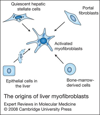
Developmentally, fibroblasts are mesenchymal in origin (Ref. Reference Parsonage18). However, it is now widely believed that myofibroblasts are derived from a number of different sources in injured adult tissues (Ref. Reference Sempowski19). In the context of the liver, there are four different cellular sources (Fig. 3). HSCs are well-characterised cells that have been shown to contribute significantly to activated myofibroblasts in various types of hepatic injury. The other potential cellular origins are bone-marrow-derived mesenchymal cells (Refs Reference Forbes20, Reference Russo21), fibrocytes (Ref. Reference Kisseleva22) and portal fibroblasts (Refs Reference Kinnman and Housset23, Reference Knittel24). Recent evidence also suggests that biliary epithelial cells and hepatocytes may contribute to the hepatic myofibroblast pool by undergoing epithelial–mesenchymal transition (EMT) (Refs Reference Zeisberg25, Reference Diaz26, Reference Robertson27, Reference Kaimori28). However, there is a need for further lineage-specific studies to explore EMT processes in liver fibrogenesis. The contribution of each cellular source to activated hepatic myofibroblasts varies according to the type of liver injury. For instance, myofibroblasts derived from portal fibroblasts – the fibroblasts surrounding the biliary tree – are important fibrogenic cells in the early stages of biliary fibrosis (Refs Reference Kinnman and Housset23, Reference Tuchweber29, Reference Tang30). In cases of rapid biliary injury, such as that seen in biliary atresia, biliary epithelial cells play an important role in biliary fibrosis via an EMT process (Ref. Reference Diaz26). It is possible that the different potential sources of fibrogenic cells in various liver diseases or different lobular regions of the diseased liver could lead to antifibrotic therapies that are specifically targeted only to certain subpopulations or diseases.
The origins of liver myofibroblasts. During the development of hepatic fibrosis, myofibroblasts are derived from a number of different sources. Hepatic stellate cells are the best characterised source. Other potential cellular origins are bone-marrow-derived mesenchymal cells, portal fibroblasts and epithelial compartments in the liver, such as cholangiocytes and hepatocytes. Biliary epithelium and hepatocytes can contribute to the activated myofibroblast pool by epithelial–mesenchymal transition.

Hepatic stellate cell transdifferentiation
After hepatic injury of any aetiology, HSCs undergo a highly regulated transdifferentiation process from quiescent cells into proliferative and fibrogenic myofibroblasts. This change in the phenotype of HSCs is also known as ‘HSC activation’ and has been divided into two phases (Ref. Reference Friedman2). First, there is an initiation phase, which renders the cells susceptible to cytokines, growth factors and other stimuli, and occurs in parallel with transcriptional alterations in early response genes. Following this, perpetuation occurs – a phase in which the differentiated phenotype of the cell is amplified by soluble and insoluble mediators (Ref. Reference Friedman2). Initiation of HSC transdifferentiation is largely due to paracrine stimulation, whereas the perpetuation of the differentiated state involves autocrine as well as paracrine loops (Fig. 2). Ultimately HSC transdifferentiation is characterised by the development of activated hepatic myofibroblasts with proliferative, contractile, migratory, fibrogenic and inflammatory properties (Ref. Reference Friedman2).
Current knowledge suggests that the initial signals driving HSC transdifferentiaion are produced by injured cells, pathogens, inflammatory mediators and alterations in the local ECM. Injured hepatocytes, Kupffer cells and other nonparenchymal cells produce necrotic cell debris, apoptotic bodies (Refs Reference Chi and Flavell31, Reference Canbay, Friedman and Gores32, Reference Watanabe33), reactive oxygen and nitrogen species (ROS and RNS) (Refs Reference Maher34, Reference Urtasun and Nieto35), as well as other active mediators, all of which signal through specific activation pathways and can be sensed by HSCs. In addition, early alterations in the mechanical stiffness of the whole tissue precede matrix deposition and can trigger transdifferentiation of HSCs (Ref. Reference Georges36).
Pattern-recognition receptors, which recognise conserved pathogen or host-associated molecular patterns, are expressed by different cells of the immune system and act as a first line of defence against injury or infection (Ref. Reference Meneghin and Hogaboam10). Recent advances in the field have identified the expression of pattern-recognition receptors such as toll-like receptor (TLR) 4 and TLR9 in hepatic myofibroblasts (Refs Reference Watanabe33, Reference Paik37). Subsequently, it has been shown that DNA from apoptotic hepatocytes, which are rich in cytosine-phosphate-guanine (CpG), can activate TLR9 in HSCs and contribute to the activated phenotype in these cells (Ref. Reference Watanabe33). This observation may explain why apoptosis of hepatocytes results in activation of HSCs and development of hepatic fibrosis (Ref. Reference Canbay38, Reference Canbay39). In addition, a recent study using TLR4 mutant mice has confirmed a role for TLR4 in liver fibrogenesis (Ref. Reference Seki40). It therefore appears that activation of several pattern-recognition receptors can act as a primary signal for HSC transdifferentiation (Ref. Reference Meneghin and Hogaboam10). In addition, HSCs have recently been shown to act as professional antigen-presenting cells (Ref. Reference Winau41). The ability of these cells to process and present antigens to immune cells can itself help to activate immune-dependent mechanisms, which subsequently influence the transdifferentiation process in HSCs. However, the impact of various immune cells on transdifferentiation of HSCs warrants further study. It is clear, therefore, that there is a network of multiple signals, as well as different cell types, that are influenced by liver injury and drive transdifferentiation in quiescent HSCs.
Maintaining the activated phenotype
Transdifferentiation of quiescent HSCs to a myofibroblast phenotype does not appear to be a transient event. Instead, the activated myofibroblasts state is maintained by paracrine signalling (e.g. ROS and RNS) from other types of liver cell, including hepatocytes, cholangiocytes and Kupffer cells (Refs Reference Friedman2, Reference Urtasun and Nieto35, Reference Thurman42). HSCs themselves develop new autocrine pathways, including those involving transforming growth factor β (TGF-β) (Refs Reference Liu43, Reference Meyer, Bachem and Gressner44), angiotensin II (Refs Reference Bataller45, Reference Bataller46), platelet-derived growth factor (PDGF) (Ref. Reference Pinzani47), monocyte chemoattractant protein 1 (MCP1) (Refs Reference Sprenger48, Reference Marra49) and vascular endothelial growth factor (VEGF) (Ref. Reference Ankoma-Sey50), which maintain the activated state of the myofibroblasts and drive fibrogenesis. One of the critical events in this regard, is that HSCs upregulate new membrane receptors and signalling proteins that prime these cells to respond to inflammatory mediators and growth factors, such as interleukin 6 (Ref. Reference Nieto51), TGF-β (Refs Reference Liu43, Reference Meyer, Bachem and Gressner44) and PDGF (Ref. Reference Pinzani47). The alteration in ECM components during this phase actively regulates HSC behaviour. Different matrix-associated molecules, such as TIMP1, integrins and other adhesion molecules, contribute to HSC survival and perpetuation of the activated myofibroblast phenotype (Ref. Reference Murphy52, Reference Zhou53). Constant signals from a stable chronic infection and/or injury through pattern-recognition receptors may also directly regulate the survival and function of HSC-derived myofibroblasts (Ref. Reference Meneghin and Hogaboam10).
MMP/TIMP production by HSCs
Activated hepatic myofibroblasts proliferate and migrate at the sites of liver injury, secreting large amounts of ECM and regulating ECM degradation. They express a combination of MMPs and their specific TIMPs. In the early phases of liver injury, HSCs transiently express MMP3 (stromelysin 1), MMP13 (collagenase 3) and uroplasminogen activator, and exhibit a matrix-degrading phenotype, all of which enables them to migrate toward the site of injury (Ref. Reference Benyon and Arthur54). In the later stages of liver injury and HSC activation, the expression pattern changes, and the cells express a combination of MMPs, which have the ability to degrade normal liver matrix while inhibiting degradation of the fibrillar collagens that accumulate in liver fibrosis. This new expression pattern is characterised by the combination of pro-MMP2 and membrane type 1 (MT1) MMP expression, which drives pericellular generation of active MMP2 and local degradation of subendothelial matrix, facilitating replacement with high-density interstitial matrix (Ref. Reference Benyon and Arthur54). In addition, there is a marked increase in the expression of TIMP1, leading to a more global inhibition of degradation of fibrillar liver collagens by interstitial collagenases (MMP1/MMP13). As a consequence, normal matrix homeostasis is severely disrupted in favour of the net deposition of fibrillar collagen, non-collagen ECM molecules and integrin ligands.
Neuroendocrine differentiation and cancer: implications for wound healing
Neuroendocrine phenotype
Neuroendocrine cells are scattered in various organs of the body and have endocrine phenotypes that share some of the structural, functional and metabolic properties of neurons. Advances in molecular pathology have led to the identification of several markers in these cells. Neuroendocrine cells typically contain secretory granules, called large dense-core vesicles because of their characteristic appearance upon electron microscopy. In addition to peptides, these granules also contain one or more chromogranin/secretogranin proteins. They express markers such as chromogranin A, glycolipid A2-B4, S-100 protein, neural cell adhesion molecule, neuron-specific enolase and synaptophysin (Refs Reference Alvaro55, Reference Roskams56, Reference Roskams57). These markers have different specificities and sensitivities in identifying the neuroendocrine phenotype of a cell; many participate in various intracellular excretory activities of cells, such as packaging of hormones and neuropeptides, or modulation of exocytosis and neurotransmitter release in synapses.
Parathyroid-hormone-related peptide was previously identified as the factor responsible for the syndrome of humoral hypercalcaemia of malignancy and is also a classical neuroendocrine peptide that is involved in growth, differentiation and angiogenesis in various tissues (Ref. Reference Roskams and Desmet58). In addition, a wide range of neuropeptides are produced by neuroendocrine cells or tissues, including serotonin, neurotrophins, endogenous opioid peptides, somatostatin, cannabinoids and calcitonin (Refs Reference Alvaro55, Reference Roskams57, Reference Marzioni59, Reference Mosca60, Reference Marzioni, Fava and Benedetti61). These factors commonly regulate growth and survival in various cells or tissues.
Little is known of the functional role of neuroendocrine cells in many organs. It has been suggested that they probably serve a paracrine or local regulatory role. Neuroendocrine differentiation has been found in a subgroup of various carcinomas, including prostate, breast, stomach, colorectal and non-small-cell lung cancer (Refs Reference Bosman62, Reference di Sant'Agnese and Cockett63, Reference Amorino and Parsons64, Reference Walker65, Reference Nesland66, Reference Eren, Celikel and Güllüoğlu67, Reference Atasoy68). In many of these tumours, neuroendocrine differentiation has adverse prognostic effects, suggesting an integral role for neuroendocrine factors in the regulation of the malignant phenotype (Refs Reference Bosman62, Reference di Sant'Agnese and Cockett63, Reference Amorino and Parsons64). The detailed molecular mechanisms underlying the observed behaviour are mainly undefined but the neuroendocrine cells of the tumour probably play a significant role during tumour growth, angiogenesis and metastasis (Refs Reference Bosman62, Reference Amorino and Parsons64, Reference DeMorrow69, Reference Chang70). The histogenesis and origins of neuroendocrine cells in the tumour environment are not clear. However, it may involve a local transdifferentiation process (Ref. Reference Bosman62).
Cancers have been described as wounds that do not heal (Ref. Reference Dvorak71), suggesting that both tumours and wounds may be part of a continuum. Indeed, there are similarities between tumour stroma generation and wound healing. This includes various features that can regulate growth, differentiation or angiogenesis in both the wound and tumour environment. In fact, persistent injuring stimuli result in inflammation and continuous wound healing – two mechanisms that lead to accentuated scar tissue formation or fibrosis and have frequently been linked to cancer formation (Refs Reference Dvorak71, Reference Lin and Karin72). We have recently coined the phrase ‘hepatic inflammation–fibrosis–cancer axis’ to reflect this phenomenon (Ref. Reference Elsharkawy and Mann73).
During chronic hepatic injury, different types of liver cells acquire a neuroendocrine phenotype. Given the putative role of the neuroendocrine system in the regulation of growth and survival in various conditions, and the shared similarities between tumours and wounds, the neuroendocrine compartment of the inflamed liver might be expected to regulate cell growth, migration and angiogenesis during wound healing. In fact, there is increasing evidence that local neuroendocrine factors do indeed influence wound healing and fibrogenesis in many organs (Refs Reference Xu5, Reference Bigliardi-Qi6, Reference Ruddell74, Reference Ebrahimkhani75, Reference Yang76, Reference Micera77).
Neuroendocrine compartments in the chronic-injured liver
In the diseased liver, atypically proliferating cholangiocytes acquire a neuroendocrine phenotype (Ref. Reference Alvaro55). These cells, also known as reactive bile ductules, have been identified as one of the major contributors to the production of various neuroendocrine factors in diverse liver pathologies, and appear at interface zones where maximal cell death and inflammation occurs (Ref. Reference Alvaro55). Proliferation of cholangiocytes precedes the development of cirrhosis in most liver diseases, including chronic cholestasis and biliary cirrhosis, alcoholic liver disease and chronic toxic liver injuries. This cholangiocyte component of the local neuroendocrine system in the injured liver has been widely studied in recent years (Refs Reference Alvaro55, Reference Roskams57, Reference Marzioni59, Reference Marzioni, Fava and Benedetti61).
Hepatic progenitor cells, also known as liver oval cells, lie within or immediately adjacent to the canal of Herring (Ref. Reference Libbrecht and Roskams78) and express neuroendocrine proteins, such as chromogranin-A, neural-cell-adhesion molecule, parathyroid-hormone-related peptide, S-100 protein, neurotrophins and neurotrophin receptors (Refs Reference Roskams56, Reference Roskams57, Reference Roskams79). These cells are activated to proliferate upon chronic liver damage in situations where the proliferation of hepatocytes is inhibited, which includes non-alcoholic steatohepatitis, chronic cholestatic liver disease, chronic alcoholic hepatitis and viral hepatitis. Progenitor cells differentiate into hepatocytes and contribute to the pool of reactive bile ductules during the course of liver disease. In addition to progenitor cells and reactive bile ductules, small hepatocytes in periportal regions express chromogranin A (Ref. Reference Roskams79). These newly formed intermediate hepatocytes are observed in chronic conditions where there is activation of progenitor components (Ref. Reference Roskams79).
HSCs, the main players in the liver wound-healing process, share a number of different markers with neuroendocrine cells and the cells of the nervous system (Ref. Reference Geerts80). They express synaptophysin, which is one of the factors primarily correlated with neuroendocrine differentiation (Refs Reference Roskams56, Reference Cassiman81, Reference Schurmann82). HSCs also express neutrophins and neural cell adhesion molecules (Ref. Reference Cassiman83). Generation of neuroendocrine factors by HSCs may help to maintain the activated myofibroblast phenotype through autocrine loops. However, what is potentially more central to the process is that upon transdifferentiation HSCs upregulate many receptors of neuroendocrine factors, which renders them susceptible to neuroendocrine regulation in wound healing. This issue will be discussed in more detail below.
Which factors trigger neuroendocrine differentiation in the injured liver?
Neuroendocrine differentiation in the liver is often associated with the presence of cellular stress and inflammation (Refs Reference Alvaro55, Reference Haley84). For instance, ductular reactions in cholangiocytes are usually accompanied by periductal inflammation, which is presumed to trigger the reaction (Refs Reference Alvaro55, Reference Alvaro, Gigliozzi and Attili85, Reference Lazaridis, Strazzabosco and LaRusso86). The possible role of hepatic inflammation in the development of neuroendocrine differentiation is also supported by studies of cancer (Refs Reference Chang70, Reference Haley84). It has been shown that interleukin 6 (IL-6) and tumour necrosis factor (TNF) α – two important cytokines in the liver – regulate both growth and neuroendocrine differentiation in different types of experimental cells, as well as human cancer cells (Refs Reference Chang70, Reference Haley84). Several recent in vitro studies with cancer cells have demonstrated that IL-6 can promote neuroendocrine differentiation through different intracellular signal-transduction pathways, including the phosphatidylinositol 3-kinase, signal transducers and activators of transcription 3 (STAT3), and mitogen-activated protein kinase (MAPK) pathways (Refs Reference Chang70, Reference Qiu, Ravi and Kung87, Reference Qiu88). However, there is clearly a need to study how much of this data can be extrapolated to liver inflammation and fibrosis.
In addition, signals derived from the ECM can also influence neuroendocrine functions in bile duct epithelial cells. It has been reported that interaction of these cells with the surrounding matrix – in particular collagen type IV and matrix heparan sulfate proteoglycan perlecan – induces neuroendocrine differentiation (Ref. Reference Roskams56). This is consistent with the fact that reactive bile ductules are always surrounded by basement membrane containing these ECM components. Therefore, the neuroendocrine phenotype in cholangiocytes and other cellular populations in the liver can be regulated by signals from both the matrix and inflammatory factors. However, the specific cellular and molecular players that promote neuroendocrine differentiation in the liver remain to be identified.
Neuroendocrine regulation of myofibroblast differentiation and wound healing
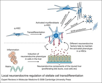
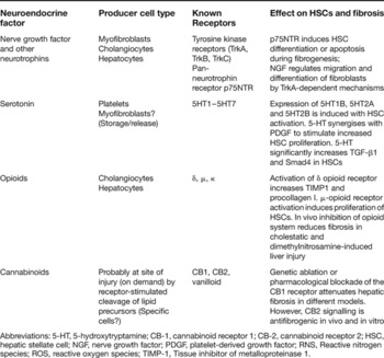
As discussed earlier, transdifferentiation of HSCs into activated myofibroblasts is one of the pivotal events during wound healing and fibrogenesis in the injured liver, and can be subdivided into an initiation phase and a perpetuation phase. After liver injury, inflammatory cells may directly contribute to this transdifferentiation; but they are not likely to provide all the growth factors necessary for maintaining or accentuating the activated phenotype at later stages. However, induction of neuroendocrine differentiation in cholangiocytes and oval cells in response to inflammation and/or other mechanisms can contribute to maintenance of the activated state of HSCs. This may occur in parallel to the expression of various neuroendocrine receptors during HSC differentiation, which renders these cells responsive to regulation (Fig. 4). Many of these factors can further affect hepatic wound healing by influencing neoangiogenesis, inflammation and hepatocyte regeneration. It should also be considered that HSCs have their own neuroendocrine features that may contribute to their fibrogenic activities via autocrine mechanisms. Here we discuss some of the recently identified neuroendocrine factors that regulate the production and function of HSC-derived myofibroblasts in the liver (Table 1). It is interesting that many medications that could potentially modulate the effect of neuroendocrine factors and prevent fibrogenesis are already in routine clinical practice in other contexts and have established safety records, making them very attractive candidates for rapid translation into clinical trials. However, it is important to stress that the regulation of HSC biology by these factors has not yet been fully defined and may be more complex than initially anticipated. Furthermore, there is no evidence that neuroendocrine factors regulate the functions of HSC-derived myofibroblasts exclusively. In fact, the role of these factors in the differentiation of other cellular sources of liver myofibroblasts is possible and warrants further study.
Local neuroendocrine regulation of stellate cell transdifferentiation. Normal hepatic structure is disrupted following injury to the liver, subsequently, quiescent stellate cells (q-HSCs) transdifferentiate into activated myofibroblasts (a-HSCs). At the same time, inflammation and altered matrix components induce neuroendocrine differentiation in proliferating bile ducts, oval cells and even HSCs. These cells produce various neuropeptides – endocannabinoids, serotonin, opioids, neutrophins – which contribute to maintenance and amplification of the differentiated phenotype in activated myofibroblasts. Neuroendocrine factors contribute to various aspects of profibrogenic activity in activated myofibroblasts, such as contraction, migration, proliferation and extracellular matrix production.

Local neuroendocrine regulation of hepatic fibrosis

Abbreviations: 5-HT, 5-hydroxytryptamine; CB-1, cannabinoid receptor 1; CB-2, cannabinoid receptor 2; HSC, hepatic stellate cell; NGF, nerve growth factor; PDGF, platelet-derived growth factor; RNS, Reactive nitrogen species; ROS, reactive oxygen species; TIMP-1, Tissue inhibitor of metalloproteinase 1.
Neutrophins and their receptors
Neutrophins were originally reported to regulate growth and development in the nervous system. A rapidly growing body of evidence now suggests that neutrophins also play profound roles in various non-neuronal tissues. The neurotrophin family consists of nerve growth factor (NGF), brain-derived neurotrophin (BDNF), neurotrophin 3 and neurotrophin 4/5. The neutrophin receptors are high-affinity neurotrophin tyrosine kinase receptors TrkA, TrkB and TrkC, as well as the ‘low’-affinity pan-neurotrophin receptor p75NTR, which belongs to the TNF-receptor superfamily (Table 1). Activated myofibroblasts, cholangiocytes and hepatocytes secrete NGF and express the receptors for different neutrophins (Refs Reference Cassiman83, Reference Gigliozzi89, Reference Oakley90). A recent study has shown that HSCs derived from p75-knockout mice showed significantly reduced differentiation into activated myofibroblasts, as well as reductions in the protein expression of smooth muscle actin and collagen I (Ref. Reference Passino91). This effect was ameliorated by transfection with the intracellular portion of p75, demonstrating a critical role for this receptor in HSC activation. Interestingly, an exon-3 knockout of p75, which retains an intact intracellular domain but lacks the structural requirements for interaction with its ligand, supports the activation of HSCs and the development of liver fibrosis in a predictable manner after carbon tetrachloride (CCl4) intoxication (Iredale, J.P. and Kendall, T.J., University of Edinburgh, UK, pers. commun.). It has been suggested that p75 signalling through Rho promotes HSC differentiation into active myofibroblasts (Ref. Reference Passino91). However, the regulation of HSCs by neutrophin receptors appears to be more complicated and warrants additional research. NGF induces apoptosis in activated HSCs via a p75-dependent mechanism (Ref. Reference Oakley90). p75NTR may therefore function not only as a regulator of HSC transdifferentiation but may also limit the fibrogenic response by exerting a negative influence on the life span of the activated myofibroblasts. A similar regulatory system has been observed in fibroblast–myofibroblast differentiation in other situations. Quiescent fibroblasts express TrkA constitutively, whereas myofibroblats express both TrkA and p75, and all produce NGF (Refs Reference Micera92, Reference Micera93). NGF regulates the migration and differentiation of fibroblasts by a TrkA-dependent mechanism, and it also induces apoptosis of activated myofibroblats via the p75 receptor. Thus, it appears that different temporal expression patterns of TrkA/p75 might regulate the final pathological process (Ref. Reference Micera93). Together, these data suggest an important role for neutrophins and their receptors during liver wound healing.
The serotonin system
Serotonin or 5-hydroxytryptamine (5-HT) has been recognised for more than 50 years as an effector on various types of smooth muscle cell and subsequently as an agent that enhances platelet aggregation and as a neurotransmitter in the nervous system. Despite the critical role of serotonergic mechanisms in the central nervous system, the brain actually contains very little serotonin in relative terms. About 95% of serotonin is produced in enterochromaffin cells throughout the gut. Enterochromaffin cells produce far more serotonin than is required by the gut and it overflows into the blood, where it is then taken up and concentrated in platelets, the only source of blood serotonin. After its release by platelets, serotonin is absorbed rapidly by various cell types via the specific membrane-bound serotonin transporter (Ref. Reference Gershon and Tack94).
Molecular cloning has revealed an unexpected diversity of receptor subtypes in serotonin signalling (5HT1–5HT7), which are coupled to different, but overlapping, transmembrane-signalling mechanisms (Table 1). The expression of serotonin receptors has been reported in hepatocytes, and the authors suggested a key role for serotonin in liver regeneration (Ref. Reference Lesurtel95). Although platelets are the main source of serotonin in the injured liver, proliferating cholangiocytes and HSCs can also secrete serotonin (Ref. Reference Marzioni59, Reference Ruddell74). Rat and human HSCs express the 5HT1B, 5HT1F 5HT2A 5HT2B and 5HT7 receptors, with expression of 5HT1B, 5HT2A and 5HT2B receptors induced upon HSC activation (Ref. Reference Ruddell74). Serotonin significantly increases the expression of TGF-β1 and Smad4 in HSCs (Ref. Reference Li96) and synergises with PDGF to stimulate increased HSC proliferation (Ref. Reference Ruddell74). In addition, HSCs express a functional serotonin transporter (SERT) and can potentially regulate the concentration of serotonin in the vicinity of injured liver cells (Ref. Reference Ruddell74). Therefore, serotonin may actively regulate hepatic fibrogenesis.
Endogenous opioid peptides
Opiates have been known for decades for their role in pain management. They have been shown to be produced endogenously in the body and to regulate cell growth, differentiation and survival in neuronal and non-neuronal cells. Endogenous opioid peptides act by interacting with three classical opioid receptors: the μ, δ and κ receptors (Table 1). Three endogenous families of classical opioid peptides have been identified in the body: the enkephalins, endorphins and dynorphins. Each family is derived from a distinct precursor polypeptide and has a characteristic anatomical distribution (Ref. Reference Mani97).
Studies from the 1980s in patients with primary biliary cirrhosis represent the earliest evidence of a correlation between endogenous opioids and liver disease (Ref. Reference Thornton and Losowsky98). It has been demonstrated that bile duct epithelium and hepatocytes express preproenkephalin mRNA following injury, which encodes Met- and Leu-enkephalin peptides (Refs Reference Bergasa99, Reference Swain100). Recent studies on the role of opioids in the pathophysiology of chronic liver injury demonstrate potentially novel targets for antifibrotic therapies (Refs Reference Ebrahimkhani75, Reference Mani97, Reference Marzioni101). Recently, we identified δ-opioid- receptor expression following transdifferentiation of HSCs. Activation of this receptor by different δ-opioid agonists increases expression of the genes encoding TIMP1 and procollagen I and inhibits apoptosis of activated myofibroblasts (Ref. Reference Ebrahimkhani75). Subsequent work has demonstrated that stimulation of opioid receptors activates calcium-dependent protein kinase C, extracellular-signal-regulated kinase, phosphatidylinositol 3-kinase (PKC–ERK–PI3K) signalling, which then mediates the effect of endogenous opioids on HSC proliferation and collagen synthesis. Furthermore, in vivo inhibition of the opioid system in a model of chronic cholestatic liver disease and in dimethylnitrosamine (DMN)-induced liver injury significantly prevented the development of hepatic fibrosis (Ref. Reference Ebrahimkhani75, Reference De Minicis102). It has been suggested that during chronic liver injury, HSC-derived myofibroblasts express opioid receptors rendering them susceptible to opioid peptides produced by epithelial compartments in the liver, which can then contribute to maintenance of the activated phenotype in these cells.
Endocannabinoids
The role of the endogenous cannabinoid system in the pathophysiology of liver disease has attracted a great deal of recent attention. Endogenous cannabinoids (endocannabinoids) are lipid mediators that include amides and esters of long-chain polyunsaturated fatty acids. Two arachidonic acid derivatives, arachidonoylethanolamide (anandamide) and 2-arachidonoylglycerol (2-AG) are the most biologically active endocannabinoids described so far (Ref. Reference Centonze103). These molecules bind to two G-protein-coupled receptors, CB1 and CB2, although other targets, such as vanilloid receptors have been identified (Table 1). Unlike classical neurotransmitters and neuropeptides, endocannabinoids are not stored in intracellular compartments but are produced ‘on demand’ by receptor-stimulated cleavage of lipid precursors (Ref. Reference Woods104).
Following transdifferentiation, HSCs express both CB1 and CB2 receptors (Ref. Reference Siegmund105). It has been reported that CB2 signalling in HSCs is antifibrogenic (Ref. Reference Julien106) and that stimulation of activated HSCs by anandamide or 2-AG provokes cell death by redox-sensitive mechanisms, not via cannabinoid receptors (Ref. Reference Siegmund105, Reference Siegmund107). However, epidemiological studies have demonstrated that daily cannabis smoking is an independent risk factor for rapid progression of fibrosis in chronic hepatitis C patients (Ref. Reference Hézode108). A further study, carried out in three mouse models of hepatic fibrogenesis, identified that genetic ablation or pharmacological blockade of the CB1 receptor significantly attenuated development of hepatic fibrosis (Ref. Reference Teixeira-Clerc109). This inhibition of CB1 receptor signalling was accompanied by decreased expression of TGF-β – an important profibrogenic cytokine in the injured liver – and occurred in parallel to the reduced proliferation and increased apoptosis of activated myofibroblasts in vivo and in vitro. Taken together, these studies suggest that CB1 signalling is the dominant pathway in hepatic fibrogenesis in response to factors that can activate both cannabinoid receptors (Ref. Reference Kunos110) and propose an intriguing role for this system in the regulation of hepatic wound healing. Antagonism of the CB1 receptor, along with potentiation of the CB2 receptor, may prove to be an effective antifibrotic therapy in the future. This may be realised soon, with clinical trials of the CB1 antagonist rimonabant already under way in the study of obesity.
Future perspectives and therapeutic potentials
The role of the neuroendocrine system in liver disease is an important emerging theme that has already shed new light on the molecular mechanisms that regulate hepatic wound healing and fibrogenesis. However, much remains to be learned before this new information can be exploited for therapies that promote liver regeneration and limit fibrosis. In particular, the presence of different receptors with counteracting signalling events means that fibrosis of the liver is a far from simple event. For instance, serotonin induces profibrogenic behaviour in HSCs, while at the same time accelerating hepatocyte regeneration and inhibiting cholangiocyte proliferation (Refs Reference Ruddell74, Reference Lesurtel95). An ideal strategy to inhibit fibrogenesis would be to inactivate the profibrogenic serotonin receptors and simultaneously stimulate those serotonin receptors that promote hepatocyte regeneration. However, the identities of the receptors responsible for mediating the fibrogenic and regenerative properties of serotonin are unknown. At different stages in the time course of liver disease, the distribution of neuroendocrine receptors can vary from one cell type to another in a dynamic fashion. For example, with bile duct ligation, δ-opioid-receptor expression gradually diminishes in cholangiocytes (Ref. Reference Marzioni111). By contrast, the same receptor is not expressed in quiescent HSCs but is expressed following transdifferentiation in response to cholestasis (Ref. Reference Ruddell74). Such opposite trends for expression of δ-opioid receptor in these two liver cell types may account for the heterogenic effects of endogenous opioid peptides on cholangiocytes and HSCs (Ref. Reference Marzioni111). Therefore, inhibition of the same signalling event at different time points in liver disease may result in distinct outcomes.
In future, we need to further dissect the expression and function of different neuroendocrine factors and the related receptors in each cell population of the liver with precise molecular biology approaches. For example, the increasing availability of cell-specific knockouts of different serotonin receptors will enable a careful dissection of the role of the various receptors in vivo. Furthermore, there is an urgent need to confirm the results in human cells and both normal and diseased human tissues.
Serotonin antagonists and reuptake inhibitors are used routinely for the management of psychiatric disorders such as depression. Naltrexone (a nonselective opioid-receptor blocker) is clinically administered for the treatment of cholestatic pruritis in primary biliary cirrhosis and also in the management of addiction and alcohol dependence (Refs Reference Oo and Neuberger112, Reference Johnson113). CB1 antagonists are currently under trial for the treatment of obesity (Ref. Reference Lillo114). The antagonist used in a recent study on the role of CB1 in liver fibrosis in mice is SR141716A (rimonabant) (Ref. Reference Hézode108); this drug is currently an available therapeutic option. Although there have been no studies on the role of these drugs in the progression of human liver disease, their availability in clinical practice makes it likely that clinical trials of these agents in chronic fibrogenic hepatic disease will not be far off.
Portal hypertension, variceal bleeding and systemic arterial vasodilatation are among the main reasons for morbidity and mortality in cirrhotic patients. It has been demonstrated that some of the neuroendocrine factors, such as endogenous cannabinoids and opioids, contribute to the systemic vasodilatory state seen in cirrhosis (Ref. Reference Iwakiri and Groszmann115). Therefore, modulation of these systems can potentially extend the life of cirrhotic individuals, not only by slowing or reversing fibrogenesis, but also by improving the associated hyperdynamic circulatory state. CB1 receptor activation has been linked to obesity and energy homeostasis, and its inhibition can exert enhanced antifibrotic effects by modulation of metabolic risk factors (Ref. Reference Gary-Bobo116). An important study has shown that serotonin plays a role in the pathogenesis of steatohepatitis, and therefore might represent a novel target for the prevention and treatment of nonalcoholic steatohepatitis (NASH) and its associated fibrosis (Ref. Reference Nocito117). Opioid-receptor blockade by naltrexone is an approved strategy for treatment of alcohol dependence by reducing alcohol craving and relapse in heavy drinkers, and it may also prove to be a more effective antifibrotic candidate in alcoholic patients.
Conclusion
It is clear that local neuroendocrine systems play a critical role in the development of hepatic fibrosis. Various modulators of different components of this system are available clinically and can be potentially useful in patients with liver disease. However, to achieve further successful therapeutic goals through neuroendocrine targeting in hepatic fibrosis, we need to consider the timing of treatment in the course of disease and the duration of the therapy, as well as cell- and receptor-specific targeting. In addition, appropriate pharmacokinetics and safety margins of the drugs should be considered when treating patients with liver disease.
Acknowledgements and funding
We recognise many outstanding published contributions of investigators in the field that could not be included in this review owing to space limitations. We would like to express our thanks to the peer reviewers of the article. Funding in the authors’ laboratory is provided by the Wellcome Trust, National Institute on Alcohol Abuse and Alcoholism (NIAAA), European Association for the Study of the Liver (EASL), Medical Research Council (MRC) and British Liver Trust. M.R.E. is an EASL Sheila Sherlock Research Fellow and A.M.E. is a Wellcome Trust Clinical Research Fellow.