Introduction
Foot-and-mouth disease (FMD) is an acute febrile, vesicular, highly contagious infectious disease of domestic and wild cloven-hooved animals and is caused by the Foot-and-mouth disease virus (FMDV). As one of the most economically devastating diseases of domestic livestock, FMD remains enzootic in many regions of the world, posing a serious threat to animal production and economics, and greatly limiting the international trade in animals and animal products. Specifically, FMDV occurs as seven immunologically distinct serotypes (O, A, C, Asia I, SAT1, SAT2 and SAT3), with numerous subtypes of each serotype reflecting significant genetic variability. There are no cross-reactions among the seven serotypes, and no protection among different subtypes within the same serotype. These multiple serotypes contribute to the difficulties in conducting laboratory diagnoses and control in the field. As a result, FMD is listed as one of the animal diseases legally reported by the Office International des Epizooties (OIE), also known as the World Organisation for Animal Health.
In recent years, FMD has remained endemic in many regions of Asia and Europe, causing significant economic losses. Owing to the complicated antigenic structure of FMDV, FMDV, in nature, is antigenically diverse. The high variation in FMDV makes it difficult to eliminate in cattle and sheep. Vaccinated or susceptible animals, such as sheep, goats and cattle, that are infected with FMDV usually go on to become carriers (Huang et al., Reference Huang, Lee and Tu2003). In FMD endemic areas, carrier animals are frequently found. If exposed to the infectious virus, vaccinated or asymptomatic convalescent animals can cause an outbreak of FMD, so it is presumed that carrier animals are likely to play a role in the spread of FMDV and pose a particular threat to susceptible contact animals, although this has yet to be proven. The mechanisms of persistent FMDV infection are unknown, although it has been suggested that virus gene mutation leading to antigenic variation is one of the key factors (Gebauer et al., 1988; Diea et al., 1990; Martin Hernandez et al., 1994). Research has revealed that A serotype FMDV shows significant antigenic variation, this being particularly the case in the A serotype strain prevalent in South American countries (Weddel et al., Reference Weddel, Yansura and Dowbenko1985).
FMDV is a positive-sense single-stranded RNA virus that belongs to the Aphthovirus genus of the Picornaviridae family. As in all highly unstable RNA virus genomes, FMDV genomes show great potential for variation and mutation, in particular in the structure protein VP1, capable of inducing neutralizing antibodies. The two regions of VP1 showing the highest mobility correspond to (1) the G–H loop around residues 141–161, which is the major antigenic site of the virus and (2) the C-terminal region comprising residues 200–213, which is a minor viral antigenic site. The nonstructural proteins 3AB and 3B of FMDV have hydrophobic units and the virus anchors in the biomembrane, leading to viral RNA synthesis. 3A is a highly conservative peptide composed of 153 amino acids, besides its carboxyl terminus variable. It was shown that 3A gene absence might be related to host range (Knowles et al., 2001), for example in swine. Following initial modification of VPg to VpgpU or VpgpUpU, 3B(VPg) acts as the primer for RNA synthesis inside a membrane-associated replication complex. The FMDV 3C protease is responsible for most of the cleavages within the polyprotein coding sequence. In addition to cleaving the viral polyprotein, it has been shown that FMDV 3C modifies certain cellular proteins. Histone H3 has been shown to be cleaved by this protease (Falk et al., 1990). It has been found that 3C protease also cleaves the translation initiation factors eIF4A and eIF4GI within FMDV-infected cells (Belsham et al., 2000). A decrease in FMDV 3C protease expression was likely associated with persistent viral infection or latent infection (Zhang, Reference Zhang2002). The overall objective of this study was to find the potential relationship between persistent FMDV infection and virus VP1 and 3ABC gene variation in order to elucidate the mechanism of persistent FMDV infection. Chinese yellow cattle were artificially infected with FMDV O/Akesu/58 strain to develop persistent infection models and obtain isolates, using reverse-transcriptase polymerase chain reaction (RT-PCR) to amplify and analyse the VP1 and 3ABC genes.
Materials and methods
Virus strains and experimental animals
FMDV O/Akesu/58 strain was isolated from infected bovine tongue epithelium in Xinjiang Uygur Autonomous Region in 1958, identified as the O serotype, and kept in the Department of Virology, Lanzhou Veterinary Research Institute, Chinese Academy of Agricultural Sciences (CAAS). The sequences of nine reference strains were downloaded from GenBank: Asia1/YNBS/58 (AY390432), Asia1/IND63/72 (AY304994), O/OMIII (AY359854), O/Campos/Brazil/58 (AJ320488), O/China/99 (AF506822), O/Akesu/58 (AF511039), O/Yunlin/Taiwan/97 (AF308157), O/JAP/2000 (AB079061) and A22/Azerbaijan/USSR/65 (X74812). FMDV O/OA58-BHK/2004 strain (passaged 8 to 10) BHK-21 cells, inoculated with the O/Akesu/58 strain adapted to proliferate, and its genome sequence, were obtained by our laboratory. The experimental animals were five 2-year-old Chinese yellow cattle from the FMD-free region of Gansu Province. After being isolated for two weeks, the animals' serum and oesophageal–pharyngeal fluid (O/P fluid) were collected and the samples analysed by RT-PCR and liquid block phase-ELISA (LBP-ELISA) assays. The five cattle were confirmed to be FMDV-negative.
Chinese yellow cattle, persistently infected FMDV and its isolates
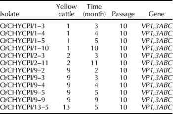
The cattle were inoculated intradermally on the tongues with 1.0×10⁴ ID50/ml O/Akesu/58 strain. A week after first displaying visible clinical signs, they developed into asymptomatic convalescent animals. O/P fluid samples were then collected (using a probang) from the five yellow cattle every month. The O/P fluids were treated and inoculated in baby hamster kidney primary cells (BHK-21) to proliferate. The BHK-21 cells with typical cytopathic effect (CPE) were collected and kept at −20°C. After centrifugation, the cell supernatants were used for RNA extraction. The 12 isolates from the persistently infected cattle were recorded every month (Table 1).
The cattle persistent infection isolates at different times

Primer design
The primers for reverse transcription (RT) and polymerase chain reaction (PCR) were designed according to the FMDV genomic sequences reported previously. The primers were synthesized by Takara Biotechnology Co., Ltd (Dalian, China).
VP31(+): 5′-CACAAATGTACAGGGATGGGT-3′
NK61(−): 5′-GACATGTCCTCCTGCATCTG -3′
201(+): 5′-ACGAGAAAGTGTCGAGTCACC-3′
205(−): 5′-TGTACGCGCTCTTCAACATCTC-3′
Viral RNA extraction and RT-PCR
The VP1 and 3ABC genes of the 12 isolates and O/OA58-BHK/2004 were sequenced and analysed. The cells were frozen and thawed repeatedly; after centrifugation, the cell supernatants were used for RNA extraction according to RNeasy Mini Kit (QIAGEN, Germany) procedures. Complementary DNA (cDNA) synthesis of VP1 and 3ABC from extracted RNA was performed using downstream primers NK61 and 205, respectively, and Avian myeloblastosis virus reverse transcriptase (Takara). In brief, a 20-μl reaction mixture contained 4 μl 5×AMV buffer, 4 μl dNTPs (2.5 nmol/l), 1 μl reverse transcription AMV (5 U/μl), 1 μl downstream primers (50 μmol/l) and 1 μl RNA template. The mixture was incubated for 10 min at 70°C to denature the RNA, then incubated for 60 min at 42°C. The first-strand cDNA was then subjected to PCR amplification using primer pairs NK61 and VP31, 201 and 205 for VP1 and 3ABC, respectively. The 50-μl reaction volume contained 5 μl 10×EX Taq-buffer, 4 μl dNTPs (2.5 mmol/l), 1 μl forward and reverse primers, respectively, 1 μl EX Taq DNA polymerase (5 U/μl, Takara), 3 μl cDNA template and 35 μl sterile double-distilled H2O. The amplification programme for VP1 was 30 cycles at 94°C for 60 s, 54°C for 60 s, 72°C for 90 s, then a final phase of 72°C for 8 min. The amplification programme for 3ABC was 35 cycles at 94°C for 60 s, 56°C for 90 s, 72°C for 2 min, then a final phase of 72°C for 10 min. PCR products were electrophoresed on 1.0% (w/v) agarose gels and purified using an agarose gel DNA extraction kit (Takara). The PCR products were purified and cloned into the pGEM-T easy vector (Promega, Madison, WI, USA), then transformed into Escherichia coli (JM109 or DH5α). The recombinant plasmids were extracted using the MiniBEST Plasmid Purification Kit (Takara) and identified by electrophoresis, EcoR I enzyme cleavage and PCR.
Sequencing and analysis
The identified correct FMDV subgenomic fragments of VP1 and 3ABC were subjected to sequencing by Takara Biotechnology (Dalian) Co., Ltd. Analysis of the sequence identity, divergence and phylogenetic relationship was carried out using DNASTAR (www.dnastar.com) and CLUSTALXL (www.dnastar.com), and compared with the parental infection strain O/Akesu/58 and O/OA58-BHK/2004. The neighbouring-joining (NJ) tree was constructed based on the result of alignment by MEGA 2.1.
Results
RT-PCR of the VP1 and 3ABC genes
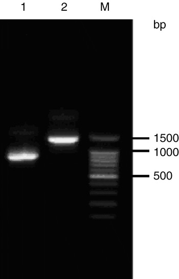
Electrophoresis of PCR products revealed that ~750 bp and ~1400 bp gene products were present (Fig. 1), indicating that the amplified VP1 and 3ABC gene fragments were within the specific target band.
Agarose gel electrophoresis of PCR products of VP1 and 3ABC. 1, VP1; 2, 3ABC; M, DNA marker.

Identification of the recombinant plasmids
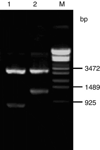
The target plasmids were screened according to the size of the DNA bands in agarose gel. The recombinant plasmids were then incubated with EcoR I for 2 h at 37°C (Fig. 2). The size of the recombinant fragments was determined by electrophoresis. Screened plasmids were further identified by PCR. Finally, the positive plasmids were selected for sequencing by Takara Biotechnology Co., Ltd.
Identification of the recombinant plasmid by restriction endonuclease digestion. Lanes: 1, restriction enzyme digestion of the VP1 recombinant plasmid; 2, restriction enzyme digestion of the 3ABC recombinant plasmid; M, DNA marker.

Alignment of the nucleotide sequence and deduced amino-acid sequence of the VP1 gene
The VP1 gene in all the yellow cattle persistent infection isolates and O/OA58-BHK/2004 were 639 nucleotides in length, and encoded 213 amino acids with no deletion or insertion. The nucleotide and amino-acid sequence identity percentages of the VP1 gene in different time isolates were above 98% and, when compared with O/OA58-BHK/2004, the nucleotide and amino-acid sequence identity percentages were 94%. However, when compared with their parental infection strain O/Akesu/58, the nucleotide sequence identity percentages were only 85%, and the amino-acid sequence identity percentages were 90%. Moreover, there were 102 nucleotide mutations in the VP1 gene of these isolates, including 82 nucleotide mutations consistent with O/OA58-BHK/2004 and four nucleotide transversion mutations A121→G, G247→A, A525→G, A631→G, with the relevant amino-acid mutations K41→E, E83→K and K211→E. Sixteen nucleotide mutations were absent in the O/OA58-BHK/2004 strain: T167→C, G192→A, C279→G, G285→A, A288→G, A291→G, T309→C, C318→A, T339→C, C348,349→T, T354→A, C363→A, C372→T, G381→A and C629→A, with relevant amino-acid mutation I56→T and A210→E.
Alignment of the nucleotide sequence and deduced amino-acid sequence of the 3ABC gene
The 3ABC gene in all the yellow cattle persistent infection isolates and O/OA58-BHK/2004 were 1311 nucleotides in length, and encoded 437 amino acids. The nucleotide and amino-acid sequence identity percentages of the 3ABC gene of the isolates were above 98%. Compared with O/OA58-BHK/2004, the nucleotide and amino-acid sequence identity percentages were 99%; however, compared with their parental infection strain O/Akesu/58, the nucleotide sequence identity percentages were only 90% and the amino-acid sequence identity percentages were 97%. There were 13 nucleotide transversion mutations in the 3ABC gene of the isolates: G21→A, G142→A, T226→C, T237→A, A256→G, A303→G, A444→G, A459→G, A749→G, A856→G, A863→G, C1017→T and C1029→T. These nucleotide mutations caused amino-acid mutations I76→T, Q84→R, K250→R, M286→V and D288→G. There were no stable nucleotide mutations in the 3ABC gene of the isolates other than O/OA58-BHK/2004.
The nucleotides of the isolates compared with FMDV reference strains
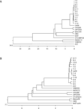
In order to analyse VP1 and 3ABC genetic variation during persistent FMDV infection, the phylogenetic trees of VP1 and 3ABC were constructed on the basis of sequence analysis and comparison of the isolates and reference sequences. The phylogenetic tree of VP1 (Fig. 3A) showed that the VP1 gene in all the isolates belonged to the same gene cluster, with the closest genetic relationship. Moreover, the VP1 gene of the isolates and O/OA58-BHK/2004 had a closer genetic relationship, belonging to the same gene cluster. However, the VP1 gene of the isolates had a more distant genetic relationship with its parental strain O/Akesu/58, and even more so with O/Yunlin/Taiwan/97.
Phylogenetic tree of VP1(A) and 3ABC(B).

The phylogenetic tree of 3ABC (Fig. 3B) showed that the 3ABC gene of the isolates and O/OA58-BHK/2004 had a closer genetic relationship, having a more distant genetic relationship with O/Akesu/58 strain. When compared with swine host strain O/Yunlin/Taiwan/97, the 3ABC gene of the isolates and A22/Azerbaijan/USSR/65 and Asia I/YNBS/58 and O/IND63/72 had a very close genetic relationship.
Discussion
In recent years, persistent FMDV infection has usually been found in the cell culture model and natural infection isolates (Weddel et al., Reference Weddel, Yansura and Dowbenko1985; Escarmis et al., Reference Escarmis, Toja, Medina and Domingo1992; Zhang and Kitching, Reference Zhang and Kitching2001). Toja and colleagues (1999) found that the nucleotide sequences of the different passages of cell culture infected with FMDV had great divergence with 2.8×10⁻³ to 7.7×10⁻³ nucleotide substitutions in L, P1, P2, P3 and 5′ untranslated regions (UTR). However, the persistently infected cattle and sheep FMDV isolates that could not be eliminated probably resulted in FMDV antigenic variation. The purpose of this research was to study the genetic variation of VP1 and 3ABC isolates from the yellow cattle persistently infected with the strain O/Akesu/58. The results found that their isolation at eight months post-infection was usual, with high secretion levels, and with correlates to serotype and host range (Alexandersen et al., Reference Alexandersen, Zhang and Donaldson2002). In this study, nine strains were isolated from the yellow cattle at five months post-persistent infection. However, three isolates were obtained after five months post-infection. Only one persistent infection strain was isolated at eleven months post-infection. The research showed that the yellow cattle infected with O/Akesu/58 could maintain their carrier state for eleven months, with high secretion virus levels at five months post-infection.
The results revealed that the homology of the VP1 gene nucleotide sequences of the different time isolates were above 98% compared with O/OA58-BHK/2004, and the homology nucleotide and amino-acid sequences were above 94%. The results highlighted that variation of the VP1 gene in both early and late isolates was minor and that the VP1 gene remained stable in the carrier state. It was presumed that the development of persistent FMDV infection could be related to the biological characteristics of the initially infected yellow cattle strains. Nucleotide analysis of the isolates showed that the mutations were not random, and some were continuously transversing in a fixed position, leading to corresponding amino-acid mutations K41→E, E83→K and K211→E; however, 16 nucleotide mutations were absent in the O/OA58-BHK/2004 strain, with their corresponding relevant amino-acid mutations I56→T and A210→E, including two amino-acid (210 and 211) mutations located on the VP1 protein lineal epitopes. The 82 nucleotide variations in all the isolates and O/OA58-BHK/2004 might have an effect in the isolate-adapted BHK-21 cells. However, only those in persistent infection isolates might be a necessary variation of FMDV persistently infected yellow cattle, and the constant transversion could be caused by immune suppression. The analysis showed that the structure and antigens of the VP1 protein produced minor change in the early infection stage; whether those variations lead to the virus evading the system's immune response requires further study. Variation of the virus gene targeted at escaping the humoral immune response is concluded not to be the key factor in persistent FMDV infection. Haydon and colleagues (2001) demonstrated, in a persistent FMDV infection cell model, the same conclusion. In contrast, Weddel and colleagues (1985) found the antigen of A serotype persistent FMDV infection isolates had a distinct effect. It was deduced that the development of persistent FMDV infection had a closer relationship with the biological characteristic of the virus itself than with antigenic gene variation in the virus.
3ABC gene nucleotide analysis of the isolates showed that the nucleotide and amino-acid sequence identity percentages of the 3ABC gene of the isolates were above 98%. The nucleotide mutations were mainly transversions and located in T226→C, T237→A, A303→G, A459→G and A863→G, leading to amino acid mutations I76→T, and D288→G. Fixed nucleotide mutation was not found in the isolates, and the homology of both nucleotides and amino acids was high compared with the parental infection strain O/Akesu/58 and the adapted BHK-21 cell strain. O/OMIII was the O/Akesu/58 adapted mouse strain and its 3A coding region was absent in 131–146. In the same region, O/OA58-BHK/2004 showed variation, with seven mutations out of 16 amino acids. However, there were no mutations in the 3A region of all the persistent infection isolates. It was concluded that the 3A region of O/Akesu/58 was more closely related to the host range and not related to persistent FMDV infection.
According to the phylogenetic tree of the VP1 gene of the isolates and the reference strains, as the antigenic gene, the VP1 of all the isolates demonstrated a certain variation and had no regularly close and/or distant relationship, but the genetic relationship was very close. Moreover, in recent years there has been neither a mutation tendency for the isolates of the prevalent Pan Asia strain (O/China/99) nor for A serotype and Asia I serotype FMDV. The phylogenetic tree of the 3ABC gene isolates showed information about the host range, but not the mutation tendency for the persistent infection isolates related to the prevalent swine FMDV strain (O/Yunlin/Taiwan/97), and their genetic relationship was distant (Zhang et al., Reference Zhang, Lv and Wang2004).
In particular, during persistent FMDV infection in yellow cattle, a tendency for continual variation of the isolates of the VP1 and 3ABC gene was not found. The VP1 gene of the isolates showed 16 nucleotide mutations having no significance in the first and second month of infection, and nucleotide transversion in later stages with stable antigenicity. Compared to the VP1 gene, the 3ABC gene remained highly stable, without absence or insertion in the early or late stages. So, the development of persistent FMDV infection could have a relationship with the biological characteristic of the initial infectious virus. As for the duration of yellow cattle persistent infection FMDV, the antigenicity of the isolates remained unchanged and it was posited that the FMDV persistent infection might not be directly related to the antigenicity variation of the virus or the correlative gene beyond the VP1 and 3ABC gene of FMDV. It was shown that the change in the B–C loop of the structure protein VP2 might be related to persistent FMDV infection (Horsington and Zhang, Reference Horsington and Zhang2007). This study of the mechanism of FMDV infection and virus gene variation has been of use in gathering important information regarding latent infection.