Introduction
Aberrant integration of information in the brain has long been considered a central pathophysiological mechanism to explain the clinical manifestations of schizophrenia (Bleuler, Reference Bleuler1911; Kraepelin, Reference Kraepelin and Robertson1919), resurging later on as the disconnection hypothesis (Friston, Reference Friston1998). Consistent with that view, over the past few decades convergent data from post-mortem and in vivo brain imaging studies have provided compelling evidence that psychosis is associated with white matter (WM) abnormalities in the brain (Ellison-Wright & Bullmore, Reference Ellison-Wright and Bullmore2009; Bora et al. Reference Bora, Fornito, Radua, Walterfang, Seal, Wood, Yücel, Velakoulis and Pantelis2011; Kochunov & Hong, Reference Kochunov and Hong2014; Mighdoll et al. Reference Mighdoll, Tao, Kleinman and Hyde2015).
Diffusion tensor imaging (DTI) allows examination of WM fibers by quantifying the diffusion of water in brain tissue and the anisotropy of this diffusion movement (Jones, Reference Jones2008). Anisotropy measures (such as fractional anisotropy, FA) reflect the degree of water diffusion directionality, whereas diffusivity indices (such as trace, TR, or mean diffusivity, MD) provide an estimate of the displacement of water molecules in a medium (Beaulieu, Reference Beaulieu2002; Jones, Reference Jones2008; Jones et al. Reference Jones, Knösche and Turner2013). Four meta-analyses of DTI studies found schizophrenia spectrum disorders to be consistently associated with FA reductions in fronto-limbic-striatal WM (Ellison-Wright & Bullmore, Reference Ellison-Wright and Bullmore2009; Bora et al. Reference Bora, Fornito, Radua, Walterfang, Seal, Wood, Yücel, Velakoulis and Pantelis2011; Patel et al. Reference Patel, Mahon, Wellington, Zhang, Chaplin and Szeszko2011; Yao et al. Reference Yao, Lui, Liao, Du, Hu, Thomas and Gong2013), specially involving the cingulum bundle, corpus callosum (CC), and inferior fronto-occipital and longitudinal fasciculi (IFOF and ILF, respectively). Similar patterns of DTI alterations have been shown in patients with first-episode psychosis (FEP), i.e. patients presenting any psychotic disorder in its very first episode (Bora et al. Reference Bora, Fornito, Radua, Walterfang, Seal, Wood, Yücel, Velakoulis and Pantelis2011; Yao et al. Reference Yao, Lui, Liao, Du, Hu, Thomas and Gong2013), although to a lesser extent than that observed in patients with chronic schizophrenia. Milder WM abnormalities have also been reported in subjects presenting prodromal psychotic symptoms and/or at increased genetic risk for developing psychosis (Carletti et al. Reference Carletti, Woolley, Bhattacharyya, Perez-Iglesias, Fusar-Poli, Valmaggia, Broome, Bramon, Johns, Giampietro, Williams, Barker and McGuire2012; Katagiri et al. Reference Katagiri, Pantelis, Nemoto, Zalesky, Hori, Shimoji, Saito, Ito, Dwyer, Fukunaga, Morita, Tsujino, Yamaguchi, Shiraga, Aoki and Mizuno2015). However, a great variability of findings is observed across different investigations (Samartzis et al. Reference Samartzis, Dima, Fusar-Poli and Kyriakopoulos2014). A number of confounding factors might influence DTI indices and, thus, could at least partly account for this heterogeneity, including illness course/chronicity, substance misuse and previous exposure to antipsychotic (AP) medication (Bora et al. Reference Bora, Fornito, Radua, Walterfang, Seal, Wood, Yücel, Velakoulis and Pantelis2011; Ho et al. Reference Ho, Andreasen, Ziebell, Pierson and Magnotta2011; Cookey et al. Reference Cookey, Bernier and Tibbo2014).
Another potential source of bias in the interpretation of the above neuroimaging results is the possibility that some of the brain alterations observed in psychosis are in fact state-dependent and related to a specific illness phase. Psychotic patients most often alternate between acute episodes of prominent positive symptoms and phases of symptom remission (Lieberman et al. Reference Lieberman, Perkins, Belger, Chakos, Jarskog, Boteva and Gilmore2001). Although structural MRI and DTI findings are commonly interpreted as reflecting static or progressive brain abnormalities (DeLisi, Reference DeLisi2008; Olabi et al. Reference Olabi, Ellison-Wright, McIntosh, Wood, Bullmore and Lawrie2011; Kochunov & Hong, Reference Kochunov and Hong2014), some investigations challenge this notion and suggest that at least some of the brain changes seen in acute psychosis are potentially reversible after symptom amelioration. Two longitudinal morphometric MRI studies have reported reversal of volumetric deficits in the superior temporal gyrus of FEP subjects after 1 year of clinical remission (Keshavan et al. Reference Keshavan, Haas, Kahn, Aguilar, Dick, Schooler, Sweeney and Pettegrew1998; Schaufelberger et al. Reference Schaufelberger, Lappin, Duran, Rosa, Uchida, Santos, Murray, McGuire, Scazufca, Menezes and Busatto2011). More recently, Katagiri et al. (Reference Katagiri, Pantelis, Nemoto, Zalesky, Hori, Shimoji, Saito, Ito, Dwyer, Fukunaga, Morita, Tsujino, Yamaguchi, Shiraga, Aoki and Mizuno2015) followed-up subjects with prodromal psychotic symptoms and found a region of FA increase over time in the CC of those individuals who did not convert to full-blown psychosis. Also, the longitudinal increase in FA significantly correlated with the improvement in sub-threshold positive symptoms observed at follow-up (Katagiri et al. Reference Katagiri, Pantelis, Nemoto, Zalesky, Hori, Shimoji, Saito, Ito, Dwyer, Fukunaga, Morita, Tsujino, Yamaguchi, Shiraga, Aoki and Mizuno2015), suggesting that changes in DTI indices might somehow reflect neurobiological processes related to acute psychosis. In this regard, no DTI study to date has been specifically designed to investigate whether microstructural WM changes are stable or state-dependent at the early stages of psychosis, i.e. varying according to the acute v. remitted phase of the disorder.
In the present DTI investigation, using a longitudinal design, we studied a group of treatment-naïve patients presenting their first episode of non-affective psychosis. FEP patients were initially evaluated at the acute psychotic phase (medication-free), and were then started on a semi-naturalistic AP treatment and were re-scanned as soon as they achieved 1 month of remission of core psychotic symptoms. We aimed to conduct a voxelwise analysis of anisotropy and diffusivity measures in order to examine the relationship between WM tissue organization changes and variations in the clinical state of patients (acute phase v. recovery phase). We hypothesized that at least some of the DTI abnormalities observed at baseline in the WM of FEP patients are state-dependent, i.e. potentially reversible following symptom remission. We predicted that FEP patients would present regions of reduced anisotropy and/or increased diffusivity in brain WM in relation to HC at baseline, as well as that FEP patients would exhibit a reversal of such initial abnormalities over time. Also, we predicted that reversal of WM abnormalities would be correlated to reduction of symptom scores.
Materials and methods
Participants and clinical assessment
FEP patients aged 16–50 years were referred to the Institute of Psychiatry, University of São Paulo, after active contact with mental health services from the metropolitan area of Sao Paulo city. At study entrance, a clinical interview and the Structured Clinical Interview (SCID) (First et al. Reference First, Spitzer, Gibbon and Williams1995) for Diagnostic and Statistical Manual for Mental Disorders (DSM-IV; American Psychiatry Association, 1994) were carried out for all cases, as well as the Alcohol Use Disorders Identification Test (Saunders et al. Reference Saunders, Aasland, Babor, de la Fuente and Grant1993) and the South Westminster Questionnaire (Menezes et al. Reference Menezes, Johnson, Thornicroft, Marshall, Prosser, Bebbington and Kuipers1996). Handedness was assessed using the Edinburgh inventory (Oldfield, Reference Oldfield1971). The clinical interview comprised questions regarding duration of untreated psychosis (DUP), general medical history, including information on previous use of psychotropic medications, and history of head trauma.
Only patients fulfilling DSM-IV criteria for any non-affective FEP enduring less than 6 months (i.e. schizophreniform disorder, brief psychotic disorder, delusional disorder and psychotic disorder not otherwise specified) and free of substance use disorders were included. Patients with psychotic disorders due to a general medical condition or substance-induced psychosis were excluded. Also, patients fulfilling criteria for other DSM-IV disorder (except mild anxiety disorders, such as specific phobia) were excluded.
Healthy volunteers free of mental disorders and with no history of psychotic or mood disorders among first-degree relatives were recruited through advertisement in the local community. HC were also assessed using the same clinical instruments.
Exclusion criteria for both groups were: (a) previous intake of any psychopharmacological drugs other than benzodiazepines; (b) history of substance dependence or abuse; (c) presence any medical disorders that could affect the central nervous system; (d) mental retardation; (e) history of head trauma with loss of consciousness; and (e) contraindications for MRI scanning.
Local ethics committees approved the study, and all subjects provided written informed consent.
Study design
On the same day of MRI scanning, FEP subjects were assessed for symptom severity with the Positive and Negative Syndrome Scale (PANSS; Kay et al. Reference Kay, Fiszbein and Opler1987). After that, patients were started on a semi-naturalistic AP treatment regimen based on the recommendations from the International Psychopharmacology Algorithm Project (IPAP; www.ipap.org).
FEP subjects were then clinically evaluated on a weekly basis by an experienced psychiatrist using the PANSS until achieving clinical remission. We adopted the remission criteria proposed by Andreasen et al. (Reference Andreasen, Carpenter, Kane, Lasser, Marder and Weinberger2005), with the exception of the length of time. Specifically, patients should present no more than mild symptom severity (PANSS items score <4) in all of the following core items of the PANSS: delusions, conceptual disorganization, hallucinatory behavior, blunted affect, social withdrawal, lack of spontaneity, mannerisms/posturing, and unusual thought content. Once the remission criteria were fulfilled, FEP patients were evaluated every other week until completing a month of sustained remission. At this point, a second MRI scanning session was performed.
In order to control for random effects related to scan acquisition, a subsample of HC with similar characteristics to that of FEP subjects evaluated at follow-up were also scanned a second time, after a follow-up interval close to that of the FEP group.
Image data acquisition
All subjects underwent MRI scanning using a 1.5 T Siemens Espree system (Siemens, Erlangen, Germany).
The DTI sequence was acquired using cardiac gating, a 12-channel head coil and parallel imaging. DTI was based on an echo-planar image (EPI) acquisition and consisted of one image without diffusion gradient (b = 0 s/mm2) plus diffusion-weighted images (DWI) acquired along 64 non-collinear directions (b = 1000 s/mm2) using the following parameters: TR = 8000 ms, TE = 110 ms, NEX = 2, FOV = 240 mm, matrix = 120 × 120 pixels, slice thickness = 2.7 mm (no gap between slices), voxel size = 2.0 × 2.0 × 2.7 mm, resulting in 50 slices covering the whole brain.
The imaging protocol also included a T1 and T2-weighted sequences, and a fluid attenuated inversion recovery (FLAIR) sequence. An expert neuroradiologist inspected all individual images aiming to identify silent brain lesions and artifacts.
Processing of neuroimaging data
The diffusion tensor images were reconstructed from the DWI data using multivariate linear fitting (Pierpaoli & Basser, Reference Pierpaoli and Basser1996). FA and TR images were computed from the tensor image for each subject. The FA value, which measures the degree of anisotropy of the diffusion at a voxel, is computed from the variance of the three eigenvalues of the diffusion tensor about their means. The TR value, which measures the mean diffusivity, is computed by adding the eigenvalues of the diffusion tensor (Jones, Reference Jones2008). FA and TR images were then aligned to a common template space via the deformable registration method DRAMMS (Ou et al. Reference Ou, Sotiras, Paragios and Davatzikos2011), available at https://www.cbica.upenn.edu/sbia/software/dramms/download.html, using a standard DTI template, named EVE (Wakana et al. Reference Wakana, Jiang, Nagae-Poetcher, van Zijl and Mori2004). Aligned FA and TR images were masked to include only values in WM voxels using the template WM mask, and both maps were smoothed with a 4 mm full-width at half-maximum (FWHM) linear Gaussian filter. Such choice of filter width followed the ‘rule of thumb’ developed for PET and fMRI studies, which states that the smoothing kernel should be at least 2–3 times the voxel dimension (Worsley et al. Reference Worsley, Evans, Marrett and Neelin1992). We avoided the use of larger smoothing kernels as these have been shown to reduce sensitivity and specificity to detect localized WM abnormalities, with partial volume effects that produce widespread averaging of DTI measures across different WM bundles and reduce power to differentiate separate anatomical tracts (Smith et al. Reference Smith, Jenkinson, Johansen-Berg, Rueckert, Nichols, Mackay, Watkins, Ciccarelli, Cader, Matthews and Behrens2006; Snook et al. Reference Snook, Plewes and Beaulieu2007; Jones & Cercignani, Reference Jones and Cercignani2010; Van Hecke et al. Reference Van Hecke, Leemans, De Backer, Jeurissen, Parizel and Sijbers2010).
Statistical analyses
Voxel-based between-group comparisons were carried out using the General Linear Model (GLM). These comparisons were carried out for: baseline DTI data between the FEP and HC groups; follow-up DTI data between the FEP and the HC that underwent the second scanning session; and rate-of-change maps between the FEP and HC subjects that underwent the second scanning session. The latter maps were generated for each and every subject by voxelwise subtraction of the baseline image values from the follow-up image values, and then division of the results by the baseline image values. The rationale for using relative instead of absolute values lays in the fact that relative values convey information of the magnitude of change, allowing comparability on changes in measures of different natures (for instance, DTI metrics and symptom scores).
Additionally, we run correlational analyses between imaging maps and clinical variables (DUP, total PANSS and PANSS sub-items) at baseline. Also, rate-of-change maps were correlated to the following variables: interval between scans, AP load (calculated as proposed by Andreasen et al. Reference Andreasen, Pressler, Nopoulos, Miller and Ho2010), and rate of change in PANSS scores across time (i.e. PANSS score at baseline minus PANSS score at follow-up, divided by PANSS score at baseline).
For the voxelwise between-group comparisons of DTI data at baseline and follow-up, we considered significant any clusters that surpassed the statistical threshold of p < 0.05, corrected for multiple comparisons (false-discovery rate, FDR). For the between-group comparison of rate-of-change maps, we applied a less rigid threshold of p < 0.001 uncorrected for multiple comparisons for clusters located specifically in WM regions where abnormalities in FEP relative to HC had been detected at baseline. Our choice to apply the latter, less conservative statistical threshold was intended to avoid type II errors in the search for confirmation of the hypothesis of reversal of FA abnormalities in treatment-naïve FEP patients associated with improvement of psychotic symptoms over time. Statistical thresholds of p < 0.001, uncorrected, have been commonly employed in previous voxel-based case-control DTI studies of psychiatric disorders (Ashtari et al. Reference Ashtari, Cottone, Ardekani, Cervellione, Szeszko, Wu, Chen and Kumra2007; Schlösser et al. Reference Schlösser, Nenadic, Wagner, Güllmar, von Consbruch, Köhler, Schultz, Koch, Fitzek, Matthews, Reichenbach and Sauer2007; Szeszko et al. Reference Szeszko, Robinson, Ashtari, Vogel, Betensky, Sevy, Ardekani, Lencz, Malhotra, McCormack, Miller, Lim, Gunduz-Bruce, Kane and Bilder2008; Gao et al. Reference Gao, Jiao, Qi, Zhong, Lu, Xiao, Lu, Xu, Zhang, Liu, Yang, Lu and Su2013; Li et al. Reference Li, Li, Zhu, Qin, Zheng, Chang, Zhang, Wang, Wang, Wang and Wang2013; Lu et al. Reference Lu, Wei, Gao, Wu, Liao, Zhang, Li, Li and Li2013; Tha et al. Reference Tha, Terae, Nakagawa, Inoue, Kitagawa, Kako, Nakato, Akter Popy, Fujima, Zaitsu, Yoshida, Ito, Miyamoto, Koyama and Shirato2013; Cheng et al. Reference Cheng, Xu, Yu, Nie, Li, Luo, Li, Liu, Bai, Shan, Xu and Xu2014; Filippi et al., Reference Filippi, Canu, Gasparotti, Agosta, Valsecchi, Lodoli, Galluzzo, Comi and Sacchetti2014; Qiu et al. Reference Qiu, Zhu, Zhang, Nie, Feng, Meng, Wu, Huang, Zhang and Gong2014; Spalletta et al. Reference Spalletta, Piras, Fagioli, Caltagirone and Piras2014; Lei et al. Reference Lei, Li, Deng, Li, Huang, Ma, Wang, Guo, Li, Jiang, Zhou, Hu, McAlonan and Li2015; Melicher et al. Reference Melicher, Horacek, Hlinka, Spaniel, Tintera, Ibrahim, Mikolas, Novak, Mohr and Hoschl2015; Itahashi et al. Reference Itahashi, Yamada, Nakamura, Watanabe, Yamagata, Jimbo, Shioda, Kuroda, Toriizuka, Kato and Hashimoto2015). Finally, we applied the more conservative statistical threshold of p < 0.05, FDR-corrected, in the correlational analyses between imaging maps and clinical variables.
Results
Demographic and clinical details
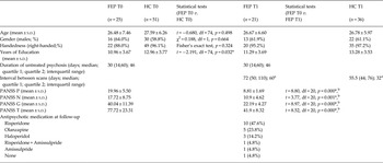
A total of 25 FEP patients and 51 HC were evaluated at baseline (see Table 1 for demographic/clinical details). There were no significant between-group differences regarding gender, age, and handedness. FEP subjects attained less years of education relative to HC, as commonly reported (Goldberg et al. Reference Goldberg, Ragland, Torrey, Gold, Bigelow and Weinberger1990).
Demographic and clinical information for patients with first-episode psychotic (FEP) and healthy controls (HC)

FEP, first-episode psychosis; HC, healthy controls; T0, data at baseline; T1, data at follow-up; s.d., standard deviation; t, t test statistics; min, minimum value; max, maximum value; df, degrees of freedom; PANSS, Positive and Negative Syndrome Scale; PANSS P, score for positive symptoms; PANSS N, score for negative symptoms; PANSS G, score for general symptoms; PANSS T, PANSS total score.
*Significant statistical difference (p < 0.05).
a Comparison between FEP and HC at follow-up: Mann–Whitney U test, p = 0.056.
b Comparisons between baseline and follow-up of PANSS scores for patients that underwent second MRI scanning (paired t test).
In total 21 patients and 36 HC completed the follow-up DTI protocol. Two patients presenting poor response to AP treatment failed to achieve remission in up to 6 months, thus they were excluded from the study; other two patients were enrolled in a pilot phase of this study, when longitudinal examination had not been included in the overall study design. A subsample of the original HC group was rescanned, taking into account the demographic characteristics of the subgroup of FEP that completed the follow-up protocol. The subgroups of FEP and HC that completed the follow-up protocol had similar clinical/demographic characteristics relative to the overall baseline groups (see Table 1). Scanning intervals of FEP and HC groups were not statistically different, although there was a trend towards a narrower interval for HC subjects (p = 0.056). At follow-up, almost half (47.6%) of patients were taking risperidone.
Baseline imaging findings
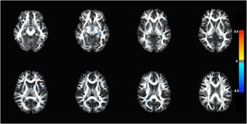
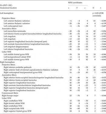
At baseline, FEP presented widespread reductions of FA in comparison with HC (p < 0.05, FDR-corrected) affecting mainly: prefrontal WM, uncinate fasciculus (UF), anterior thalamic radiation/anterior limb of internal capsule (ATR/ALIC), corticospinal tract, superior longitudinal fasciculus (SLF), IFOF and ILF bilaterally; the left fornix/stria terminalis and cingulum bundle; and the right CC/forceps major and cerebellar WM (see Fig. 1 and Table 2). Conversely, there were no significant TR differences between FEP patients and HC at the corrected p < 0.05 statistical level.
Baseline comparison between FEP and HC. FA map showing widespread reduced anisotropy in FEP, affecting mostly fronto-limbic WM and long associative, projective and commissural fasciculi (p < 0.05, FDR corrected). Blue color represents reduced FA in patients relative to HC, whereas red-yellow colors represent increased FA in patients relative to HC. Color intensity represents Student's t test statistics (i.e. the darker the color, the higher the value of the test). Results are overlaid on axial slices from JHU white matter tractography atlas (Wakana et al. Reference Wakana, Jiang, Nagae-Poetcher, van Zijl and Mori2004). Details for each significant cluster are provided in Table 2. FEP, first-episode psychosis; HC, health controls; FA, fractional anisotropy; WM, white matter; FDR, false-discovery rate.

Between group comparisons of DTI maps at baseline

FA: fractional anisotropy; FEP: first-episode psychosis; HC: Healthy Controls; WM: white matter; FDR: false discovery rate; N: number of significant voxels in each anatomical region; MNI coordinates represents location of peak voxel; t: t test statistics. For the sake of clarity, near clusters located in the same WM tract/region were merged to the most significant cluster.
No correlations between FA or TR indices and any of the clinical variables investigated at baseline (DUP and PANSS scores) survived FDR correction for multiple comparisons.
Longitudinal DTI findings
No differences in FA or TR were observed in the between-group comparisons of DTI maps at follow-up at the p < 0.05 level, FDR-corrected.
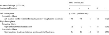
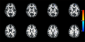
Regarding comparisons of the rate-of-changes maps, the FEP group showed three clusters of FA increase over time at the uncorrected p < 0.001 level of significance, located in the right ATR and UF/IFOF, and in the left IFOF/ILF relative to controls (see Fig. 2 and Table 3). Concerning TR, no significant differences were observed between FEP and HC in the comparison of rate-of-change maps.
Follow-up comparison between FEP and HC. FA rate-of-change map showing FA increase over time in patients, specifically in the right anterior thalamic radiation, right uncinate fasciculus/inferior fronto-occipital fasciculus, and left inferior fronto-occipital fasciculus/inferior longitudinal fasciculus (p < 0.001, uncorrected). Blue color represents reduced FA rate-of-change in patients relative to HC, whereas red-yellow colors represent increased FA rate-of-change in patients relative to HC. Color intensity represents Student's t test statistics (i.e. the darker the color, the higher the value of the test). Results are overlaid on axial slices from JHU white matter tractography atlas (Wakana et al. Reference Wakana, Jiang, Nagae-Poetcher, van Zijl and Mori2004). Details for each significant cluster are provided in Table 3. FEP, first-episode psychosis; HC, health controls; FA, fractional anisotropy; FDR, false-discovery rate.

Between-groups comparisons of DTI rate-of-changes maps

FA, fractional anisotropy; FEP, first-episode psychosis; HC, Healthy Controls; WM, white matter; FDR, false-discovery rate; N, number of significant voxels in each anatomical region; MNI coordinates represents location of peak voxel; t, t test statistics.
* p (FDR-corrected) = 0.29.
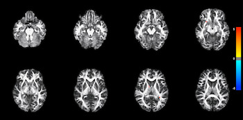
The improvement in psychotic symptoms over time was significantly correlated (p < 0.05, FDR-corrected) with FA increases in a vast number of WM tracts (see Fig. 3). As main findings, reductions in total PANSS scores were associated with longitudinal FA increases in the ATR, corticospinal tract, CC, frontotemporal WM, SLF, IFOF/ILF and UF/ILF bilaterally. We also found significant correlations between reductions in PANSS sub-items (positive, negative and general) and increases in FA in such tracts (see online Supplementary Table S1 for detailed information on correlations).
Correlation analyses between changes in FA and total PANSS scores. FA map showing clusters of negative correlation between the rates of changes in FA and total PANSS over time in FEP (p < 0.05, FDR corrected). Blue color represents negative correlations between the rate of symptoms reduction and the rate of FA increasing over time, whereas red-yellow colors represent positive correlations. Color intensity represents the Sperman's rho coefficient statistics (i.e. the darker the color, the higher the value of the test). Results are overlaid on axial slices from JHU white matter tractography atlas (Wakana et al. Reference Wakana, Jiang, Nagae-Poetcher, van Zijl and Mori2004). Details for each significant cluster are provided in online Supplementary Material/Table S1. FEP, first-episode psychosis; FA, fractional anisotropy; PANSS, positive and negative symptoms scale; FDR, false-discovery rate.

No correlations between improvement in psychotic symptoms over time and TR reduction survived correction for multiple comparisons. Also, there were no significant correlations between either FA or TR indices and AP load at the p < 0.05 level, FDR-corrected.
Discussion
The present DTI investigation employed a case-control, within-subject design in order to assess WM anisotropy and diffusivity indices in treatment-naïve FEP patients focusing on state-dependent longitudinal changes (i.e. in acute psychosis v. sustained remission of core symptoms). In line with our initial prediction, there were significant direct correlations (at a strict p < 0.05 statistical threshold, corrected) between reductions in positive, negative, general and total PANSS scores and longitudinal FA increases in some of the WM tracts known to be critically involved in the pathophysiology of psychotic disorders, such as the SLF, ILF, IFOF, UF, cingulum bundle, ATR and CC. At a less conservative statistical threshold (p < 0.001, uncorrected), we also observed significant post-treatment reversal of the baseline FA reductions that were detected in FEP patients relative to HC in some of such WM tracts. To the best of our knowledge, this is the first demonstration that some of the WM abnormalities found in untreated FEP are directly associated with the acute phase of the disease and that the remission of outbreak symptoms occurs in parallel with an apparent recovery of such WM microstructural changes.
At baseline, patients with acute FEP showed widespread FA reductions affecting mostly fronto-limbic WM and long associative, projective, and commissural fasciculi. These FA reductions are consistent with the findings of previous DTI studies in FEP (Bora et al. Reference Bora, Fornito, Radua, Walterfang, Seal, Wood, Yücel, Velakoulis and Pantelis2011; Yao et al. Reference Yao, Lui, Liao, Du, Hu, Thomas and Gong2013). Such findings provided us with a solid basis from which to investigate reversal of abnormal DTI indices after symptom remission in psychosis and correlations of rates of change in those indices v. the degree of clinical improvement.
All of the WM tracts where our FEP patients exhibited FA increases after clinical remission (left IFOF/ILF, right ATR, and right UF/IFOF) have been previously shown to be implicated in psychosis (Bora et al. Reference Bora, Fornito, Radua, Walterfang, Seal, Wood, Yücel, Velakoulis and Pantelis2011; Yao et al. Reference Yao, Lui, Liao, Du, Hu, Thomas and Gong2013). However, this is the first time that WM changes in these brain structures are shown to be specifically linked to switching from acute psychosis to remission.
The ATR is part of the ALIC, and it carries axonal fibers from thalamic nuclei to the prefrontal (PFC) and anterior cingulate cortex (ACC), being involved in executive functioning and memory (Mamah et al. Reference Mamah, Conturo, Harms, Akbudak, Wang, McMichael, Gado, Barch and Csernansky2010). Schizophrenia patients have significant impairments of memory and executive performance and longitudinal studies have demonstrated that such deficits are further impaired during the acute phase of the illness (Bates et al. Reference Bates, Liddle, Kiehl and Ngan2004; Klingberg et al. Reference Klingberg, Wittorf, Sickinger, Buchkremer and Wiedemann2008). Neuroimaging studies have shown structural and microstructural abnormalities in the ATR/ALIC of schizophrenia patients, which correlated with worse performance in executive and memory tests (Levitt et al. Reference Levitt, Kubicki, Nestor, Ersner-Hershfield, Westin, Alvarado, Kikinis, Jolesz, McCarley and Shenton2010; Reference Levitt, Alvarado, Nestor, Rosow, Pelavin, McCarley, Kubicki and Shenton2012; Mamah et al. Reference Mamah, Conturo, Harms, Akbudak, Wang, McMichael, Gado, Barch and Csernansky2010). Also, schizophrenia patients present disturbed structural and functional connectivity between frontal cortex and thalamus (Marenco et al. Reference Marenco, Stein, Savostyanova, Sambataro, Tan, Goldman, Verchinski, Barnett, Dickinson, Apud, Callicott, Meyer-Lindenberg and Weinberger2012; Kubota et al. Reference Kubota, Miyata, Sasamoto, Sugihara, Yoshida, Kawada, Fujimoto, Tanaka, Sawamoto, Fukuyama, Takahashi and Murai2013; Anticevic et al. Reference Anticevic, Cole, Repovs, Murray, Brumbaugh, Winkler, Savic, Krystal, Pearlson and Glahn2014; Wagner et al. Reference Wagner, De la Cruz, Schachtzabel, Güllmar, Schultz, Schlösser, Bär and Koch2015), and such abnormality seems to affect working memory performance (Marenco et al. Reference Marenco, Stein, Savostyanova, Sambataro, Tan, Goldman, Verchinski, Barnett, Dickinson, Apud, Callicott, Meyer-Lindenberg and Weinberger2012). Besides, a recent investigation has shown that measures of FA in ATR are correlated to positive symptoms scores (Ebdrup et al. Reference Ebdrup, Raghava, Nielsen, Rostrup and Glenthøj2016).
The associative WM tracts in which increases in FA were observed over time (IFOF, UF, and IFL) connect frontal, temporal, parietal and occipital areas, and they have been implicated in several cognitive functions, such as visuospatial processing, emotional regulation, memory and language (Catani & Thiebaut de Schotten, Reference Catani and Thiebaut de Schotten2008; Ćurčić-Blake et al. Reference Ćurčić-Blake, Nanetti, van der Meer, Cerliani, Renken, Pijnenborg and Aleman2015). Structural and functional connectivity of these networks have consistently been shown to be impaired in schizophrenia (Allen et al. Reference Allen, Modinos, Hubl, Shields, Cachia, Jardri, Thomas, Woodward, Shotbolt, Plaze and Hoffman2012; Ćurčić-Blake et al. Reference Ćurčić-Blake, Liemburg, Vercammen, Swart, Knegtering, Bruggeman and Aleman2013). For instance, reduced FA in the left IFOF, UF and ATR has been associated with auditory verbal hallucinations in schizophrenia patients (Ćurčić-Blake et al. Reference Ćurčić-Blake, Nanetti, van der Meer, Cerliani, Renken, Pijnenborg and Aleman2015; Oestreich et al. Reference Oestreich, McCarthy-Jones and Whitford2015) and microstructural abnormalities in the left UF have been linked to negative symptoms (Kitis et al. Reference Kitis, Ozalay, Zengin, Haznedaroglu, Eker, Yalvac, Oguz, Coburn and Gonul2012; Lei et al. Reference Lei, Li, Deng, Li, Huang, Ma, Wang, Guo, Li, Jiang, Zhou, Hu, McAlonan and Li2015). Interestingly to this regard, reductions in the negative PANSS sub-scores during follow-up were significantly correlated to FA increases over time in the UF/IFOF in the present investigation.
In general terms, the significant correlations observed between symptom reductions and FA increases over time in the present study corroborate our hypothesis that the observed DTI changes reflect WM abnormalities underlying the emergence of psychotic symptoms. It is important to highlight that no correlation was found in the opposite direction, i.e. FA increase over time was invariably related to symptom reductions. Also, the WM locations where we observed increased FA longitudinally have been also correlated with decreasing PANSS scores in our study. Moreover, the fact that no significant correlations were found between the longitudinal changes in FA and AP load during follow-up further suggests that the observed FA increases were primarily related to clinical improvement rather than to the exposure to AP medication.
Up until now, few longitudinal studies investigated the effects of AP treatment in the DTI indices of FEP patients, all employing fixed follow-up intervals (Wang et al. Reference Wang, Cheung, Deng, Li, Huang, Ma, Wang, Jiang, Sham, Collier, Gong, Chua, McAlonan and Li2013; Reis Marques et al. Reference Klingberg, Wittorf, Sickinger, Buchkremer and Wiedemann2014; Szeszko et al. Reference Szeszko, Robinson, Ikuta, Peters, Gallego, Kane and Malhotra2014; Ebdrup et al. Reference Ebdrup, Raghava, Nielsen, Rostrup and Glenthøj2016; Zeng et al. Reference Zeng, Ardekani, Tang, Zhang, Zhao, Cui, Fan, Zhuo, Li, Xu, Goff and Wang2016). The results of those studies were highly heterogeneous with regard to both the direction (i.e. FA increase or decrease) and topographical location of the findings in the brain. For instance, two studies reported 6 or 12 weeks of AP treatment to be associated with FA reductions over time (Wang et al. Reference Wang, Cheung, Deng, Li, Huang, Ma, Wang, Jiang, Sham, Collier, Gong, Chua, McAlonan and Li2013; Szeszko et al. Reference Szeszko, Robinson, Ikuta, Peters, Gallego, Kane and Malhotra2014), whereas two other investigations found FA increases after 6 or 12 weeks of AP exposure (Reis Marques et al. Reference Klingberg, Wittorf, Sickinger, Buchkremer and Wiedemann2014; Ebdrup et al. Reference Ebdrup, Raghava, Nielsen, Rostrup and Glenthøj2016). A third study (Zeng et al. Reference Zeng, Ardekani, Tang, Zhang, Zhao, Cui, Fan, Zhuo, Li, Xu, Goff and Wang2016) failed to find differences in FEP patients relative to controls after an 8-week trial of AP. In four out of those five investigations, no significant correlations between pre- v. post-treatment FA changes and symptom reduction in FEP patients was observed (Wang et al. Reference Wang, Cheung, Deng, Li, Huang, Ma, Wang, Jiang, Sham, Collier, Gong, Chua, McAlonan and Li2013; Szeszko et al. Reference Szeszko, Robinson, Ikuta, Peters, Gallego, Kane and Malhotra2014; Reis Marques et al. Reference Klingberg, Wittorf, Sickinger, Buchkremer and Wiedemann2014; Ebdrup et al. Reference Ebdrup, Raghava, Nielsen, Rostrup and Glenthøj2016). Interestingly, Zeng et al. (Reference Zeng, Ardekani, Tang, Zhang, Zhao, Cui, Fan, Zhuo, Li, Xu, Goff and Wang2016) observed a significant negative correlation between longitudinal changes in FA values and change in positive symptoms over time. It is important to notice that such studies were designed to evaluate DTI changes specifically related to AP exposure, in which FEP patients were re-scanned after a fixed follow-up period, regardless of their clinical status at the time of the second scanning session (i.e. with not all patients having achieved remission). Thus, these investigations may have lacked sensitivity to detect state-dependent DTI changes specifically related to the amelioration of psychotic symptoms, as investigated herein. There are other limitations that should be weighted in the interpretation of the results of such studies: three of them enrolled FEP patients who had been previously exposed to AP or other psychotropic medications and/or subjects with comorbid substance misuse (Szeszko et al. Reference Szeszko, Robinson, Ikuta, Peters, Gallego, Kane and Malhotra2014; Reis Marques et al. Reference Klingberg, Wittorf, Sickinger, Buchkremer and Wiedemann2014; Ebdrup et al. Reference Ebdrup, Raghava, Nielsen, Rostrup and Glenthøj2016; Zeng et al. Reference Zeng, Ardekani, Tang, Zhang, Zhao, Cui, Fan, Zhuo, Li, Xu, Goff and Wang2016); in Reis Marques et al. (Reference Reis Marques, Taylor, Chaddock, Dell'acqua, Handley, Reinders, Mondelli, Bonaccorso, Diforti, Simmons, David, Murray, Pariante, Kapur and Dazzan2014), HC were scanned only at baseline. Also, in three of those previous studies, some of the evaluated patients had a history of psychotic symptoms for more than a year by the time of the first scanning session (Szeszko et al. Reference Szeszko, Robinson, Ikuta, Peters, Gallego, Kane and Malhotra2014; Ebdrup et al. Reference Ebdrup, Raghava, Nielsen, Rostrup and Glenthøj2016; Zeng et al. Reference Zeng, Ardekani, Tang, Zhang, Zhao, Cui, Fan, Zhuo, Li, Xu, Goff and Wang2016), thus possibly confounding the results due to the effects of illness chronicity.
Brain anisotropy indices mostly reflect myelination and axonal membrane integrity, indirectly representing the degree of fiber density and tissue organization within WM tracts (Beaulieu, Reference Beaulieu2002; Concha et al. Reference Concha, Livy, Beaulieu, Wheatley and Gross2010; Jones et al. Reference Jones, Knösche and Turner2013). Post-mortem studies suggest that schizophrenia is associated with myelin/oligodendrocyte abnormalities (Takahashi et al. Reference Takahashi, Sakurai, Davis and Buxbaum2011), which are believed to result either from disturbed neurodevelopmental processes or as a consequence of inflammation associated with acute psychosis (Najjar & Pearlman, Reference Najjar and Pearlman2015). Oligodendroglia has a vast number of progenitor cells that can repair damaged myelin sheaths (Bartzokis, Reference Bartzokis2012). Animal studies suggest that AP can restore myelin (Xiao et al. Reference Xiao, Xu, Zhang, Wei, He, Jiang, Li, Dyck, Devon, Deng and Li2008; Zhang et al. Reference Zhang, Zhang, Wang, Jiang, Xu, Xiao, Bi, Wang, Zhu, Zhang, He, Tan, Zhang, Kong and Li2012) as well as produce inhibitory effects on activated microglia (Monji et al. Reference Monji, Kato, Mizoguchi, Horikawa, Seki, Kasai, Yamauchi, Yamada and Kanba2013). AP seem to promote myelination through inhibition of the glycogen synthase kinase-3 (GSK-3) enzyme (Bartzokis, Reference Bartzokis2012). Thus, one can hypothesize that the increases in FA observed after symptom remission in our psychosis patients could reflect myelin healing promoted by such medications, which might restore structural as well as functional connectivity in the affected WM tracts. In agreement with such reasoning, a longitudinal DTI study evaluating multiple sclerosis patients showed that the remyelination after acute lesions is mainly characterized by FA increase and stable diffusivity indices (Fox et al. Reference Fox, Cronin, Lin, Wang, Sakaie, Ontaneda, Mahmoud, Lowe and Phillips2011).
Overall, our results suggest that anisotropy abnormalities are more pronounced in acute psychosis than diffusivity changes. Few studies have assessed diffusivity measures in FEP (Ruef et al. Reference Ruef, Curtis, Moy, Bessero, Badan Bâ, Lazeyras, Lövblad, Haller, Malafosse, Giannakopoulos and Merlo2012; Lee et al. Reference Lee, Kubicki, Asami, Seidman, Goldstein, Mesholam-Gately, McCarley and Shenton2013; Filippi et al. Reference Filippi, Canu, Gasparotti, Agosta, Valsecchi, Lodoli, Galluzzo, Comi and Sacchetti2014; Zeng et al. Reference Zeng, Ardekani, Tang, Zhang, Zhao, Cui, Fan, Zhuo, Li, Xu, Goff and Wang2016). Lee et al. (Reference Lee, Kubicki, Asami, Seidman, Goldstein, Mesholam-Gately, McCarley and Shenton2013) reported widespread TR increases across the whole cerebral WM; Ruef et al. (Reference Ruef, Curtis, Moy, Bessero, Badan Bâ, Lazeyras, Lövblad, Haller, Malafosse, Giannakopoulos and Merlo2012) found a trend towards increased MD in the right SLF and middle cerebellar peduncle; and Filippi et al. (Reference Filippi, Canu, Gasparotti, Agosta, Valsecchi, Lodoli, Galluzzo, Comi and Sacchetti2014) observed increased MD bilaterally in the fornix and thalamic radiation at the same time that several other brain areas displayed reduced MD; Zeng et al. (Reference Zeng, Ardekani, Tang, Zhang, Zhao, Cui, Fan, Zhuo, Li, Xu, Goff and Wang2016) found increased MD in bilateral ATR, left IFOF and right IFL, forceps major and forceps minor. In our study, no region of between-group differences in TR survived correction for multiple comparisons. Also, no correlation between TR maps and clinical variables survived correction for multiple comparisons. In three out of four previous investigations (Ruef et al. Reference Ruef, Curtis, Moy, Bessero, Badan Bâ, Lazeyras, Lövblad, Haller, Malafosse, Giannakopoulos and Merlo2012; Lee et al. Reference Lee, Kubicki, Asami, Seidman, Goldstein, Mesholam-Gately, McCarley and Shenton2013; Zeng et al. Reference Zeng, Ardekani, Tang, Zhang, Zhao, Cui, Fan, Zhuo, Li, Xu, Goff and Wang2016), there were no correlations between diffusivity indexes and symptom scores; only Filippi et al. (Reference Filippi, Canu, Gasparotti, Agosta, Valsecchi, Lodoli, Galluzzo, Comi and Sacchetti2014) found inverse correlations between MD and symptoms. Such heterogeneity in the investigation of diffusivity indexes in FEP might be partially explained by lower sensitivity of MD to pathological damage in comparison to FA, as reported in patients with multiple sclerosis (Sbardella et al. Reference Sbardella, Tona, Petsas and Pantano2013).
It is possible that not only state-dependent, but also trait-based pathological WM findings are present in psychotic disorders. This is supported by findings of FA reductions in individuals at genetic risk for schizophrenia (Skudlarski et al. Reference Skudlarski, Schretlen, Thaker, Stevens, Keshavan, Sweeney, Tamminga, Clementz, O'Neil and Pearlson2013). We have attempted to evaluate such possibility by carrying out a cross-sectional comparison between remitted FEP patients and the HC group at follow-up. No differences in FA or TR were observed even at the uncorrected p < 0.001 level of significance in this analysis, suggesting that there were no residual DTI abnormalities after symptom remission in the FEP group. Nevertheless, caution must be exercised when interpreting such negative finding, given that a smaller number of FEP individuals were available for the cross-sectional between-group comparison carried out at follow-up.
Progression of volumetric and DTI brain changes has been documented after the onset of both schizophrenia and affective psychoses, particularly in those patients with a non-remitting course of illness (Mori et al. Reference Mori, Ohnishi, Hashimoto, Nemoto, Moriguchi, Noguchi, Nakabayashi, Hori, Harada, Saitoh, Matsuda and Kunugi2007; Hulshoff Pol & Kahn, Reference Hulshoff Pol and Kahn2008; Olabi et al. Reference Olabi, Ellison-Wright, McIntosh, Wood, Bullmore and Lawrie2011; Rosa et al. Reference Rosa, Zanetti, Duran, Santos, Menezes, Scazufca, Murray, Busatto and Schaufelberger2015; Sun et al. Reference Sun, Chen, Lee, Bezerianos, Collinson and Sim2016). Several mechanisms have been proposed to explain such findings, including progression of neurodevelopmental injuries, disturbed neuroplasticity with increased inflammation and impaired resilience to cellular insults (Haroutunian et al. Reference Haroutunian, Katsel, Roussos, Davis, Altshuler and Bartzokis2014; Najjar & Pearlman, Reference Najjar and Pearlman2015). Interesting to this regard, one large investigation combining resting-state functional MRI and molecular genetics suggested that genes implicated in NMDA-related long-term potentiation, protein kinase A (PKA) signaling, immune response/ inflammation, synaptogenesis and axon guidance mediate the altered functional connectivity observed in schizophrenia and psychotic bipolar disorder (Meda et al. Reference Meda, Ruaño, Windemuth, O'Neil, Berwise, Dunn, Boccaccio, Narayanan, Kocherla, Sprooten, Keshavan, Tamminga, Sweeney, Clementz, Calhoun and Pearlson2014). Dynamic changes in these cellular systems might underlie both our finding of state-dependent FA changes that significantly correlated with symptom improvement in FEP patients, and the findings of progressive WM abnormalities that have been observed in psychosis patients with a chronic/recurrent course of illness in previous studies. In this sense, an apparent recovery of microstructural WM damage as suggested by the findings presented herein does not challenge the notion of progressive neuropathological mechanisms in schizophrenia of poorer prognosis; instead, our results only shed light on another likely process, namely the acuteness of clinical symptoms reflecting the acuteness of tissue damage (which generally occurs in other chronic brain diseases such as multiple sclerosis, for instance).
The present investigation evaluated an enriched sample of treatment-naïve FEP patients with relatively short illness duration (DUP of up to 6 months) and free of substance use disorders. Hence, we could avoid DTI alterations related to chronicity, previous AP exposure and drug misuse. Also, patients were clinically assessed at a weekly basis until they reached a fully sustained remission, thus avoiding the risk of considering a transient clinical improvement as a sustained positive clinical response. Finally, a significant proportion of HC were also evaluated longitudinally, enabling us to control our results for variations over time not strictly related to psychosis.
Nevertheless, some limitations of our study need to be addressed. The relatively small size of the study groups increases the risks of both type I and type II statistical errors. For instance, perhaps due to the limited sample size, longitudinal between-group differences in FA were observed only at the uncorrected p < 0.001 statistical level. Although we had a clear hypothesis of FA changes associated with symptom improvement over time in FEP, the interpretation of findings significant only at an uncorrected level of statistical significance clearly warrants caution. The imperious need for caution as regards to over-interpreting positive findings in neuroimaging studies have been recently highlighted (Eklund et al. Reference Eklund, Nichols and Knutsson2016). However, the relevance of reporting such findings even at a less rigid level of significance is based on the following facts: the WM tracts where we found FA increases in FEP subjects over time at this statistical threshold were among the WM regions where we had detected significant baseline FA reductions in FEP relative to HC at a strict, FDR-corrected p < 0.05 level of significance; and the same WM tracts also displayed significant correlations between FA changes and symptom improvement over time, at the same rigid statistical threshold. Moreover, the negative results of the cross-sectional comparison between remitted FEP patients v. HC at follow-up, even at the uncorrected p < 0.001 level, further confirm the hypothesis of state-dependent DTI changes in FEP. A second limitation of our study relates to the protocol used for the acquisition of DTI data, as we acquired only one reference ‘B0’ (non-diffusion) image. Although this is a standard strategy (Mukherjee et al. Reference Mukherjee, Chung, Berman, Hess and Henry2008), it has been recently advised that 1/8th to 1/10th of images during DTI acquisitions should be B0 scans, as this provides greater accuracy in the estimation of tensors and FA values (Nir et al. Reference Nir, Jahanshad, Villalon-Reina, Toga, Jack, Weiner and Thompson2013; Lim et al. Reference Lim, Park, Jang, Park and Kim2014). Finally, we chose to report only TR values rather than other diffusivity indices such as radial diffusivity and axial diffusivity. Although all diffusion parameters are thought to be overall sensitive to tissue properties such as myelination, axonal orientation and axonal density, no DTI measure can be taken as more specific to a given property (Alexander et al. Reference Alexander, Lee, Lazar and Field2007; Jones et al. Reference Jones, Knösche and Turner2013). Studies using synthetic models of crossing fibers have shown that pathological changes to the WM microstructure may result in unpredictable changes to AD and RD measurements, unrelated to the underlying original tissue organization, thus suggesting that such diffusivity indices may not always be reliable (Wheeler-Kingshott & Cercignani, Reference Wheeler-Kingshott and Cercignani2009).
In conclusion, the findings reported in this study provide direct evidence that WM changes affecting brain areas/tracts critical to the integration of perceptual information, cognition and emotions are detectable soon after the onset of FEP and may partially reverse in direct relation to the remission of acute symptoms. Such neuroimaging results reinforce the view that WM abnormalities in large brain tracts are a key state-related neurobiological feature of acute psychotic disorders, deserving to be more closely targeted in future studies investigating the causes and brain mechanisms underlying such disorders.
Supplementary Material
The supplementary material for this article can be found at https://doi.org/10.1017/S0033291717001015
Acknowledgements
We acknowledge Fabio L. Duran for his technical support on spatial localization of our findings. We acknowledge Silvia de Vicentiis for her support on the evaluation of healthy controls. We thank all of the patients and healthy controls that participated in this investigation. This study received financial support from the Conselho Nacional de Desenvolvimento Científico e Tecnológico (CNPq, Brazil). M.H. Serpa receives a research scholarship from the Centro de Aperfeiçoamento de Pessoal de Nível Superior (CAPES, Brazil).